Quick Summary
- 3D printing creates patient-specific anatomical models, surgical guides, or custom metal implants using information from CT and MRI scans.
- Computer navigation acts like a surgical GPS, helping surgeons perform planned bone cuts with greater precision.
- These technologies are tools that assist the surgeon—they do not replace surgical expertise.
- They are particularly useful for complex bone tumors, especially around the pelvis, joints, and other anatomically challenging areas.
- Not every patient requires 3D printing or navigation. The best treatment depends on the type, size, location, and stage of the tumor.
- Successful treatment continues to depend on careful planning by a multidisciplinary sarcoma team, meticulous surgery, and appropriate rehabilitation.
Why Has Bone Tumor Surgery Changed?
The goal of bone tumor surgery has always been the same: completely remove the tumor while preserving as much normal function as possible. This can be challenging because many tumors lie close to major nerves, blood vessels, muscles, and joints.
Modern MRI (which shows soft tissues) and CT scans (which define bone anatomy) allow surgeons to understand a tumor’s exact size and extent before surgery. Traditionally, surgeons relied on two-dimensional images, anatomical landmarks, and experience to plan bone cuts. While highly effective, this approach is particularly difficult for tumors involving the pelvis, sacrum, spine, or major joints, where anatomy is complex.
Today, digital technologies convert CT and MRI scans into three-dimensional models, enabling surgeons to visualize anatomy, plan bone cuts, and even rehearse surgery before entering the operating room. These innovations do not replace surgical expertise—they help execute carefully planned operations with greater precision in selected patients.
Doctor Explains
Advanced technology does not change the basic principles of bone tumor surgery. The priority remains complete tumor removal with adequate surgical margins (a rim of healthy tissue around the tumor). Tools such as 3D printing and computer navigation simply help surgeons achieve these goals more accurately when appropriate.
Why Precision Matters
Bone tumor surgery leaves little room for error. Removing too little bone may leave microscopic tumor cells behind, increasing the risk of recurrence. Removing too much healthy bone can make reconstruction more difficult and reduce long-term function. Digital planning helps surgeons achieve the right balance between cancer control and preserving healthy tissue.
Some Bone Tumors Are Especially Challenging
Digital planning is particularly useful for tumors involving the:
- Pelvis
- Sacrum
- Hip joint
- Shoulder girdle
- Spine
- Recurrent tumors after previous surgery
In these situations, even small inaccuracies can affect reconstruction and long-term function.
Clinical Pearl
The greatest advantage of modern technology is not making surgery more complex—it makes complex surgery more predictable.
Reassurance Box
Recommending advanced technology does not necessarily mean your tumor is more serious. Likewise, conventional surgery is often the best treatment for many patients. The right approach is always the one best suited to your individual condition.
What Is 3D Printing in Bone Tumor Surgery?
3D printing, also known as additive manufacturing, is a technology that creates physical objects layer by layer from a digital design. In bone tumor surgery, it is used to produce patient-specific anatomical models, surgical guides, and custom implants. Using CT and MRI scans, surgeons create a detailed three-dimensional digital model of the patient’s bone and tumor. This model helps them study the anatomy, plan tumor removal, and choose the best method of reconstruction before surgery.
What Can Be 3D Printed?
1. Anatomical Models
Life-sized replicas of a patient’s bone and tumor help surgeons understand complex anatomy, rehearse difficult procedures, educate trainees, and explain the planned surgery to patients and families.
2. Patient-Specific Surgical Guides
Also called Patient-Specific Instruments (PSIs), these custom guides fit precisely onto an individual’s bone and help surgeons reproduce the planned bone cuts accurately during surgery.
3. Custom Implants
When standard implants are unlikely to provide an ideal fit, custom titanium implants can be designed specifically for the patient’s anatomy, particularly after removal of large or irregular bone tumors.
Table. Applications of 3D Printing in Bone Tumor Surgery
Is the Implant Actually “Printed”?
Yes—but not like a household printer. Medical implants are manufactured using specialized industrial equipment that builds the implant layer by layer from a surgeon-approved digital design. Every implant undergoes rigorous quality testing and sterilization before surgery and is developed through collaboration between surgeons, biomedical engineers, and manufacturing specialists.
Did You Know?
Surgeons can often hold a full-size 3D model of your bone, study the tumor from every angle, and even rehearse the operation before entering the operating room.
Does Every Patient Need 3D Printing?
No. Most bone tumor surgeries are successfully performed using conventional techniques and standard implants. 3D printing is usually reserved for complex situations such as pelvic tumors, unusual bone anatomy, revision surgery, large bone defects, or selected joint-preserving procedures, where personalized planning or reconstruction may provide an advantage.
Doctor Insight
Newer technology is not automatically better. The best treatment is the one that safely removes the tumor and provides the most durable reconstruction. For many patients, conventional techniques remain the most appropriate choice.
What Is Computer Navigation?
If 3D printing helps surgeons plan bone tumor surgery, computer navigation helps them perform that plan with greater precision. Also known as computer-assisted navigation or image-guided surgery, it uses CT scans, specialized software, and infrared tracking cameras to display the exact position of surgical instruments in real time. Like a GPS for surgery, it guides the surgeon relative to the bone and tumor, especially in anatomically complex areas. Importantly, navigation does not perform the operation—the surgeon remains in complete control of every decision and bone cut.
How Does Computer Navigation Work?
Before surgery, CT scans—and sometimes MRI scans—are uploaded into planning software to map the tumor and define precise bone cuts. During surgery, temporary reference markers are attached to the bone and tracked by infrared cameras. After registration, which aligns the patient’s anatomy with the preoperative images, the navigation system displays instrument position in real time, helping the surgeon reproduce the planned resection accurately.
What Is Registration?
Registration is the process of matching the patient’s actual anatomy with the preoperative 3D CT images. Like aligning your GPS with your current location, it ensures that the navigation system displays surgical instruments accurately. Because navigation depends entirely on this step, surgeons verify registration carefully before and during surgery.
Doctor Explains: Navigation is only as accurate as its registration. Experienced surgeons routinely confirm accuracy throughout the procedure.
Is Navigation the Same as Robotic Surgery?
No. Computer navigation provides real-time guidance but does not move instruments. Robotic surgery uses robotic systems to assist selected surgical tasks. In orthopaedic oncology, robotic surgery is still evolving, whereas computer navigation is the more established technology for complex bone tumor surgery.
Does Navigation Replace Surgical Experience?
No. Navigation is an advanced tool, not a substitute for surgical expertise. The surgeon still determines tumor margins, protects nerves and blood vessels, manages unexpected findings, reconstructs the bone, and responds to complications. Navigation enhances precision but cannot replace clinical judgment.
Clinical Pearl: Successful bone tumor surgery depends on three pillars:
- Careful preoperative planning
- Meticulous surgical technique
- Appropriate use of advanced technology when indicated
Why Are 3D Printing and Navigation Used Together?
While 3D printing and computer navigation are valuable individually, they are most effective when used together. Think of them as complementary steps in a single digital workflow. 3D printing helps surgeons plan the operation and create patient-specific models, cutting guides, or custom implants when needed. Computer navigation helps execute that plan accurately during surgery. Together, they improve the precision and predictability of complex bone tumor surgery.
The Digital Workflow
The treatment process typically follows six steps:
- Detailed Imaging – CT scans define bone anatomy, while MRI shows the tumor and surrounding soft tissues.
- Virtual Surgical Planning – A 3D digital model is created to map tumor boundaries and plan bone cuts.
- Design of Personalized Tools – Patient-specific cutting guides or custom implants are designed and approved by the surgeon.
- Manufacturing – Anatomical models, guides, or titanium implants are produced using medical 3D printing.
- Navigation-Assisted Surgery – Navigation guides the surgeon in accurately reproducing the planned resection.
- Reconstruction – The bone is reconstructed using standard implants, biological techniques, or custom implants.
Why Is This Combination Helpful?
Bone tumor surgery requires complete tumor removal while preserving as much healthy bone and joint function as possible. Even a few millimeters can affect surgical margins or joint preservation. Virtual planning determines the ideal bone cuts, while navigation helps reproduce them precisely. This is particularly valuable in complex areas such as the pelvis, where important nerves and blood vessels are nearby.
What Does Current Research Show?
Current evidence suggests that, in selected patients, these technologies can improve resection accuracy, achieve more consistent surgical margins, facilitate complex reconstruction, enhance implant fit, and improve surgical planning. However, they require specialized expertise, increase planning time, and are not necessary for every patient. Long-term studies are still evaluating their impact on implant survival and functional outcomes.
Doctor Insight: Technology should complement—not replace—sound surgical judgment. Conventional techniques remain the best option for many patients.
Did You Know?
Surgeons can virtually rehearse your operation, simulate bone cuts, and plan reconstruction before entering the operating room.
Reassurance Box
Recommending 3D printing or navigation does not necessarily mean your tumor is more severe. These technologies are used to personalize treatment and improve surgical precision in selected cases.
Which Patients Benefit Most from 3D Printing and Navigation?
Not every patient with a bone tumor requires 3D printing, computer navigation, or a custom implant. The decision is individualized and depends on the tumor type, size, location, relationship to nearby joints and nerves, and the complexity of reconstruction. For many patients, conventional surgical techniques remain the safest and most effective option. Advanced technologies are reserved for cases where they can meaningfully improve surgical planning, precision, or reconstruction.
Factors That Influence the Decision
Before recommending these technologies, the multidisciplinary team considers:
- Type of tumor (benign or malignant)
- Tumor size and location
- Proximity to joints, nerves, or blood vessels
- Complexity of the expected bone defect
- Suitability of a standard implant
- Whether personalized planning is likely to improve surgical accuracy
No single technology is appropriate for every patient.
Patients Most Likely to Benefit
1. Patients with Pelvic Bone Tumors
Pelvic tumors are technically challenging because of the complex anatomy and nearby nerves, blood vessels, and joints. Virtual planning, navigation, and custom implants can improve precision and reconstruction after large tumor resections.
2. Tumors Around Major Joints
For tumors near the hip, knee, shoulder, or ankle, precise planning may help preserve healthy bone and joints while maintaining safe surgical margins, leading to better long-term function.
3. Complex Bone Reconstruction
Large or irregular bone defects may not be suitable for standard implants. In selected patients, patient-specific implants provide a better anatomical fit, improved fixation, and easier reconstruction.
4. Revision Surgery
Previous surgery can distort normal anatomy and create scar tissue. Digital planning helps surgeons understand these changes and prepare a safer reconstruction strategy.
5. Selected Children and Adolescents
Because children’s bones continue to grow, careful planning may help preserve growth plates and joints. The decision depends on age, tumor location, expected growth, and reconstruction goals.
6. Rare or Unusual Bone Tumors
Tumors involving the pelvis, sacrum, scapula, or recurrent tumors often require personalized planning and reconstruction because of their complex anatomy.
Reassurance Box
If your surgeon does not recommend navigation or a custom implant, it does not mean you are receiving less advanced care. Many bone tumors are treated successfully with standard surgical techniques. The best treatment is the one that offers complete tumor removal, maximum safety, durable reconstruction, and the best long-term function.
Virtual Surgical Planning
Virtual Surgical Planning (VSP) is the process of planning bone tumor surgery before entering the operating room. Using CT scans (to define bone anatomy) and MRI scans (to show the tumor and surrounding tissues), surgeons create a highly detailed three-dimensional digital model of the patient’s anatomy. This allows them to visualize the tumor, simulate surgery, evaluate reconstruction options, and anticipate technical challenges before the first incision. VSP is particularly valuable for complex tumors where precision is critical.
Step 1. Creating a Three-Dimensional Digital Model
CT and MRI images are combined using specialized software to create an accurate 3D model of the patient’s bone and tumor. The model can be rotated, enlarged, measured, and viewed from any angle, providing a much clearer understanding than conventional two-dimensional imaging.
Step 2. Planning the Tumor Resection
The surgeon maps the tumor boundaries, determines safe surgical margins, identifies healthy bone that can be preserved, and plans the exact bone cuts before surgery.
Step 3. Designing Reconstruction
The reconstruction is planned in advance using the most suitable option, such as a standard prosthesis, biological reconstruction, donor bone (allograft), custom titanium implant, or a combination of techniques.
Step 4. Surgical Simulation
Surgeons can virtually rehearse the operation by assessing bone cuts, implant positioning, screw placement, and potential technical challenges. This preparation improves confidence and reduces uncertainty during surgery.
Figure 4. Virtual Surgical Planning for a Pelvic Bone Tumor Showing Planned Resection and Reconstruction
Doctor Insight
Successful bone tumor surgery begins long before the operation. Careful preoperative planning, multidisciplinary collaboration, and thoughtful reconstruction are as important as the surgical procedure itself.
Patient-Specific Instruments (PSIs)
After Virtual Surgical Planning, surgeons may use Patient-Specific Instruments (PSIs)—custom-made surgical guides designed to fit only one patient’s bone. Like a key fitting a single lock, the guide locks into one precise position and helps the surgeon reproduce the planned bone cuts accurately during surgery.
How Are Patient-Specific Instruments Made?
Using the patient’s CT scan, biomedical engineers create a guide that matches the unique shape of the bone. The guide is then manufactured with medical-grade 3D printing, sterilized, and prepared for surgery.
Advantages of Patient-Specific Instruments
Potential benefits include:
- Accurate reproduction of the surgical plan
- More consistent bone cuts
- Reduced reliance on intraoperative estimation
- Better precision in complex anatomy
- Easy integration into the surgical workflow
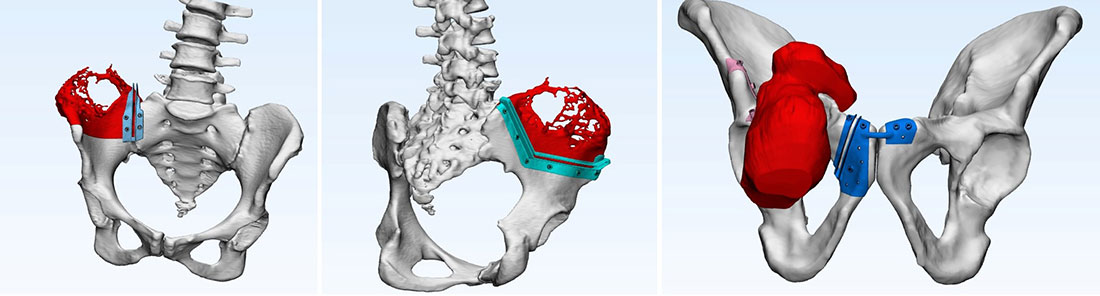
Figure 1. 3D planning and patient-specific cutting guides improve the accuracy of bone tumor removal during complex limb-salvage surgery, especially for pelvic tumors.
Limitations
PSIs are not suitable for every patient. They require manufacturing time, accurate positioning, and may need redesign if the tumor changes before surgery. Most importantly, they cannot replace careful surgical planning or clinical judgment. In some cases, surgeons use computer navigation, or combine both technologies for greater precision.
Table. Patient-Specific Instruments vs Computer Navigation
Clinical Pearl
Patient-specific instruments and computer navigation are complementary technologies that are often used together in complex bone tumor surgery.
Did You Know?
A patient-specific guide is designed to fit only one person’s bone, helping surgeons reproduce the surgical plan with remarkable accuracy.
Patient Tip
Ask your surgeon to show you your 3D digital bone model—it can help you better understand your surgery and reconstruction options.
Custom 3D-Printed Implants
After a bone tumor is removed, the resulting bone defect must often be reconstructed. The choice of reconstruction depends on the size and location of the defect, whether a joint has been preserved, and the patient’s age, activity level, and overall health. While standard modular implants work well for most patients, complex defects may require a custom 3D-printed implant designed specifically for the individual’s anatomy using CT and MRI scans. Instead of adapting the patient to the implant, the implant is tailored to fit the patient.
When Is a Custom Implant Considered?
Custom implants are reserved for complex cases where standard implants may not provide an ideal reconstruction, including:
- Large pelvic bone defects
- Joint-preserving resections
- Extensive bone loss
- Revision surgery
- Unusual anatomy
- Rare tumors involving bones such as the pelvis or scapula
For routine defects, standard implants remain the preferred option.
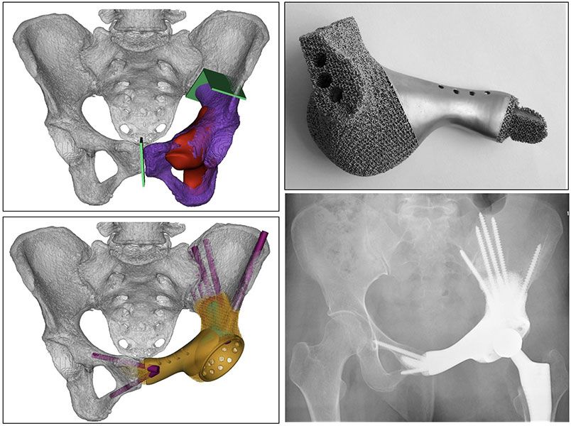
Figure 2. A customized 3D-printed implant provides personalized reconstruction of the pelvis after bone tumor surgery, helping restore anatomy and function.
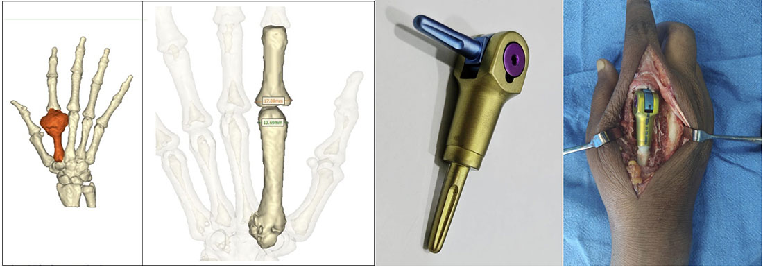
Figure 3. Customized 3D-printed implants allow precise reconstruction after bone tumor removal at rare and challenging anatomical sites.
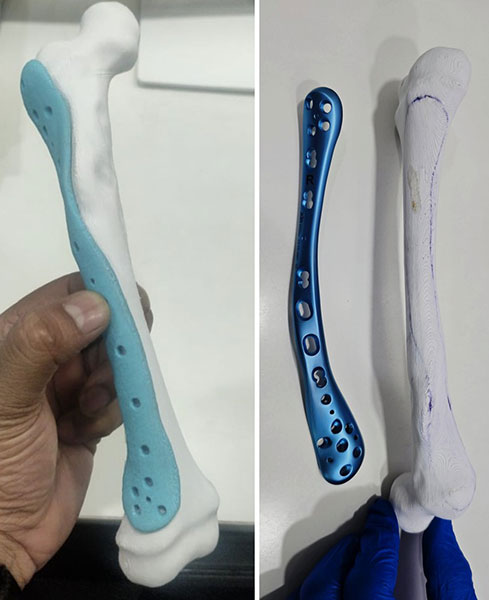
Figure 4. Patient-specific 3D planning and customized implants enable personalized reconstruction for complex bone tumors in children with deformed or fractured bones.
How Is a Custom Implant Designed?
The design process involves orthopaedic oncologists, biomedical engineers, imaging specialists, and implant manufacturers. CT and MRI scans are used to create a 3D digital model, allowing surgeons to define bone removal, implant shape, fixation points, screw placement, and joint reconstruction. The design undergoes multiple reviews before manufacturing.
What Materials Are Used?
Most custom implants are made from medical-grade titanium, which is strong, lightweight, corrosion-resistant, biocompatible, and compatible with medical imaging. Many implants also have porous surfaces that encourage bone to grow into the implant, improving long-term stability.
How Long Does Manufacturing Take?
Unlike standard implants, custom implants require several weeks for imaging review, digital planning, design approval, manufacturing, quality testing, and sterilization. Therefore, they are best suited for planned rather than emergency surgery.
How Is the Implant Inserted?
After the tumor is removed with safe surgical margins, the custom implant is positioned and fixed using specially designed screws or fixation devices. The surgeon then checks implant stability, alignment, limb length, and joint position before reconstructing the surrounding soft tissues.
How Long Do Custom Implants Last?
There is no fixed lifespan for any orthopaedic implant. Longevity depends on factors such as age, activity level, bone quality, implant design, tumor type, and complications like infection or loosening. Although many implants function well for years, some patients may eventually require revision surgery. Regular follow-up remains essential.
Table 5. Standard Modular Implants vs Custom 3D-Printed Implants
Doctor Explains
A custom implant is not automatically better than a standard implant. It is a personalized solution for selected complex cases where it is expected to improve reconstruction, while standard implants remain the best choice for many patients.
Advantages of 3D Printing and Navigation
The goal of these technologies is not to make surgery more technologically impressive—it is to improve patient care.
Although every patient is different, several potential advantages have been reported in appropriately selected cases.
Better Preoperative Planning
Perhaps the greatest benefit occurs before surgery.
Three-dimensional digital planning allows surgeons to understand complex anatomy in ways that are difficult with conventional two-dimensional imaging alone.
This preparation helps anticipate technical challenges and develop a detailed operative strategy.
Greater Surgical Precision
Navigation and patient-specific instruments help surgeons reproduce the planned operation more accurately.
This is particularly valuable when bone cuts must be performed close to joints, nerves, or blood vessels.
Improved Reconstruction
Custom implants may provide a closer anatomical match in complex reconstructions.
A better fit may facilitate implant fixation and simplify reconstruction after tumor removal.
Better Communication
Three-dimensional models can improve communication between:
- surgeons
- radiologists
- engineers
- physiotherapists
- patients
- families
Many patients find it easier to understand their condition when they can visualize a model of their own anatomy.
Educational Value
Digital models also allow surgeons to rehearse technically demanding procedures and educate residents and fellows before surgery.
Clinical Pearl
The greatest benefit of personalized technology is often improved surgical planning, rather than simply shortening the operation or making it more technologically advanced.
Limitations and Risks
Although advanced technologies have transformed many aspects of bone tumor surgery, they also have important limitations.
Understanding these limitations helps patients develop realistic expectations.
They Are Not Needed for Every Patient
Most bone tumor operations around the world are still successfully performed without custom implants or navigation.
These technologies should only be used when they provide a clear clinical benefit.
Additional Planning Time
Personalized surgery requires careful planning before the operation.
This preparation improves accuracy but also increases the time needed before surgery.
Higher Cost
Designing and manufacturing individualized implants is generally more expensive than using standard implants.
The exact cost varies depending on:
- implant complexity
- country
- healthcare system
- insurance coverage
Patients should discuss financial aspects with their treating hospital before surgery.
Limited Availability
Specialized software, engineering support, manufacturing facilities, and experienced surgical teams are required.
As a result, these technologies are primarily available in specialist musculoskeletal oncology centers.
Technology Cannot Eliminate Risk
Even with advanced planning, surgery still carries potential risks such as:
- infection
- bleeding
- nerve injury
- implant loosening
- fracture
- wound complications
- local recurrence
- need for additional surgery
Technology reduces uncertainty—it does not eliminate complications.
Long-Term Evidence Is Still Evolving
Although early and mid-term studies are encouraging, researchers continue to study the long-term durability of custom implants and their effect on functional outcomes.
As with many surgical innovations, evidence continues to evolve.
Red Flag Box
Be cautious if you are told that 3D printing or navigation guarantees a cure, completely eliminates recurrence, or makes surgery risk-free. No technology can replace meticulous surgical technique, appropriate tumor biology, and careful long-term follow-up.
Did You Know?
The success of bone tumor surgery depends far more on complete tumor removal with appropriate surgical margins than on the specific technology used during the operation.
Reassurance Box
It is completely reasonable to ask your surgeon why they recommend—or do not recommend—3D printing or navigation in your case. A personalized explanation will help you understand how these technologies fit into your overall treatment plan.
Patient Tip
Before surgery, ask your surgeon:
- Will I need a standard or custom implant?
- Why was this option chosen?
- Will navigation or patient-specific guides be used?
- How long is the expected recovery?
- When can I begin rehabilitation?
Frequently Asked Questions (FAQs)
1. What is 3D printing in bone tumor surgery?
3D printing is an advanced technology that creates patient-specific anatomical models, surgical guides, or custom titanium implants using CT and MRI scans. It helps surgeons plan complex bone tumor operations with greater precision and, in selected patients, improves reconstruction after tumor removal.
2. What is computer navigation in bone tumor surgery?
Computer navigation is a real-time guidance system that helps surgeons accurately reproduce their preoperative surgical plan. Similar to a GPS, it tracks surgical instruments relative to the patient’s anatomy, allowing more precise bone cuts while preserving healthy tissue whenever possible.
3. Does every patient with a bone tumor need a custom 3D-printed implant?
No. Most patients achieve excellent results with standard implants or conventional reconstruction techniques. Custom 3D-printed implants are reserved for selected complex cases, such as pelvic tumors, unusual bone anatomy, large bone defects, or revision surgery, where a personalized implant offers a clear advantage.
4. What are the advantages of using 3D printing and computer navigation together?
Using these technologies together allows surgeons to plan surgery in three dimensions, simulate the operation, create patient-specific guides or implants when required, and accurately execute the surgical plan. This combination may improve precision, reconstruction, and preservation of healthy bone in carefully selected patients.
5. Are 3D-printed implants safe?
Yes. Medical 3D-printed implants are manufactured from biocompatible materials, most commonly medical-grade titanium alloys. They undergo strict quality control, testing, and sterilization before being used in surgery and are designed to meet the same safety standards as conventional orthopaedic implants.
6. Which bone tumors are most likely to require 3D printing or navigation?
These technologies are most commonly considered for complex tumors involving the pelvis, sacrum, shoulder blade (scapula), spine, or tumors located close to major joints. They may also be useful for revision surgery and limb-salvage procedures requiring complex reconstruction.
7. Can 3D printing improve the success of bone tumor surgery?
Current research suggests that, in selected patients, 3D printing and navigation can improve surgical planning, increase the accuracy of bone resections, enhance implant fit, and assist complex reconstruction. However, successful outcomes still depend primarily on proper tumor removal and experienced surgical judgment.
8. How long does it take to make a custom 3D-printed implant?
The process usually takes several weeks. It includes detailed imaging, virtual surgical planning, implant design, surgeon approval, manufacturing, quality testing, and sterilization. Because of this, custom implants are generally used for planned surgeries rather than emergency procedures.
9. Are custom 3D-printed implants better than standard implants?
Not necessarily. Standard modular implants remain the best option for many patients and have an excellent long-term track record. Custom implants are designed for situations where standard implants cannot adequately reconstruct complex bone defects or unusual anatomy.
10. How long do custom 3D-printed implants last?
There is no fixed lifespan for any orthopaedic implant. Longevity depends on factors such as the patient’s age, activity level, bone quality, tumor type, implant design, and the occurrence of complications such as infection or loosening. Regular follow-up is essential regardless of the implant used.
11. Will using advanced technology reduce my chances of tumor recurrence?
The most important factor in preventing recurrence is complete tumor removal with adequate surgical margins. Technologies such as 3D printing and computer navigation help surgeons execute the planned operation more accurately but do not replace sound oncological surgical principles.
12. Should I choose a hospital that offers 3D printing and computer navigation for bone tumor surgery?
Advanced technology can be valuable for selected complex cases, but the experience of the orthopaedic oncologist and multidisciplinary bone tumor team remains the single most important factor in achieving good outcomes. These technologies are tools that support expert surgical decision-making—they do not replace it.
Medical Disclaimer
The information provided on this page is intended for educational purposes only and should not be considered a substitute for professional medical advice, diagnosis, or treatment. Every bone tumor is unique, and the decision to use technologies such as 3D printing, patient-specific instruments, computer navigation, or custom implants depends on the individual patient’s condition, imaging findings, tumor characteristics, and the treating surgeon’s clinical judgment. Treatment recommendations should always be made after evaluation by an experienced orthopaedic oncologist within a multidisciplinary bone tumor team. If you have been diagnosed with a bone tumor or are experiencing concerning symptoms, please consult your treating specialist for personalized medical advice.
References
- Bruschi A, Donati DM, Di Bella C. What to choose in bone tumour resections? Patient specific instrumentation versus surgical navigation: A systematic review. Journal of Bone Oncology. 2023;42:100503.
- Mounsef PJ, Blackman B, Sharma O, Aoude A, Bozzo A. Enhanced Functional and Surgical Outcomes With 3D Printing in Orthopedic Oncology: A Comparative Meta-Analysis Against Conventional Techniques. J Surg Oncol. 2025;132(6):1131-1142.
- Galoustian NA, et al. Intraoperative Navigation, 3D Printed Prostheses, and Conventional Techniques in Femoral and Tibial Tumor Resections: A Systematic Review. Journal of Orthopaedic Research. 2025.
- Aiba, H., Spazzoli, B., Tsukamoto, S., Mavrogenis, A. F., Hermann, T., Kimura, H., Murakami, H., Donati, D. M., & Errani, C. (2023). Current Concepts in the Resection of Bone Tumors Using a Patient-Specific Three-Dimensional Printed Cutting Guide. Current Oncology, 30(4), 3859-3870.
 +91 93558 33005