Quick Summary
Chondrosarcoma at a Glance
- Chondrosarcoma is a cancer that produces cartilage.
- It is one of the most common primary bone cancers in adults.
- It usually develops in the pelvis, thigh bone (femur), upper arm bone (humerus), shoulder blade (scapula), or ribs.
- Surgery is the main treatment for most patients.
- Conventional chondrosarcoma generally does not respond well to chemotherapy or radiotherapy.
- The tumor grade (how aggressive the cancer cells appear under the microscope) plays a major role in determining treatment and prognosis.
- Most patients can undergo limb salvage surgery, preserving the arm or leg while removing the tumor.
- Long-term follow-up is important because some tumors can recur after treatment.
Clinical Pearl
The first surgery offers the best opportunity for cure. A carefully planned operation performed by a specialist orthopaedic oncology team is one of the most important factors influencing long-term outcomes in chondrosarcoma.
Reassurance Box
Being told that you have a bone cancer can feel overwhelming. However, not all chondrosarcomas behave the same way. Many grow slowly, many can be treated successfully with surgery, and advances in limb salvage techniques now allow most patients to keep their affected limb while achieving excellent cancer control.
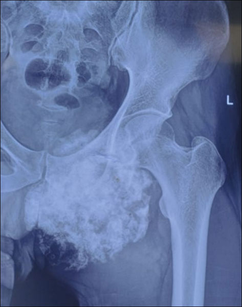
Figure 1. Chondrosarcoma of the pelvis: X-ray showing a large cartilage-forming bone tumor in the left pelvis, illustrating a typical site where chondrosarcoma can develop.
Introduction
Learning that you or someone you love may have chondrosarcoma raises many questions.
- Is it cancer?
- Can it be cured?
- Will surgery be enough?
- Will I lose my arm or leg?
- Why isn’t chemotherapy being recommended?
These are among the most common concerns patients bring to their first consultation.
One reason chondrosarcoma can be confusing is that it behaves very differently from many other cancers. While people often associate cancer treatment with chemotherapy or radiotherapy, most patients with conventional chondrosarcoma are treated primarily with surgery. This difference is not widely understood and is often explained only briefly on hospital websites.
This guide has been developed to provide clear, evidence-based answers in language that patients and families can understand. It explains how chondrosarcoma is diagnosed, why surgery is usually the most effective treatment, what recovery involves, and what to expect during long-term follow-up.
Throughout the article, medical terms are explained in plain language, common misconceptions are addressed, and practical advice is provided to help you make informed decisions about your care.
Doctor Explains
Primary bone cancer means that the cancer starts in the bone itself. This is different from metastatic bone disease, where a cancer that began elsewhere in the body (such as the breast, lung, or prostate) spreads to the bones.
Did You Know?
Although chondrosarcoma is one of the most common primary bone cancers in adults, it is still considered a rare cancer. Because of its rarity, treatment is best planned by a multidisciplinary sarcoma team experienced in managing bone tumors.
What Is Chondrosarcoma?
Chondrosarcoma is a malignant (cancerous) tumor that forms cartilage, a smooth, rubber-like tissue found throughout the body.
Cartilage normally serves important functions. It covers the ends of bones inside joints, allowing them to move smoothly, and forms part of structures such as the ribs, nose, and ear. In chondrosarcoma, cells that produce cartilage begin to grow uncontrollably and form a tumor.
Unlike many other cancers, chondrosarcoma usually develops inside the bone rather than in soft tissues. As the tumor enlarges, it gradually replaces normal bone, weakens its structure, and may extend into surrounding muscles or other nearby tissues.
Most chondrosarcomas occur in adults between 40 and 70 years of age, although certain uncommon subtypes can affect younger individuals.
Where Does Chondrosarcoma Usually Occur?
Chondrosarcoma can develop in almost any bone containing cartilage, but it most commonly affects:
- Pelvis (hip bones) – the most frequent site.
- Femur (thigh bone).
- Humerus (upper arm bone).
- Shoulder blade (scapula).
- Ribs.
- Less commonly, the spine, hands, and feet.
The location of the tumor influences:
- Symptoms.
- Surgical planning.
- Reconstruction options.
- Functional recovery.
- Risk of recurrence.
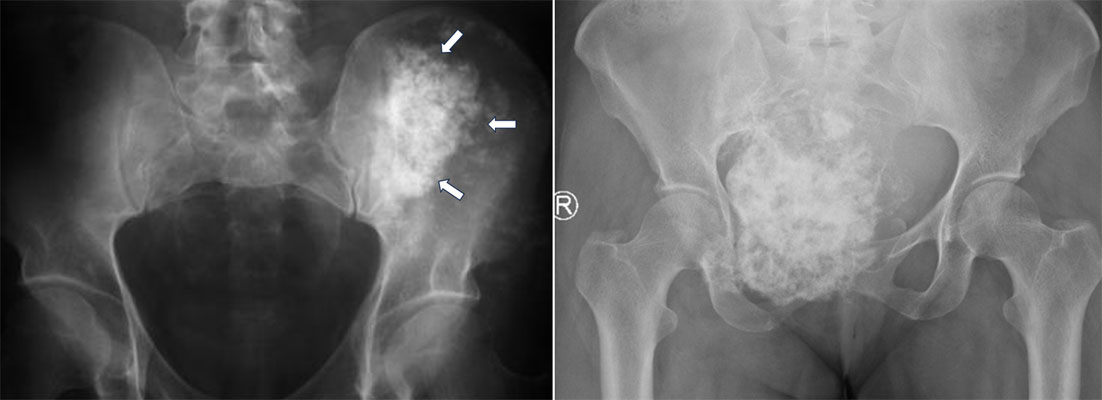
Figure 2. Chondrosarcoma of the pelvis: X-rays showing a large cartilage-forming bone tumor involving the pelvic bone.
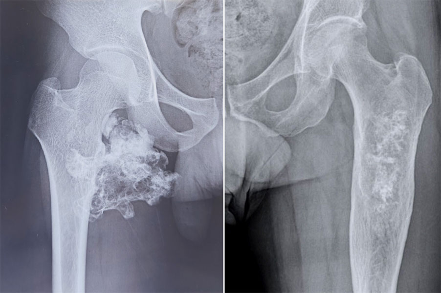
Figure 3. Chondrosarcoma of the thigh bone (femur): X-rays showing a cartilage-producing tumor involving the upper femur.
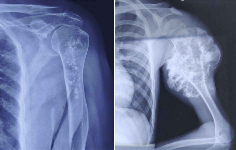
Figure 4. Chondrosarcoma of the upper arm bone (humerus): X-ray showing a cartilage-forming bone tumor involving the humerus.
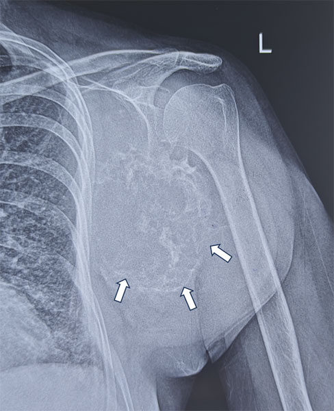
Figure 5. Chondrosarcoma of the shoulder blade (scapula): X-ray showing a destructive cartilage-forming tumor involving the scapula.
Who Is Most Likely to Develop Chondrosarcoma?
Most patients have no identifiable cause for developing chondrosarcoma.
However, certain factors are associated with an increased risk:
- Increasing age.
- Pre-existing benign cartilage tumors such as enchondromas (non-cancerous cartilage tumors inside the bone).
- Rare inherited conditions such as Ollier disease and Maffucci syndrome, in which multiple cartilage tumors develop.
- Previous radiation exposure (uncommon).
Importantly, most people with chondrosarcoma have no family history of bone cancer, and nothing they did caused the disease.
Patient Tip
Many patients worry that they may have done something to cause the tumor. In most cases, chondrosarcoma develops without any identifiable lifestyle or environmental cause, and there is nothing you could have done to prevent it.
Is Chondrosarcoma Always Aggressive?
No.
One of the defining features of chondrosarcoma is that its behaviour varies considerably.
Some tumors grow slowly over many years and have a relatively low risk of spreading, while others are much more aggressive and require more extensive treatment.
Doctors estimate how aggressive a tumor is by assessing its grade—a measure of how abnormal the cancer cells appear under the microscope. Tumor grading will be explained later in this guide because it plays a central role in treatment planning and prognosis.
Doctor Insight
Not all chondrosarcomas should be viewed in the same way. Two patients may both be diagnosed with chondrosarcoma but require very different treatments because their tumors differ in grade, location, size, and biological behaviour. This is why individualized treatment planning is essential.
Why Early Specialist Assessment Matters
Many patients first present with persistent bone pain or a swelling that is mistaken for arthritis, a sports injury, or a benign bone lesion. Because chondrosarcoma often grows slowly, symptoms may develop gradually and can be overlooked for months.
Although most patients are not diagnosed in an emergency, delaying specialist evaluation may allow the tumor to enlarge, making surgery more complex and potentially reducing the options for limb preservation.
If imaging suggests a cartilage-producing bone tumor, referral to an orthopaedic oncologist before biopsy or surgery is strongly recommended. Careful planning from the beginning helps ensure that the biopsy, diagnosis, and definitive treatment follow established oncological principles.
Why Chondrosarcoma Is Different from Other Bone Cancers
One of the most important things to understand about chondrosarcoma is that it is not treated like most other bone cancers.
Many patients have heard of osteosarcoma or Ewing sarcoma, where chemotherapy is usually an essential part of treatment. It is therefore natural to wonder why chemotherapy is not being recommended for them.
The answer lies in the unique biology of chondrosarcoma.
Unlike many cancers, conventional chondrosarcoma is relatively resistant to both chemotherapy and radiotherapy. This means that medicines and radiation, which are highly effective for some cancers, usually do not destroy conventional chondrosarcoma cells effectively.
As a result, complete surgical removal of the tumor with an adequate margin of healthy tissue is the cornerstone of treatment.
Understanding this principle helps explain why your orthopaedic oncologist places so much emphasis on careful surgical planning.
Quick Summary
Conventional chondrosarcoma is different because:
- It usually grows from cartilage-producing cells.
- It generally responds poorly to chemotherapy.
- Radiotherapy has a limited role in most patients.
- Surgery offers the best chance of cure.
- The quality of the first surgery has a major influence on long-term outcomes.
Doctor Explains
Chemotherapy is treatment using medicines that destroy rapidly dividing cancer cells.
Radiotherapy uses carefully targeted high-energy radiation to kill cancer cells.
Although these treatments are highly effective for many cancers, conventional chondrosarcoma cells are naturally less sensitive to them. Consequently, surgery remains the primary treatment for most patients.
Why Is Surgery the Main Treatment?
The goal of surgery is straightforward:
Remove the entire tumor in one piece together with a surrounding rim of normal tissue.
This surrounding rim is called a surgical margin.
A surgical margin is the layer of healthy tissue removed along with the tumor to reduce the chance that microscopic cancer cells remain behind.
Removing the tumor completely provides the greatest chance of long-term disease control and minimizes the risk of the cancer returning in the same location.
Unlike some other cancers, there is usually no effective medical treatment that can compensate for incomplete surgery in conventional chondrosarcoma.
This is why surgical planning is so meticulous.
What Makes Chondrosarcoma Surgery Different?
Before surgery, the multidisciplinary team carefully reviews:
- Your symptoms.
- X-rays.
- MRI scans.
- CT scans, when needed.
- Biopsy findings.
- Tumor grade.
- Tumor location.
- Relationship to nearby nerves, blood vessels, and joints.
Only after all this information has been integrated is the surgical plan finalized.
Every operation is individualized because no two tumors are exactly alike.
Clinical Pearl
For conventional chondrosarcoma, the first operation provides the greatest opportunity for complete tumor removal. Revision surgery after an inadequate initial procedure is often more complex and may reduce the options for limb preservation.
Why Doesn’t Chemotherapy Usually Work?
This is one of the questions patients ask most frequently.
To answer it, it helps to understand how chemotherapy works.
Most chemotherapy drugs are designed to target rapidly dividing cells.
Conventional chondrosarcoma cells generally divide more slowly than the cells found in osteosarcoma or Ewing sarcoma. In addition, the cartilage-rich environment surrounding these tumors limits the penetration of many chemotherapy drugs.
As a result, chemotherapy has not consistently demonstrated meaningful benefit for most patients with conventional chondrosarcoma.
For this reason, international treatment guidelines do not recommend routine chemotherapy for conventional chondrosarcoma after complete surgical removal.
Are There Exceptions?
Yes.
Not all chondrosarcomas behave in the same way.
Some uncommon subtypes, such as:
- Mesenchymal chondrosarcoma
- Dedifferentiated chondrosarcoma
may respond differently and can require chemotherapy as part of treatment.
These uncommon tumors will be discussed later in this guide.
Did You Know?
The recommendation not to use chemotherapy for conventional chondrosarcoma is not because treatment is being withheld. It is because decades of research have shown that surgery is usually far more effective for this particular disease.
Does Radiotherapy Have a Role?
For most patients with conventional chondrosarcoma, radiotherapy is not the primary treatment.
However, there are important situations in which radiotherapy may be considered.
These include:
- Tumors that cannot be safely removed completely because of their location.
- Certain tumors involving the skull base or spine, where complete surgery may not be possible.
- Selected patients with residual disease after surgery.
- Rare situations where surgery is not feasible because of other medical conditions.
Modern techniques, such as proton beam therapy and high-precision radiotherapy, continue to be studied and may benefit carefully selected patients treated at specialist centres.
Your treatment team will discuss whether radiotherapy has a role in your individual situation.
Doctor Insight
Surgery and radiotherapy should not be viewed as competing treatments. Rather, radiotherapy is used selectively when surgery alone cannot achieve the desired oncological outcome or when anatomical constraints limit complete tumor removal.
Why the First Surgery Matters So Much
In musculoskeletal oncology, there is a saying:
“You only get one chance to do the first operation properly.”
This is particularly true for chondrosarcoma.
If the initial operation removes the tumor completely with appropriate margins, the likelihood of long-term local control is significantly improved.
Conversely, if part of the tumor is left behind or the tumor is removed without proper planning, microscopic cancer cells may remain in the surrounding tissues. This can increase the risk of local recurrence, meaning the tumor grows back in the same area.
Recurrent tumors are often more difficult to treat because:
- Previous surgery alters the normal anatomy.
- Scar tissue develops around important structures.
- Wider resections may be required.
- Reconstruction becomes more complex.
- Limb salvage may become more challenging.
For these reasons, suspected chondrosarcoma should ideally be managed at a specialist sarcoma centre from the beginning.
Patient Tip
If you have been told that you may have chondrosarcoma, avoid undergoing surgery before a definitive diagnosis has been established. Asking whether your case has been reviewed by a multidisciplinary sarcoma team is entirely appropriate and can help ensure that treatment is planned correctly from the outset.
Types of Chondrosarcoma
Although all chondrosarcomas produce cartilage, they are not all the same disease.
Under the microscope, some tumors grow slowly and behave in a relatively indolent (less aggressive) manner, while others grow rapidly and have a higher likelihood of spreading. Identifying the exact subtype is therefore an essential part of diagnosis because it influences treatment, prognosis, follow-up, and sometimes the need for additional therapies.
The subtype is determined by combining information from:
- Your symptoms and clinical examination.
- Imaging studies such as MRI and CT scans.
- Findings from the biopsy. (For a detailed explanation of how biopsy is performed and why it is carefully planned, see Bone Biopsy.)
- Microscopic examination by a specialist musculoskeletal pathologist.
Fortunately, most patients have conventional chondrosarcoma, which behaves differently from the rarer subtypes discussed below.
There are five major types of chondrosarcoma:
- Conventional chondrosarcoma (most common)
- Clear cell chondrosarcoma
- Mesenchymal chondrosarcoma
- Dedifferentiated chondrosarcoma
- Secondary chondrosarcoma
Each subtype has distinct biological behaviour and requires an individualized treatment approach.
Conventional Chondrosarcoma
Conventional chondrosarcoma accounts for approximately 85–90% of all chondrosarcomas, making it by far the most common subtype.
It develops from cartilage-producing cells inside the bone and usually affects adults between 40 and 70 years of age.
These tumors most frequently arise in:
- Pelvis
- Femur (thigh bone)
- Humerus (upper arm bone)
- Ribs
- Scapula (shoulder blade)
Conventional chondrosarcomas may grow slowly or more aggressively depending on their tumor grade.
Importantly, most conventional chondrosarcomas are treated primarily with surgery, as discussed earlier. Chemotherapy is generally ineffective for this subtype.
Clinical Pearl
When patients hear the term “chondrosarcoma,” it almost always refers to conventional chondrosarcoma unless another subtype has been specifically identified.
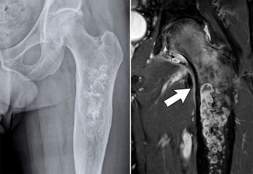
Figure 6. Conventional chondrosarcoma of the proximal femur: X-rays showing a cartilage-forming tumor with characteristic mineralization within the bone.
Clear Cell Chondrosarcoma
Clear cell chondrosarcoma is an uncommon subtype that usually grows slowly.
It often develops near the ends of long bones, particularly around the:
Because it grows gradually, symptoms may be present for months or even years before the diagnosis is made.
Despite its relatively indolent behaviour, clear cell chondrosarcoma is still malignant (cancerous) and requires complete surgical removal. Inadequate treatment may allow the tumor to recur locally, sometimes many years later.
Long-term follow-up is therefore important even after successful surgery.
Did You Know?
💡 Some patients with clear cell chondrosarcoma report pain for several years before the diagnosis is established because the tumor often enlarges slowly.
Mesenchymal Chondrosarcoma
Mesenchymal chondrosarcoma is a rare but high-grade subtype.
A high-grade tumor contains cancer cells that appear very abnormal under the microscope and usually behave more aggressively than low-grade tumors.
Unlike conventional chondrosarcoma, mesenchymal chondrosarcoma often affects younger adults and may occasionally occur in children.
It can arise in:
- Bone
- Soft tissues outside the skeleton
Because this subtype behaves differently, treatment often includes:
- Surgery
- Chemotherapy
- Occasionally radiotherapy
This is one of the important exceptions to the general principle that chemotherapy has little role in chondrosarcoma.
Doctor Explains
If your doctor recommends chemotherapy for mesenchymal chondrosarcoma, this does not contradict the statement that chemotherapy is ineffective for most conventional chondrosarcomas. Different subtypes have different biological characteristics, and treatment is tailored accordingly.
Dedifferentiated Chondrosarcoma
Dedifferentiated chondrosarcoma is another uncommon but highly aggressive subtype.
The word dedifferentiated means that part of the tumor has changed into a much more aggressive form of cancer.
Under the microscope, two distinct components are seen:
- A conventional cartilage-producing tumor.
- A second high-grade cancer growing alongside it.
Patients with dedifferentiated chondrosarcoma usually require more intensive treatment than those with conventional disease.
Management often involves:
- Extensive surgery.
- Consideration of chemotherapy.
- Careful staging to determine whether the disease has spread.
- Close postoperative surveillance.
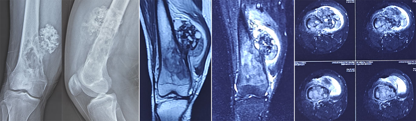
Figure 7. Dedifferentiated chondrosarcoma: X-rays and MRI showing an aggressive cartilage tumor with areas of bone destruction and soft-tissue extension.
Doctor Insight
Dedifferentiated chondrosarcoma highlights why obtaining an accurate biopsy is so important. The treatment plan depends not only on identifying chondrosarcoma but also on recognising its specific subtype. This requires close collaboration between the orthopaedic oncologist, radiologist, and pathologist.
Secondary Chondrosarcoma
Most chondrosarcomas develop de novo, meaning they arise as a new tumor without a pre-existing lesion.
However, some develop from an existing benign cartilage tumor.
These are called secondary chondrosarcomas.
The most common conditions associated with secondary chondrosarcoma are:
- Enchondroma – a benign cartilage tumor inside the bone.
- Osteochondroma – a benign bony growth covered by cartilage.
Rare inherited disorders such as:
- Ollier disease
- Maffucci syndrome
also increase the lifetime risk of developing secondary chondrosarcoma.
Fortunately, most enchondromas and osteochondromas never become cancerous.
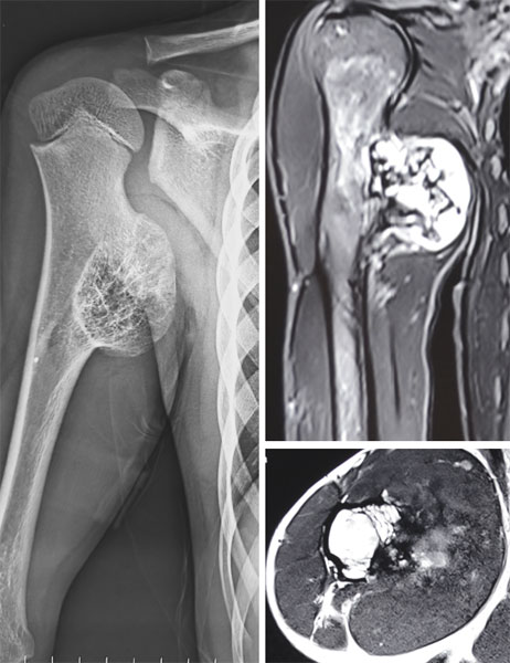
Figure 8A. Secondary chondrosarcoma arising in an osteochondroma: Imaging showing malignant transformation of a pre-existing osteochondroma into chondrosarcoma.
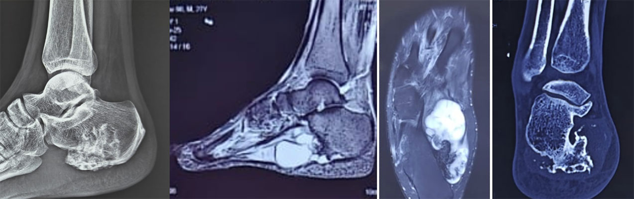
Figure 8B. Secondary chondrosarcoma arising from osteochondroma: Imaging demonstrating the cartilage tumor developing from a pre-existing benign bone growth.
Reassurance Box
Many patients become anxious after learning that chondrosarcoma can arise from a benign cartilage tumor. It is important to remember that the vast majority of enchondromas and osteochondromas remain benign throughout life. Your doctor will recommend treatment only if there are clinical or imaging features suggesting malignant transformation.
Symptoms of Chondrosarcoma
The symptoms of chondrosarcoma often develop gradually.
Unlike many other cancers, patients rarely become seriously unwell in the early stages. Instead, the tumor usually causes symptoms by slowly enlarging and weakening the affected bone.
Because these symptoms are common to many non-cancerous conditions, diagnosis is sometimes delayed.
The most frequent symptom is persistent pain, particularly pain that:
- Gradually becomes worse over time.
- Is unrelated to a recent injury.
- Persists despite rest or pain medication.
- Begins to interfere with daily activities or sleep.
Some patients notice a swelling or lump near the affected bone, while others first present after a pathological fracture—a fracture that occurs because the bone has been weakened by disease rather than a significant injury. (To understand how doctors assess the risk of fractures in weakened bones, see Assessment of Risk of Pathological Fracture.)
Tumors arising in the pelvis may cause deep buttock or groin pain, whereas tumors near joints can reduce movement or cause stiffness.
When chondrosarcoma develops close to major nerves, patients may occasionally experience:
- Numbness.
- Tingling.
- Weakness in the affected limb.
These symptoms are uncommon but should be assessed promptly.
Patient Tip
Keep a record of when your pain began, what makes it worse, and whether it limits your daily activities. This information helps your doctor understand how the symptoms have evolved and guides decisions about imaging and further investigations.
Clinical Pearl
Persistent, progressive bone pain deserves investigation. While most causes are not cancer, symptoms that continue to worsen without an obvious explanation should not be dismissed, particularly in adults with a cartilage-producing bone lesion.
How Is Chondrosarcoma Diagnosed?
Receiving a diagnosis of chondrosarcoma involves much more than a single test.
Unlike many common illnesses, chondrosarcoma cannot usually be diagnosed from an X-ray, MRI, or biopsy alone. Instead, doctors combine information from your symptoms, physical examination, imaging studies, and biopsy findings to arrive at the most accurate diagnosis.
This process is known as clinicoradiological-pathological correlation, meaning that the clinical features, radiology (imaging), and pathology (microscopic examination of tissue) must all agree before treatment is planned.
This careful approach helps avoid misdiagnosis and ensures that the most appropriate treatment is recommended.
Diagnosing chondrosarcoma usually involves:
- Clinical examination
- X-rays
- MRI scan
- CT scan (when required)
- Biopsy
- Histopathological examination
- Multidisciplinary team review
Each step provides unique information, and no single investigation is sufficient on its own.
Clinical Evaluation
Diagnosis begins with a detailed medical history and physical examination.
Your doctor will ask about:
- When the pain started.
- Whether the pain is becoming worse.
- Night pain or pain at rest.
- Swelling or a visible lump.
- Previous injuries.
- Previous benign cartilage tumors.
- Any family history of bone tumors.
During the examination, the doctor will assess:
- The location of tenderness.
- Size of any swelling.
- Joint movement.
- Muscle strength.
- Function of nearby nerves.
- Blood supply to the affected limb.
Although the examination alone cannot diagnose chondrosarcoma, it provides valuable clues that help determine which investigations are needed.
Doctor Explains
A clinical examination means more than simply checking where the pain is located. Your doctor is also assessing how the tumor affects nearby muscles, joints, nerves, and blood vessels. This information is important for planning both investigations and surgery.
Plain X-rays: The First Imaging Test
For most patients, the first investigation is a plain X-ray.
Although additional scans are almost always required, X-rays often provide the first indication that a cartilage-producing tumor is present.
Typical features that may suggest chondrosarcoma include:
- Areas where normal bone has been replaced by tumor.
- Characteristic calcification, meaning deposits of calcium within the cartilage matrix that create distinctive patterns on the X-ray.
- Expansion of the bone.
- Thinning or destruction of the outer layer of bone (cortex).
- Extension into surrounding soft tissues in more advanced tumors.
Certain calcification patterns are highly suggestive of a cartilage-producing tumor, but they cannot reliably distinguish a benign enchondroma from chondrosarcoma. Additional imaging and biopsy are usually required.
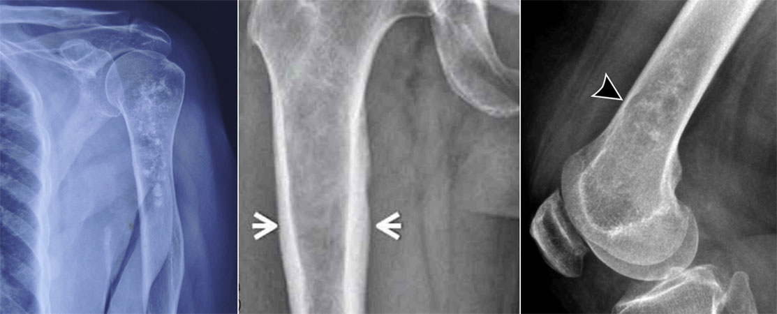
Figure 9. X-ray features of chondrosarcoma: X-rays showing typical bone destruction and abnormal cartilage mineralization associated with chondrosarcoma.
Clinical Pearl
Every suspected bone tumor should have high-quality X-rays before advanced imaging is performed. X-rays often provide important diagnostic information that complements MRI rather than being replaced by it.
MRI: The Most Important Imaging Test
Once a bone tumor is suspected, the next investigation is usually an MRI (Magnetic Resonance Imaging) scan.
MRI uses a powerful magnetic field and radio waves—not radiation—to produce highly detailed images of bones, muscles, cartilage, nerves, and blood vessels.
For chondrosarcoma, MRI is the single most important imaging study because it shows:
- The exact size of the tumor.
- Whether it extends beyond the bone.
- Involvement of nearby muscles.
- Relationship to nerves and blood vessels.
- Whether nearby joints are affected.
- The safest pathway for performing a biopsy.
This information is essential for planning limb salvage surgery and determining whether complete removal of the tumor is feasible.
For this reason, MRI should almost always be performed before the biopsy. A biopsy can cause bleeding and inflammation around the tumor, making MRI interpretation more difficult. (Learn more in our detailed guide on Bone Biopsy.)
Doctor Insight
MRI is not performed simply to “confirm” the diagnosis. Its primary role is to define the extent of the tumor and guide surgical planning. A well-performed MRI often determines whether limb salvage surgery is possible.
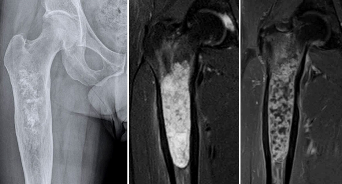
Figure 10. MRI of chondrosarcoma of the femur:MRI demonstrates the extent of the tumor within the femur and its relationship with surrounding soft tissues.
CT Scan: Looking More Closely at Bone
A CT (Computed Tomography) scan uses X-rays to create detailed cross-sectional images of the body.
CT is particularly useful for evaluating:
- Mineralization within the tumor.
- Cortical destruction.
- Complex anatomy of the pelvis, ribs, or spine.
- Surgical planning for pelvic tumors.
Compared with MRI, CT provides a clearer picture of bone architecture and calcification patterns.
CT scans are also commonly performed to examine the lungs, as the lungs are the most frequent site where high-grade chondrosarcoma can spread.
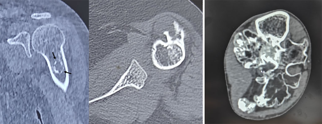
Figure 11A. CT scan for evaluation of chondrosarcoma: CT imaging provides detailed information about bone destruction, tumor mineralization and the extent of the bone tumor.
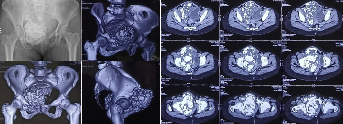
Figure 11B. CT scan of pelvic chondrosarcoma: CT shows cortical bone destruction and characteristic calcification within the cartilage-forming tumor.
Did You Know?
💡 MRI is usually better for evaluating soft tissues, whereas CT provides superior detail of bone. The two investigations complement each other rather than competing.
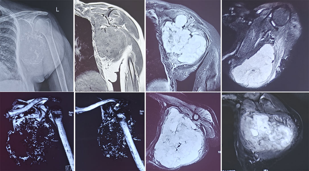
Figure 12. Imaging of chondrosarcoma of the scapula: X-ray, CT and MRI demonstrate the bone tumor and its extent within the shoulder region.
Is PET-CT Needed?
PET-CT (Positron Emission Tomography–Computed Tomography) combines metabolic imaging with CT scanning.
It is not routinely required for every patient with chondrosarcoma.
Your doctor may recommend PET-CT in selected situations, such as:
- Uncertain diagnosis.
- Suspected recurrent disease.
- Assessment of aggressive subtypes.
- Evaluation for metastatic disease when indicated.
Most patients with conventional chondrosarcoma can be accurately assessed using X-rays, MRI, CT, and biopsy.
Why Is a Biopsy Necessary?
Although imaging studies can strongly suggest chondrosarcoma, they cannot reliably determine the exact subtype or tumor grade.
A biopsy involves removing a small sample of tissue so that it can be examined under the microscope by a pathologist.
The biopsy helps answer several critical questions:
- Is the tumor benign or malignant?
- What subtype of chondrosarcoma is present?
- What is the tumor grade?
- Are additional molecular tests required?
Whenever possible, the biopsy should be performed by—or in consultation with—the same specialist team that will carry out the definitive surgery. This allows the biopsy pathway to be planned so it can be removed during surgery, reducing the risk of local recurrence. (A detailed discussion of biopsy planning, techniques, and recovery is available in Bone Biopsy.)
🚨 Red Flag Box: Never Proceed Directly to Surgery Without a Diagnosis
If imaging suggests a bone tumor:
- Do not undergo curettage (“scraping out” the lesion) without a confirmed diagnosis.
- Avoid excision of a suspected bone tumor before appropriate imaging and biopsy.
- Ensure that the biopsy is carefully planned, ideally by the team responsible for definitive treatment.
An unplanned procedure can complicate future surgery and may reduce options for limb preservation.
Histopathology: Confirming the Diagnosis
After the biopsy, the tissue is examined by a musculoskeletal pathologist—a doctor who specializes in diagnosing bone and soft tissue tumors.
The pathologist evaluates:
- The appearance of the tumor cells.
- The amount and type of cartilage produced.
- Cellular atypia (how abnormal the cells appear).
- Mitotic activity (how actively the cells are dividing).
- Features that determine the tumor grade.
Additional investigations, such as immunohistochemistry (using antibodies to identify specific proteins) or molecular testing (looking for characteristic genetic changes), may be required in selected cases.
The pathology findings are then interpreted alongside the imaging studies before a final diagnosis is made.
Enchondroma vs Atypical Cartilaginous Tumor (ACT) vs Chondrosarcoma: Why the Difference Matters
One of the greatest challenges in musculoskeletal oncology is distinguishing between a benign enchondroma, an atypical cartilaginous tumor (ACT), and a low-grade chondrosarcoma.
These three conditions all arise from cartilage-producing cells and often look similar on imaging and even under the microscope. Yet, their treatment and long-term outlook can be very different.
For patients, this can be confusing. It is not uncommon to receive one opinion suggesting observation while another recommends surgery. Understanding why this happens can help you have a more informed discussion with your healthcare team.
Although they share many similarities:
- Enchondroma is a benign (non-cancerous) cartilage tumor.
- Atypical Cartilaginous Tumor (ACT) is a locally aggressive cartilage tumor with a very low risk of spreading.
- Chondrosarcoma is a malignant (cancerous) tumor that can invade surrounding tissues and, depending on its grade, may spread to other parts of the body.
Making the correct diagnosis requires careful interpretation of clinical findings, imaging, pathology, and sometimes follow-up over time.
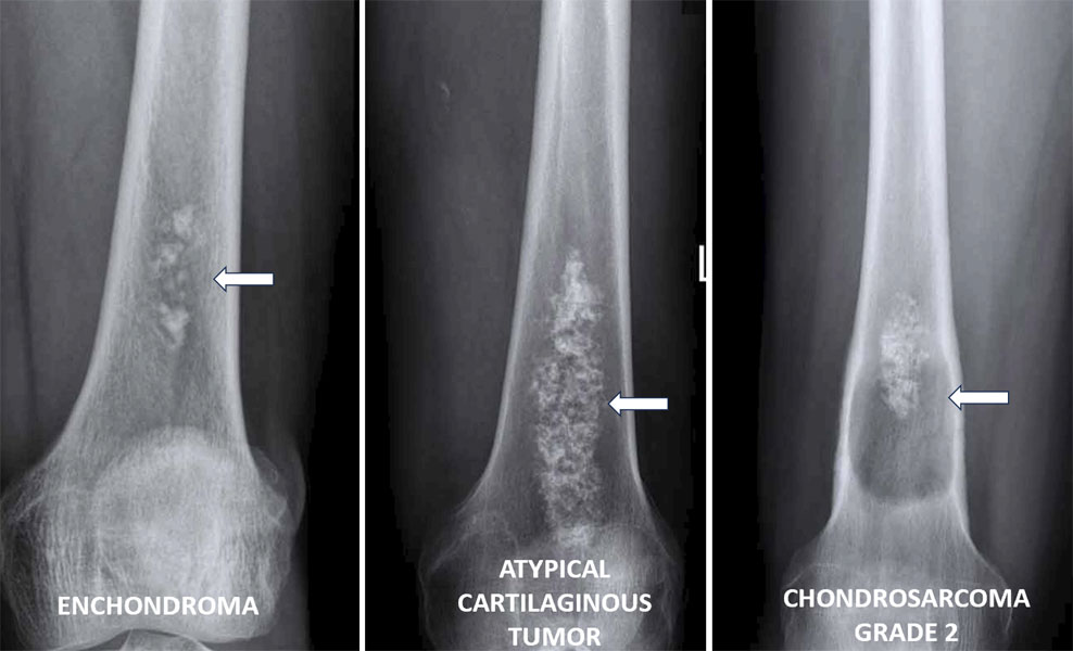
Figure 13. Enchondroma vs atypical cartilaginous tumor vs chondrosarcoma: X-rays illustrate the spectrum of cartilage tumors and the features that help distinguish benign from malignant lesions.
Doctor Explains
A benign tumor grows locally and does not spread to distant organs.
A malignant tumor has the ability to invade surrounding tissues and, in some cases, spread to other parts of the body (metastasize).
What Is an Enchondroma?
An enchondroma is a benign cartilage tumor that develops inside the bone.
It is one of the most common benign bone tumors and is frequently discovered incidentally, meaning it is found during imaging performed for an unrelated reason, such as evaluating a sports injury or arthritis.
Most enchondromas:
- Cause no symptoms.
- Remain stable throughout life.
- Never become cancerous.
- Do not require surgery.
Instead, many patients only need periodic clinical review and imaging to ensure the lesion remains unchanged.
Where Do Enchondromas Commonly Occur?
Enchondromas most often arise in:
- Small bones of the hand.
- Humerus (upper arm).
- Femur (thigh bone).
- Tibia (shin bone).
They can occur at almost any age but are commonly diagnosed in young and middle-aged adults.
Reassurance Box
If you have been diagnosed with an enchondroma, remember that most enchondromas never transform into cancer. Surgery is not routinely required unless the lesion becomes symptomatic, weakens the bone significantly, or shows features suggestive of malignant change.
What Is an Atypical Cartilaginous Tumor (ACT)?
An Atypical Cartilaginous Tumor (ACT) occupies the middle ground between a benign enchondroma and a conventional low-grade chondrosarcoma.
ACT is a locally aggressive cartilage-producing tumor. This means it can slowly enlarge and damage the surrounding bone, but it has an extremely low risk of spreading (metastasizing) when it occurs in the limbs.
The term ACT is now preferred by the World Health Organization (WHO) for tumors that were previously called Grade 1 chondrosarcoma in the long bones of the arms and legs.
This change in terminology reflects their biological behaviour—they behave much less aggressively than higher-grade chondrosarcomas.
However, the same microscopic appearance in the pelvis, scapula, spine, or axial skeleton is still classified as Grade 1 chondrosarcoma because tumors in these locations tend to behave more aggressively.
This distinction is important and often misunderstood.
Did You Know?
💡 The same tumor may be called ACT in the femur but Grade 1 chondrosarcoma in the pelvis because the expected biological behaviour differs according to its location.
What Is Low-Grade Chondrosarcoma?
A low-grade chondrosarcoma is a malignant cartilage-producing tumor that grows slowly but has the potential to:
- Invade surrounding bone.
- Extend into soft tissues.
- Recur after treatment.
- Rarely spread to distant organs.
Although low-grade tumors are less aggressive than intermediate- or high-grade chondrosarcomas, they still require appropriate oncological management.
Treatment depends on several factors, including:
- Tumor location.
- Size.
- Symptoms.
- Imaging characteristics.
- Multidisciplinary team assessment.
Why Is It Sometimes Difficult to Tell Them Apart?
This is one of the most challenging areas in bone tumor diagnosis—even for experienced specialists.
No single investigation can reliably distinguish all enchondromas from low-grade chondrosarcomas.
Instead, doctors look for a combination of features.
Clinical Clues
Patients with chondrosarcoma are more likely to have:
- Persistent pain unrelated to activity.
- Progressive worsening of symptoms.
- Pain at night.
- Increasing swelling.
An asymptomatic lesion discovered incidentally is more likely to represent an enchondroma, although exceptions occur.
Imaging Clues
Radiologists carefully assess several imaging features.
Features that may suggest chondrosarcoma include:
- Endosteal scalloping.
- Cortical destruction.
- Soft tissue extension.
- Bone expansion.
- Progressive increase in size on serial imaging.
Endosteal scalloping refers to erosion of the inner surface of the cortex (outer layer of bone) by the growing cartilage tumor.
Mild scalloping may occur in benign lesions, but deep endosteal scalloping affecting more than two-thirds of the cortical thickness raises concern for malignancy.
MRI also helps determine whether the tumor extends beyond the bone into surrounding muscles or other soft tissues.
Pathology Clues
Under the microscope, the differences may be subtle.
The pathologist evaluates:
- Cellularity (number of tumor cells).
- Nuclear atypia (how abnormal the nuclei appear).
- Bone permeation.
- Pattern of cartilage formation.
Even experienced musculoskeletal pathologists sometimes find the distinction difficult when evaluating tissue alone.
This is why pathology findings must always be interpreted alongside imaging.
Why the Multidisciplinary Approach Is Essential
Because there is no single “gold standard” test, diagnosis often depends on discussion within a multidisciplinary sarcoma team (MDT).
For example:
- The radiologist may identify imaging features suggesting an aggressive lesion.
- The pathologist may report findings compatible with a low-grade cartilage tumor.
- The orthopaedic oncologist considers the patient’s symptoms and examination findings.
Only after combining all of this information is the final diagnosis established.
This integrated approach significantly improves diagnostic accuracy and helps avoid both overtreatment and undertreatment.
How Does Treatment Differ?
The management of these lesions reflects their biological behaviour.
Enchondroma vs ACT vs Low-Grade Chondrosarcoma
Treatment decisions are individualized and depend on tumor location, symptoms, imaging findings, and multidisciplinary assessment.
Doctor Insight
One of the greatest advances in musculoskeletal oncology has been recognizing that not every cartilage lesion requires immediate surgery. Equally important is identifying those lesions that should not simply be observed. The challenge lies in selecting the right treatment for the right patient.
🚨 Red Flag Box: Features That Warrant Specialist Evaluation
Seek assessment by an orthopaedic oncologist if a cartilage lesion is associated with:
- Persistent or worsening pain.
- Progressive enlargement on serial imaging.
- Deep endosteal scalloping.
- Cortical destruction.
- Soft tissue extension.
- A pathological fracture.
- Uncertainty regarding the diagnosis.
These features do not automatically mean cancer, but they require careful evaluation by a specialist sarcoma team.
Why a Second Opinion Can Be Helpful
Because differentiating these cartilage tumors is sometimes challenging, obtaining a second opinion at a specialist bone tumor centre can be valuable, particularly when:
- Surgery is being considered.
- Imaging and biopsy findings do not agree.
- The diagnosis remains uncertain.
- The tumor is located in the pelvis, spine, or shoulder girdle.
A second opinion often involves review of both the imaging and the biopsy slides by specialists who manage cartilage tumors regularly.
Understanding Tumor Grade: Why It Matters More Than Tumor Size
After confirming that a patient has chondrosarcoma, one of the next questions is:
“How aggressive is my tumor?”
The answer depends largely on the tumor grade.
Tumor grade describes how abnormal the cancer cells appear under the microscope and how aggressively they are expected to behave.
It is important not to confuse tumor grade with tumor stage.
- Grade describes the biological behaviour of the tumor.
- Stage describes how far the cancer has spread within the body.
Two patients may have tumors of similar size but completely different grades—and therefore require different treatments and have different prognoses.
Quick Summary
Tumor grade helps doctors estimate:
- How quickly the tumor is likely to grow
- The risk of local recurrence
- The chance of spreading to other organs
- The type of surgery required
- The intensity of long-term follow-up
Doctor Explains
Think of tumor grade as the tumor’s personality, not its size.
A small high-grade tumor may behave more aggressively than a much larger low-grade tumor.
How Is Tumor Grade Determined?
Tumor grade is determined by a musculoskeletal pathologist after examining tissue obtained during the biopsy or, in some cases, after the entire tumor has been removed.
The pathologist assesses several microscopic features, including:
- Cellularity (how densely packed the tumor cells are).
- Nuclear atypia (how abnormal the cell nuclei appear).
- Mitotic activity (how frequently the cells are dividing).
- Pattern of bone infiltration.
- Areas of tumor necrosis (dead tumor tissue).
No single feature determines the grade. Instead, all findings are interpreted together, along with the imaging studies.
The Three Main Grades of Conventional Chondrosarcoma
Grade 1 (Low Grade)
Grade 1 tumors resemble normal cartilage more closely than higher-grade tumors.
They usually:
- Grow slowly.
- Rarely spread to distant organs.
- Have an excellent prognosis after appropriate surgery.
- Are now often referred to as Atypical Cartilaginous Tumors (ACT) when located in the long bones of the arms and legs.
Although low-grade tumors are the least aggressive form of chondrosarcoma, they still require specialist evaluation because treatment depends on their location and imaging features.
Grade 2 (Intermediate Grade)
Grade 2 tumors show more abnormal cellular features and tend to behave more aggressively.
Compared with Grade 1 tumors, they have:
- Faster growth.
- Greater bone destruction.
- Higher risk of local recurrence.
- Increased likelihood of spreading to the lungs.
Wide surgical excision is usually recommended.
Grade 3 (High Grade)
Grade 3 tumors contain markedly abnormal, rapidly dividing cells.
These tumors:
- Grow quickly.
- Frequently extend beyond the bone.
- Have the highest risk of metastasis.
- Require aggressive surgical management and close postoperative surveillance.
Although Grade 3 chondrosarcomas are uncommon, they account for a disproportionate number of disease-related deaths.
Tumor Grades at a Glance
Does a Higher Grade Mean a Poor Prognosis?
Not necessarily.
Tumor grade is one of the most important prognostic factors, but it is not the only one.
Doctors also consider:
- Tumor location.
- Tumor size.
- Whether complete removal is possible.
- Surgical margins.
- Tumor subtype.
- Presence or absence of metastasis.
- Overall health of the patient.
Many patients with intermediate- or even high-grade chondrosarcoma achieve long-term disease control when the tumor is diagnosed early and completely removed.
Clinical Pearl
Tumor grade influences treatment—but it does not determine your outcome by itself. Prognosis depends on several factors, including successful surgery, tumor location, and whether the disease has spread.
Can the Tumor Grade Change?
Occasionally, yes.
A biopsy samples only a small portion of the tumor. In some cases, the final examination of the entire surgical specimen may reveal areas of higher-grade disease that were not captured in the biopsy sample.
For this reason, the definitive tumor grade is sometimes assigned only after surgery.
Did You Know?
💡 A biopsy provides a highly reliable diagnosis, but because it examines only part of the tumor, the final pathology report after surgery may occasionally refine the tumor grade.
Why Grade Matters Throughout Your Treatment
Tumor grade influences almost every stage of management, including:
- The type of surgery recommended.
- The width of surgical margins required.
- Whether staging investigations such as CT of the chest are needed.
- The frequency of follow-up visits.
- The intensity of long-term surveillance.
Rather than viewing the grade as simply a number, it is helpful to think of it as a guide that helps your medical team tailor treatment to the biology of your tumor.
Reassurance Box
Learning that your tumor has been “graded” can be intimidating. Remember that the grade is not a prediction of what will happen to you. It is a tool that helps your healthcare team recommend the most appropriate treatment and follow-up based on the best available evidence.
Treatment of Chondrosarcoma
The treatment of chondrosarcoma is highly individualized. There is no single treatment plan that is suitable for every patient. The recommended approach depends on several factors, including:
- Tumor subtype
- Tumor grade
- Tumor location
- Tumor size
- Whether the cancer has spread (metastasized)
- Your age, general health, and functional needs
For most patients with conventional chondrosarcoma, surgery is the most important treatment and offers the best chance of cure. Unlike osteosarcoma or Ewing sarcoma, chemotherapy and radiotherapy have only a limited role in conventional chondrosarcoma because these tumors are relatively resistant to these treatments.
Treatment planning is best carried out by a multidisciplinary sarcoma team, where orthopaedic oncologists, radiologists, pathologists, medical oncologists, radiation oncologists, and rehabilitation specialists work together to develop an individualized treatment plan.
Quick Summary
Treatment depends on the biology of the tumor rather than simply its size.
For most patients:
- Low-grade tumors (ACT/Grade 1): Observation or surgery, depending on the location, symptoms, and imaging findings.
- Grade 2 and Grade 3 conventional chondrosarcoma: Wide surgical excision is the standard treatment.
- Mesenchymal chondrosarcoma: Surgery combined with chemotherapy, and occasionally radiotherapy.
- Dedifferentiated chondrosarcoma: Surgery remains the cornerstone of treatment, with chemotherapy considered in selected patients.
Treatment Goals
Before recommending treatment, your surgeon will discuss the goals of management. These usually include:
- Completely removing the tumor.
- Preventing local recurrence.
- Preserving limb function whenever possible.
- Relieving pain.
- Maintaining independence and quality of life.
- Minimizing treatment-related complications.
The balance between these goals differs from one patient to another. For example, treatment for a small Grade 1 lesion in the humerus is very different from that for a large pelvic chondrosarcoma.
Surgery
For conventional chondrosarcoma, surgery is the primary treatment because it provides the highest likelihood of long-term disease control.
The exact operation depends on:
- The bone involved.
- Tumor size.
- Relationship to joints.
- Involvement of major nerves and blood vessels.
- Tumor grade.
- Previous surgery, if any.
The aim is to remove the tumor en bloc, meaning in one piece, together with an adequate margin of healthy tissue. Removing the tumor in one piece minimizes the risk of leaving microscopic disease behind.
Detailed information regarding surgical techniques, reconstruction options, rehabilitation, and recovery is discussed in our dedicated guide on Limb Salvage Surgery.
Limb Salvage Surgery
Today, more than 90% of patients with extremity chondrosarcoma can undergo limb salvage surgery, allowing preservation of the arm or leg while achieving appropriate cancer clearance.
Depending on the location of the tumor, reconstruction may involve:
- Endoprosthetic replacement (metallic artificial joint or bone replacement)
- Biological reconstruction using bone grafts
- Allograft reconstruction (donor bone)
- Combination techniques
The choice of reconstruction depends on the patient’s age, tumor location, expected function, and surgeon’s assessment.
(Learn more about these procedures in our comprehensive article on Limb Salvage Surgery.)
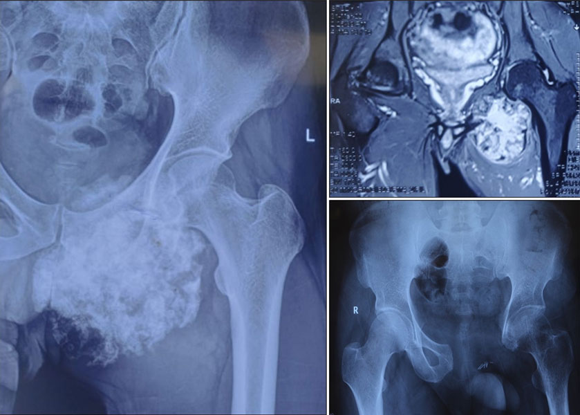
Figure 14. Pelvic chondrosarcoma treated with wide surgical resection: Imaging and surgical photographs demonstrate removal of the tumor with wide margins.
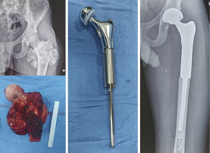
Figure 15. Proximal femur chondrosarcoma treated with limb-salvage surgery: Wide tumor removal followed by hip reconstruction using a proximal femur endoprosthesis.
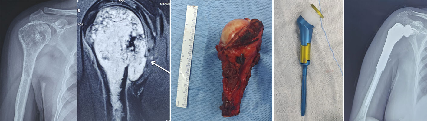
Figure 16. Chondrosarcoma of the upper humerus treated with limb-salvage surgery: Wide resection of the tumor followed by shoulder reconstruction with an endoprosthesis.
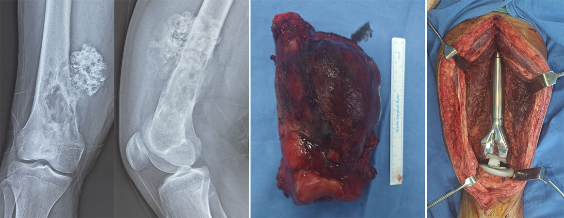
Figure 17. Chondrosarcoma of the lower femur treated with limb-salvage surgery: Wide tumor removal followed by knee reconstruction using a distal femur endoprosthesis.
When Is Amputation Necessary?
Advances in imaging, surgical techniques, and reconstruction have dramatically reduced the need for amputation.
However, amputation may still be the safest option in selected situations, including:
- Extensive involvement of major nerves or blood vessels.
- Massive contamination from previous unplanned surgery.
- Tumors that cannot be completely removed while preserving a functional limb.
- Recurrent tumors where limb salvage is no longer feasible.
Although this possibility can be distressing, the decision is made only when it offers the best chance of complete tumor removal and long-term survival.
Reassurance Box
Many patients worry that a diagnosis of bone cancer automatically means losing a limb. Fortunately, this is no longer true. With modern limb salvage techniques, most patients with chondrosarcoma affecting the arms or legs can be treated without amputation.
Does Every Patient Need Surgery?
No.
Not every cartilage-producing tumor requires immediate surgery.
Patients with:
- Stable enchondromas.
- Selected atypical cartilaginous tumors (ACT).
- Small asymptomatic lesions with benign imaging features.
may be managed with careful observation, involving periodic clinical review and imaging.
The decision between observation and surgery depends on:
- Symptoms.
- Changes on serial imaging.
- Tumor location.
- Risk of fracture.
- Diagnostic certainty.
Observation should always be undertaken under the supervision of a clinician experienced in bone tumors.
Treatment of Different Types of Chondrosarcoma
Recovery After Treatment
Recovery after treatment for chondrosarcoma depends on several factors, including the location of the tumor, the type of reconstruction performed, your general health, and the extent of surgery. Every patient’s recovery is different, and rehabilitation is an important part of achieving the best possible functional outcome.
Detailed information about rehabilitation protocols, physiotherapy, weight-bearing, and functional recovery is available in our Limb Salvage Surgery guide.
What to Expect After Surgery
Immediately after surgery, your healthcare team will focus on:
- Pain control.
- Wound healing.
- Prevention of blood clots and infections.
- Early physiotherapy.
- Gradual return to daily activities.
The duration of hospital stay varies depending on the complexity of the operation and the type of reconstruction.
Rehabilitation
Rehabilitation aims to help you regain:
- Joint movement.
- Muscle strength.
- Balance and coordination.
- Independence in daily activities.
The rehabilitation programme is individualized and may continue for several months after surgery.
Returning to Daily Activities
Most patients gradually return to routine activities, although the timeline varies depending on the operation performed.
Your surgeon will advise when it is safe to:
- Walk without support.
- Drive.
- Return to work.
- Resume recreational activities.
High-impact sports are generally discouraged after major limb reconstruction to reduce the risk of implant failure or fracture.
Follow-up After Treatment
Even after successful treatment, regular follow-up is essential because chondrosarcoma can recur.
Follow-up visits typically include:
- Clinical examination.
- X-rays of the operated bone.
- Periodic chest imaging to detect lung metastases in higher-grade tumors.
- Additional MRI or CT scans when clinically indicated.
The frequency of follow-up depends on the tumor grade, subtype, and time since treatment.
Clinical Pearl
Successful treatment does not end with surgery. Regular follow-up is essential for detecting recurrence early, monitoring reconstruction, and maintaining long-term function.
Can Chondrosarcoma Come Back?
Yes. Chondrosarcoma can recur, even after successful treatment. A recurrence means that the tumor returns after it has been treated.
Recurrence may occur:
- Locally, where the original tumor was removed.
- At distant sites, most commonly in the lungs (metastasis).
The risk of recurrence depends on several factors and varies considerably from one patient to another.
What Increases the Risk of Recurrence?
The most important factors include:
- Higher tumor grade.
- Incomplete tumor removal or inadequate surgical margins.
- Large tumors.
- Tumors arising in the pelvis, spine, or other anatomically complex locations.
- Certain aggressive subtypes, such as dedifferentiated and mesenchymal chondrosarcoma.
Conversely, patients with completely excised low-grade tumors generally have an excellent chance of long-term local control.
Clinical Pearl
The most effective way to reduce the risk of recurrence is complete removal of the tumor during the first operation.
How Is Recurrence Detected?
Most recurrences are identified during routine follow-up before they cause significant symptoms.
Your doctor may recommend:
- Clinical examination.
- X-rays of the operated bone.
- Chest CT or chest X-ray, depending on the tumor grade.
- MRI when local recurrence is suspected.
Between scheduled visits, you should seek medical attention if you notice:
- New or increasing pain.
- A new lump or swelling.
- Reduced function of the operated limb.
- Persistent cough or unexplained breathlessness.
These symptoms do not necessarily indicate recurrence, but they should be evaluated promptly.
🚨 Red Flag Box: Contact your treating team if you develop:
- Progressive pain near the previous surgical site.
- A new swelling or lump.
- A pathological fracture.
- Persistent cough lasting several weeks.
- Unexplained weight loss or worsening fatigue.
Can Recurrent Chondrosarcoma Be Treated?
Yes. Treatment depends on:
- Whether the recurrence is local or metastatic.
- Tumor grade.
- Previous treatment.
- Overall health.
- Whether complete surgical removal is still possible.
Whenever feasible, surgical removal remains the preferred treatment for local recurrence.
Prognosis
One of the first questions patients ask is:
“What are my chances of being cured?”
The prognosis for chondrosarcoma varies widely because it depends on the biological behaviour of the individual tumor rather than the diagnosis alone.
Many patients—particularly those with low-grade conventional chondrosarcoma—achieve long-term cure following complete surgical removal.
Factors That Influence Prognosis
The most important prognostic factors include:
- Tumor grade (the single most important factor).
- Tumor subtype.
- Tumor size.
- Tumor location.
- Surgical margins.
- Presence or absence of metastasis at diagnosis.
- Local recurrence.
Age alone is generally less important than the biological characteristics of the tumor.
Is Chondrosarcoma Curable?
In many patients, yes.
The likelihood of cure is highest when:
- The tumor is diagnosed early.
- It has not spread.
- Complete surgical removal is achieved.
- Appropriate follow-up is maintained.
Higher-grade tumors carry a greater risk of recurrence and metastasis, but successful treatment is still possible, particularly when managed at an experienced sarcoma centre.
Reassurance Box
A diagnosis of chondrosarcoma does not automatically mean a poor outcome. Many patients return to active, independent lives after treatment. Your prognosis depends on the specific characteristics of your tumor rather than the name of the disease alone.
Frequently Asked Questions (FAQs)
1. What is chondrosarcoma?
Chondrosarcoma is a malignant (cancerous) bone tumor that produces cartilage. It most commonly affects adults and usually develops in the pelvis, femur, humerus, ribs, or shoulder blade.
2. Is chondrosarcoma a common cancer?
No. Chondrosarcoma is a rare cancer, but it is one of the most common primary bone cancers in adults.
3. What causes chondrosarcoma?
In most patients, the exact cause is unknown. Rarely, it develops from pre-existing cartilage tumors such as enchondromas or osteochondromas, or in people with conditions like Ollier disease or Maffucci syndrome.
4. What are the first symptoms of chondrosarcoma?
The most common symptom is persistent, gradually worsening bone pain. Some patients also develop swelling, a lump, stiffness around a joint, or a pathological fracture.
5. Can chondrosarcoma be mistaken for arthritis or a sports injury?
Yes. Because symptoms often develop slowly, chondrosarcoma may initially be mistaken for arthritis, tendon problems, or a muscle injury.
6. How is chondrosarcoma diagnosed?
Diagnosis is based on a combination of clinical examination, X-rays, MRI, CT scans, biopsy, and histopathological examination. No single test is sufficient on its own.
7. Why is MRI important?
MRI shows the exact size of the tumor, its relationship to nearby muscles, joints, nerves, and blood vessels, and helps plan both the biopsy and surgery.
8. Is a biopsy always necessary?
In most cases, yes. A biopsy confirms the diagnosis, identifies the subtype of chondrosarcoma, and determines the tumor grade.
9. Why should the biopsy be performed at a specialist centre?
A poorly planned biopsy can complicate definitive surgery. Ideally, the biopsy should be performed by—or in consultation with—the team that will carry out the final operation.
10. Does every patient with chondrosarcoma need surgery?
Most patients do. However, selected patients with Atypical Cartilaginous Tumors (ACT) or benign cartilage lesions may only require observation.
11. Why isn’t chemotherapy routinely used?
Conventional chondrosarcoma is relatively resistant to chemotherapy. Surgery provides the best chance of cure. Chemotherapy is mainly used for rare subtypes such as mesenchymal chondrosarcoma.
12. Is radiotherapy effective for chondrosarcoma?
Radiotherapy has a limited role in conventional chondrosarcoma but may be recommended for selected patients, particularly when complete surgical removal is not possible.
13. Can the tumor spread to other parts of the body?
Yes. Higher-grade chondrosarcomas can spread, most commonly to the lungs. Low-grade tumors have a much lower risk.
14. Can chondrosarcoma come back after surgery?
Yes. Local recurrence can occur, especially in high-grade tumors or when the tumor is not completely removed. Regular follow-up is therefore essential.
15. What is the difference between an enchondroma and chondrosarcoma?
An enchondroma is a benign cartilage tumor, whereas chondrosarcoma is malignant. Distinguishing between them sometimes requires careful evaluation by a specialist sarcoma team.
16. What does tumor grade mean?
Tumor grade describes how aggressive the cancer cells appear under the microscope. Higher-grade tumors generally have a greater risk of recurrence and metastasis.
17. Will I lose my arm or leg?
Not necessarily. With modern techniques, most patients with tumors affecting the arms or legs can undergo limb salvage surgery, avoiding amputation.
18. Is chondrosarcoma hereditary?
Most cases are not inherited. Only a small proportion occur in association with inherited conditions such as Ollier disease or Maffucci syndrome.
19. Can I return to work after treatment?
Many patients return to work after recovery. The timing depends on the type of surgery, rehabilitation, and the physical demands of the job.
20. How long will I need follow-up?
Follow-up usually continues for several years because recurrence may occur long after treatment. The schedule depends on the tumor grade, subtype, and treatment received.
21. Is chondrosarcoma curable?
Yes. Many patients with localized conventional chondrosarcoma are cured with complete surgical removal of the tumor.
22. Should I seek a second opinion?
A second opinion is reasonable, particularly if the diagnosis is uncertain, surgery is being considered, or the tumor is located in a complex anatomical area such as the pelvis or spine.
Myths vs Facts
Key Takeaways
- Chondrosarcoma is a malignant cartilage-producing bone tumor that most commonly affects adults.
- Conventional chondrosarcoma is biologically different from osteosarcoma and Ewing sarcoma because surgery—not chemotherapy—is the cornerstone of treatment.
- Accurate diagnosis requires careful correlation of clinical findings, imaging, and biopsy results.
- The tumor grade is one of the most important factors influencing treatment and prognosis.
- Most tumors affecting the arms and legs can be treated with limb salvage surgery, preserving the affected limb.
- Chemotherapy and radiotherapy are reserved for selected situations and are not routinely used for conventional chondrosarcoma.
- Complete removal of the tumor during the first operation offers the best chance of long-term cure.
- Regular follow-up is essential because recurrence can occur even after successful treatment.
- Most patients with localized conventional chondrosarcoma achieve excellent outcomes following appropriate treatment by a specialist multidisciplinary sarcoma team.
References
International Guidelines
- National Comprehensive Cancer Network (NCCN). NCCN Clinical Practice Guidelines in Oncology: Bone Cancer. Latest available version.
- ESMO–EURACAN–GENTURIS Clinical Practice Guidelines. Bone Sarcomas: Diagnosis, Treatment and Follow-up. Annals of Oncology.
- WHO Classification of Soft Tissue and Bone Tumours. 5th Edition. International Agency for Research on Cancer (IARC); 2020.
- British Sarcoma Group Guidelines for the Management of Bone Sarcomas.
- . Gelderblom H, Hogendoorn PCW, Dijkstra SD, et al. The Clinical Approach Towards Chondrosarcoma. The Oncologist. 2008.
- Murphey MD, Walker EA, Wilson AJ, et al. From the Archives of the AFIP: Imaging of Primary Chondrosarcoma. Radiographics.
- Gazendam, A., Popovic, S., Parasu, N., & Ghert, M. (2023). Chondrosarcoma: A Clinical Review. Journal of Clinical Medicine, 12(7), 2506.
Medical Disclaimer
The information provided in this article is intended for educational purposes only and should not be considered a substitute for professional medical advice, diagnosis, or treatment.
Every patient with chondrosarcoma is unique. Treatment recommendations depend on multiple factors, including the tumor subtype, grade, location, imaging findings, biopsy results, and the patient’s overall health. Decisions regarding diagnosis and treatment should always be made in consultation with a qualified orthopaedic oncologist or multidisciplinary sarcoma team.
While every effort has been made to ensure the accuracy and currency of the information presented, medical knowledge and treatment recommendations continue to evolve. Patients should discuss their individual circumstances with their treating healthcare professionals before making any medical decisions.
If you have persistent bone pain, a suspicious bone lesion, or have been diagnosed with a bone tumor, seek evaluation at a specialist bone tumor or sarcoma centre.
About the Author
Dr. Love Kapoor, MS (Orthopaedics), Fellowship in Musculoskeletal Oncology (AIIMS, New Delhi), Former Assistant Professor, Orthopaedic Oncology, AIIMS, New Delhi.
Orthopaedic Oncologist specializing in bone tumors, soft tissue sarcomas, limb salvage surgery, bone reconstruction, mega prosthesis surgery, and metastatic bone disease.
Meet Your Orthopaedic Oncologist
Choosing treatment for a bone or soft tissue tumor often involves complex decisions. An orthopaedic oncologist specializes in diagnosing and treating tumors affecting the bones, joints, and soft tissues while working closely with medical oncologists, radiation oncologists, radiologists, pathologists, plastic surgeons, vascular surgeons, physiotherapists, and rehabilitation specialists.
Dr. Love Kapoor is an orthopaedic oncologist with dedicated fellowship training in musculoskeletal oncology. His clinical practice focuses on:
- Primary bone tumors
- Soft tissue sarcomas
- Limb salvage surgery
- Bone reconstruction
- Mega prosthesis surgery
- Management of metastatic bone disease
- Complex revision surgery after tumor reconstruction
His approach emphasizes:
- Accurate diagnosis before treatment
- Multidisciplinary decision-making
- Evidence-based care
- Limb preservation whenever oncologically safe
- Personalized treatment planning
- Long-term functional recovery
If you have been diagnosed with a bone tumor, have been advised to undergo amputation, or would like a second opinion regarding limb salvage surgery, consultation with an experienced orthopaedic oncologist can help you better understand your treatment options.
 +91 93558 33005