Quick Summary
- Osteosarcoma is a primary bone cancer that begins inside the bone.
- It most commonly affects adolescents and young adults, especially during periods of rapid growth.
- It most often develops around the knee (distal femur or proximal tibia) or the upper arm (proximal humerus).
- Persistent bone pain and swelling should always be evaluated.
- Diagnosis requires imaging and a biopsy.
- Treatment usually combines chemotherapy and surgery.
- Many patients are candidates for limb salvage surgery rather than amputation.
- Early diagnosis and treatment at an experienced musculoskeletal oncology centre can improve outcome.
Being told that you or someone you love may have osteosarcoma can be overwhelming. It is natural to have many questions:
- Is this really cancer?
- Can it be cured?
- Will I lose my leg or arm?
- Will I need chemotherapy?
- What happens next?
These concerns are completely understandable.
The reassuring news is that modern treatment for osteosarcoma has advanced considerably. Improvements in chemotherapy, imaging, surgical planning, limb reconstruction, rehabilitation, and multidisciplinary cancer care have transformed outcomes for many patients. Today, many individuals can be treated with limb salvage surgery, allowing preservation of the affected limb while achieving effective cancer control. Every treatment plan, however, is individualized based on the location of the tumor, its stage, and the patient’s overall health.
This comprehensive guide explains every stage of the osteosarcoma journey—from recognising the earliest symptoms and understanding the diagnosis to chemotherapy, surgery, rehabilitation, survivorship, and long-term follow-up. Whether you are a patient, family member, caregiver, or healthcare professional, our goal is to provide clear, evidence-based information that helps you make informed decisions with confidence.
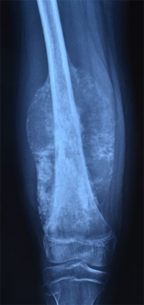
Figure 1. Osteosarcoma of the thigh bone (femur)
What is Osteosarcoma?
Osteosarcoma is the most common primary malignant bone tumor, meaning it starts within the bone itself rather than spreading from another organ. It develops when immature bone-forming cells, called osteoblasts, begin to grow in an uncontrolled manner. These abnormal cells produce immature bone tissue, known as osteoid, which is a characteristic feature of osteosarcoma.
The disease most commonly affects children, adolescents, and young adults, particularly during periods of rapid bone growth. It typically arises in the metaphysis, the wider part of a long bone near the growth plate, most often around the knee.
Although osteosarcoma is uncommon, it is one of the most important bone cancers to recognize because early diagnosis and timely treatment can make a significant difference. Persistent bone pain that does not improve, swelling around a bone or joint, or a pathological fracture (a fracture occurring through a weakened bone) should never be ignored.
Treatment is usually planned by a multidisciplinary team, which may include an orthopaedic oncologist, medical oncologist, musculoskeletal radiologist, pathologist, radiation oncologist, physiotherapist, specialist nurses, and rehabilitation experts. Working together, this team develops an individualized treatment plan that balances complete tumor removal, preservation of limb function, and long-term quality of life.
Clinical Pearl
The most successful treatment of Osteosarcoma begins with an accurate diagnosis. Imaging should be completed before biopsy whenever possible, and both biopsy and surgery should ideally be planned by the team responsible for definitive treatment.
Doctor Insight
One of the most common misconceptions is that every Osteosarcoma requires amputation. Fortunately, this is no longer true for many patients. Advances in surgical techniques, chemotherapy, and reconstruction now allow limb preservation in appropriately selected cases. The primary goal, however, always remains complete and safe removal of the tumor, because effective cancer control is the foundation of successful treatment.
Patient Tip
📝 Start a medical folder from the beginning. Keep copies of your X-rays, MRI scans, CT scans, biopsy reports, blood test results, and treatment summaries. Having these records readily available can make future consultations, second opinions, and follow-up visits much easier.
Did You Know?
💡 Osteosarcoma is rare. Most teenagers and adults with persistent bone pain do not have osteosarcoma. However, pain that is persistent, progressively worsening, associated with swelling, or occurs at night should always be evaluated by a healthcare professional.
If You Have Just Been Diagnosed
Receiving a diagnosis—or even the possibility—of osteosarcoma can be frightening. It is normal to feel anxious, confused, or overwhelmed.
At this stage, remember these important points:
- You are not expected to make every treatment decision immediately.
- Additional investigations are often needed before the diagnosis and treatment plan are finalized.
- Modern treatment has improved significantly over the past few decades.
- Many patients are able to undergo limb salvage surgery, avoiding amputation.
- Your care is usually planned by a multidisciplinary team with expertise in bone tumors.
- It is appropriate to ask questions and to seek a second opinion if you are uncertain about your treatment options.
Try to take one step at a time. The first priority is establishing an accurate diagnosis and understanding the stage of the disease. Once this information is available, your healthcare team can recommend the treatment approach that is most appropriate for your individual situation.
Remember: Every person’s osteosarcoma is different. Treatment recommendations are based on the specific characteristics of your tumor, your overall health, and your personal goals—not on someone else’s experience.
Clinical Pearl
Osteosarcoma is uncommon, but it is the most common primary bone cancer. Because it often affects young people, persistent bone pain should not automatically be attributed to sports injuries or “growing pains.”
Doctor Explains
Patients often ask, “Is osteosarcoma the same as bone cancer?”
The answer is yes, but with an important distinction.
“Bone cancer” is a broad term that includes several different cancers arising in bone. Osteosarcoma is one specific type of primary bone cancer. Other primary bone cancers include Ewing sarcoma and chondrosarcoma, each with different biological behavior and treatment approaches.
Did You Know?
💡 Nearly half of all osteosarcomas occur around the knee, making persistent pain or swelling in this region particularly important to investigate when symptoms are unexplained.
Where Does Osteosarcoma Commonly Occur?
Osteosarcoma can develop in almost any bone, but it has a strong preference for the rapidly growing ends of long bones. It typically arises in the metaphysis, the wider part of a long bone near the growth plate, most often around the knee. As osteosarcoma grows, it often spreads within the marrow cavity (soft tissue inside bones where blood cells are produced) before extending through the outer layer of the bone. As the tumor enlarges, it may lift the periosteum (thin membrane covering the outer surface of the bone), producing characteristic imaging findings such as Codman’s triangle, which can sometimes be seen on X-rays.
Around the Knee
Approximately 50–60% of osteosarcomas occur around the knee.
The two most common locations are:
- Distal femur (the lower end of the thigh bone)
- Proximal tibia (the upper end of the shin bone)
These regions experience rapid growth during adolescence, which may partly explain why they are the most frequent sites.
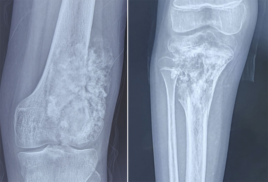
Figure 2. Osteosarcoma around the knee: X-rays showing osteosarcoma involving the lower end of the thigh bone (femur) and upper end of the shin bone (tibia), one of the most common locations for osteosarcoma.
Upper Arm
The proximal humerus (the upper end of the arm bone near the shoulder) is another common location.
Patients may notice pain while lifting the arm or performing overhead activities.
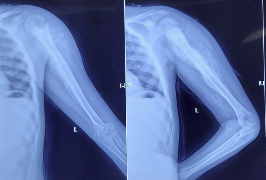
Figure 3. Osteosarcoma of the upper arm bone (humerus): X-ray showing a destructive bone tumor involving the humerus.
Pelvis
Osteosarcoma of the pelvis is less common but can be more challenging to diagnose because symptoms often develop gradually and the tumor may become quite large before it is detected.
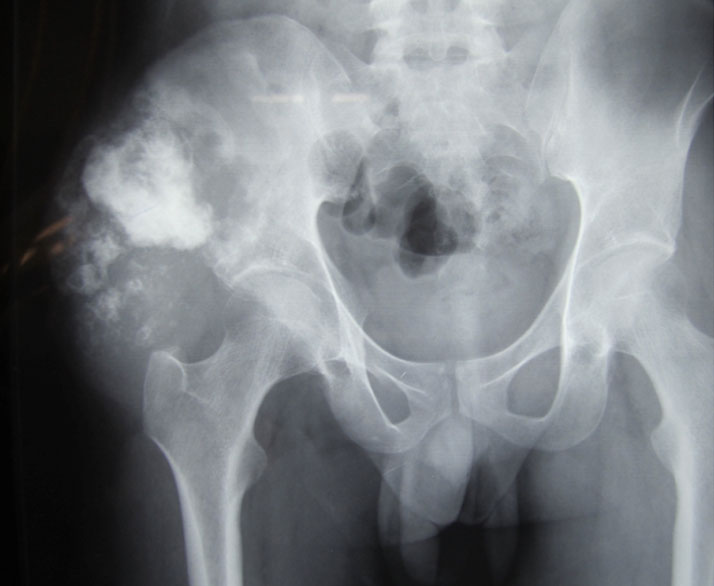
Figure 4. Osteosarcoma of the pelvis: X-ray showing a large osteosarcoma involving the pelvic bone.
Other Bones
Less commonly, osteosarcoma may occur in:
- Femoral shaft
- Tibial shaft
- Fibula
- Radius
- Ulna
- Jaw (mandible or maxilla)
- Spine (rare)
- Small bones of the hands and feet (very rare)
Clinical Pearl
Persistent pain around the knee in an adolescent that lasts more than a few weeks—especially if it occurs at night or is associated with swelling—should be investigated rather than assumed to be a sports injury or growing pain.
- Persistent Bone Pain
- Bone Tumor Diagnosis
- MRI for Bone Tumors
What are the various types of Osteosarcoma?
Osteosarcoma is not a single disease. It includes several subtypes that differ in their appearance under the microscope, biological behavior, and treatment approach.
Fortunately, the majority of patients have conventional high-grade osteosarcoma, which has well-established treatment protocols.
Conventional High-Grade Osteosarcoma
This is the most common subtype and accounts for approximately 80–90% of cases.
It usually affects children, adolescents, and young adults and is treated with a combination of chemotherapy and surgery.
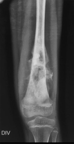
Figure 5. Conventional osteosarcoma: X-ray showing a destructive bone lesion with abnormal new bone formation, a characteristic feature of osteosarcoma.
Telangiectatic Osteosarcoma
A rare subtype containing blood-filled spaces.
Because it can resemble an aneurysmal bone cyst on imaging, a biopsy is essential to establish the correct diagnosis.
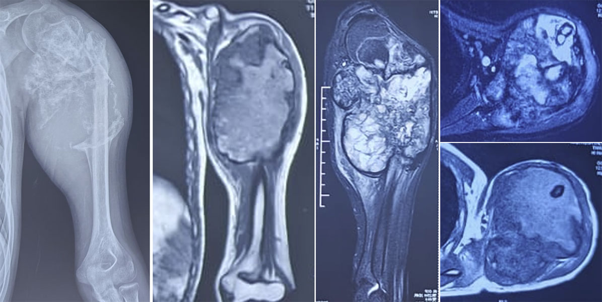
Figure 6. Telangiectatic osteosarcoma of the upper arm: MRI demonstrates the extent of the tumor within the humerus and surrounding soft tissues.
Internal Link: → Aneurysmal Bone Cyst
Parosteal Osteosarcoma
A low-grade tumor that develops on the outer surface of the bone.
It usually grows more slowly than conventional osteosarcoma and often has an excellent prognosis when completely removed.
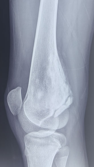
Figure 7. Parosteal osteosarcoma: Imaging showing osteosarcoma arising from the outer surface of the bone.
Periosteal Osteosarcoma
An intermediate-grade surface osteosarcoma that arises from the periosteum, the outer covering of the bone.
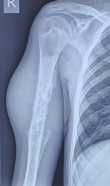
Figure 8. Periosteal osteosarcoma: X-ray showing a surface-based osteosarcoma arising from the outer layer of the bone.
Secondary Osteosarcoma
This develops in association with another bone disorder or after previous radiation therapy.
It is more commonly seen in older adults.
Doctor Insight
Although these subtypes have different biological characteristics, the diagnosis should never be based on imaging alone. The exact subtype can only be confirmed through careful examination of biopsy tissue by an experienced musculoskeletal pathologist.
Bone Tumor Biopsy
Causes and Risk Factors of Osteosarcoma
One of the first questions patients and families ask is:
“Why did this happen?”
For most people with osteosarcoma, there is no single identifiable cause. In the majority of cases, the disease develops sporadically, meaning it occurs by chance without anything the patient did or did not do. It is not caused by an injury, exercise, diet, or lifestyle.
Researchers believe osteosarcoma develops when genetic changes occur within osteoblasts, the bone-forming cells, causing them to grow and divide uncontrollably. Exactly why these changes occur remains an active area of research.
Who Is Most at Risk?
Although osteosarcoma is rare, certain factors are associated with a higher risk.
Rapid Bone Growth
Osteosarcoma most commonly occurs during adolescence, particularly during periods of rapid growth. This is one reason it frequently affects the bones around the knee, which grow quickly during puberty.
Previous Radiation Therapy
People who have previously received high-dose radiation therapy to a bone have a small increased risk of developing osteosarcoma years later. This is uncommon and usually occurs after treatment for another cancer.
Certain Inherited Genetic Syndromes
A small proportion of patients have inherited conditions that increase the risk of osteosarcoma. These include:
- Li-Fraumeni syndrome
- Hereditary retinoblastoma
- Rothmund-Thomson syndrome
- Bloom syndrome
- Werner syndrome
These conditions are rare, and most patients with osteosarcoma do not have an inherited genetic disorder.
Pre-existing Bone Disorders
Certain long-standing bone diseases can occasionally increase the risk of osteosarcoma in adults, particularly:
- Paget’s disease of bone
- Previous bone infarction (rare)
- Chronic osteomyelitis (very uncommon)
These account for only a small proportion of cases.
What Does Not Cause Osteosarcoma?
Many patients worry that something they did caused the disease. Current evidence does not support these concerns.
There is no evidence that osteosarcoma is caused by:
- Sports or exercise
- Minor injuries or falls
- Mobile phones
- Diet or specific foods
- Vitamin deficiencies
- Stress
- Routine medications
- Vaccinations
An injury may draw attention to a tumor because imaging is performed after the accident, but the injury itself does not cause the cancer.
Did You Know?
💡 More than 90% of patients with osteosarcoma have no identifiable risk factor. In most cases, the disease develops without any warning signs or preventable cause.
Did I Do Something to Cause This?
This is one of the most common questions patients and parents ask.
The answer is almost always no.
Nothing you ate, any exercise you performed, or an injury you sustained has been shown to cause osteosarcoma. For most people, the disease develops because of complex genetic changes within bone cells that occur by chance. It is important not to blame yourself or your child—there is usually nothing that could have been done to prevent it.
Clinical Pearl
A history of trauma should never exclude the possibility of osteosarcoma. Persistent pain after an injury that fails to improve deserves further evaluation, particularly if imaging shows an abnormal bone lesion.
Doctor Insight
One of the most common reasons for delayed diagnosis is that early osteosarcoma is mistaken for a sports injury, particularly in active adolescents. While sports injuries are far more common than bone cancer, pain that persists beyond the expected recovery period—or progressively worsens despite treatment—should prompt further investigation.
Patient Tip
If your pain has been treated as a muscle strain or sports injury but continues for several weeks, ask whether further imaging, such as an X-ray or MRI, is appropriate rather than continuing repeated courses of pain medication.
- Persistent Bone Pain — for patients with ongoing symptoms.
- Bone Tumor Diagnosis — to understand the next diagnostic steps.
- MRI for Bone Tumors — explaining the role of MRI.
- Bone Tumor Biopsy — why biopsy is required before treatment.
What are the symptoms of Osteosarcoma?
The symptoms of osteosarcoma usually develop gradually over several weeks or months. In the early stages, they can resemble more common conditions such as a sports injury, muscle strain, or growing pains, particularly in children and adolescents. This similarity is one reason why diagnosis may sometimes be delayed.
Although symptoms vary depending on the location and size of the tumor, persistent, localized bone pain is the most common presenting symptom.
Persistent Bone Pain
Pain is the earliest and most frequent symptom of osteosarcoma.
It typically:
- Begins gradually
- Is localized to one area of the bone
- Becomes progressively worse over time
- May occur during activity initially but later at rest
- Often becomes more noticeable at night
Unlike a minor injury, the pain usually does not resolve with rest and tends to become more persistent.
Swelling or a Lump
As the tumor enlarges, swelling may develop over the affected bone.
Patients or parents may notice:
- A firm swelling
- A visible lump
- Tenderness over the area
- Warmth (occasionally)
Swelling often appears weeks after the onset of pain.
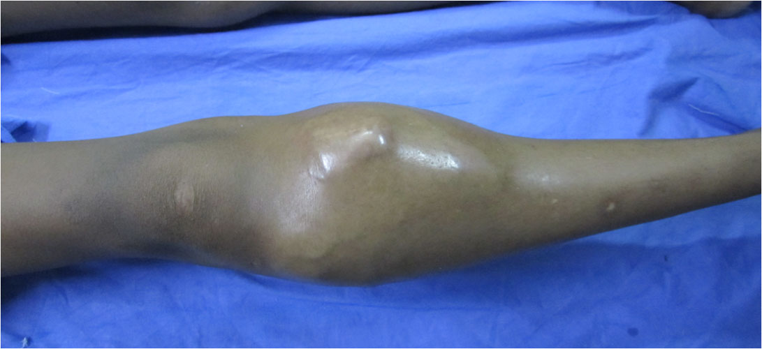
Figure 9. Painful swelling as a presentation of osteosarcoma: A visible swelling around the affected limb may be one of the early symptoms of bone cancer.
Reduced Joint Movement
When the tumor is located near a joint, movement may become uncomfortable or restricted.
For example:
- Difficulty bending the knee
- Shoulder stiffness
- Pain while climbing stairs
- Difficulty squatting or kneeling
These symptoms occur because the surrounding muscles and soft tissues become irritated by the growing tumor.
Limping or Difficulty Using the Limb
Tumors affecting the lower limb may cause:
- Limping
- Reduced weight-bearing
- Difficulty running
- Avoidance of sports or physical activities
Young children may simply refuse to use the affected limb without clearly describing the pain.
Pathological Fracture
Occasionally, osteosarcoma weakens the bone sufficiently that it breaks after minimal trauma. This is known as a pathological fracture.
Although uncommon as the first presentation, a fracture through an abnormal bone should always prompt investigation for an underlying cause.
Pathological Fractures in Bone Tumors
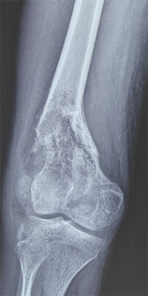
Figure 10. Pathological fracture due to osteosarcoma: A weakened bone affected by osteosarcoma can fracture even after minor injury or normal activity.
When Should You See an Orthopaedic Oncologist?
Arrange prompt medical assessment if you experience:
- Bone pain lasting more than 4–6 weeks
- Pain that continues despite treatment
- Night pain that regularly interrupts sleep
- Swelling over a bone or near a joint
- An unexplained limp
- An abnormal bone lesion on an X-ray
- A fracture occurring after minimal trauma
These symptoms do not necessarily indicate osteosarcoma, but they should not be ignored.
Clinical Pearl
Pain that persists beyond the expected recovery period after an injury deserves reassessment. While most sports injuries improve with time, osteosarcoma-related pain typically becomes more persistent and progressive.
Doctor Insight
One of the most common reasons for delayed diagnosis is that adolescents remain active despite pain, assuming it is related to sports or exercise. Similarly, parents and healthcare providers may initially attribute symptoms to overuse injuries because these are far more common than bone tumors. Maintaining a low threshold for imaging when symptoms persist can help avoid unnecessary delays.
Patient Tip
Keep a simple symptom diary noting:
- When the pain started
- Whether it is improving or worsening
- Activities that increase pain
- Whether pain occurs at night
- Any swelling or limitation of movement
This information can help your doctor understand how the symptoms have evolved.
| Anchor Text |
| persistent bone pain |
| pathological fracture |
| abnormal bone lesion |
| MRI |
| orthopaedic oncologist |
Clinical Evaluation
Diagnosing osteosarcoma begins with a careful clinical assessment. While imaging and biopsy are essential, the patient’s history and physical examination often provide the first clues that a bone tumor may be present.
Medical History
Your doctor will ask detailed questions about your symptoms, including:
- When did the pain begin?
- Is the pain getting worse?
- Does it occur at night or at rest?
- Is there associated swelling?
- Has there been any recent injury?
- Has the pain affected walking, sports, or daily activities?
- Is there a personal or family history of cancer or inherited genetic disorders?
These questions help distinguish osteosarcoma from more common conditions such as sports injuries or infection.
Physical Examination
The examination focuses on both the affected limb and your overall health.
Your doctor will assess:
- The location and size of any swelling
- Tenderness over the bone
- Joint movement
- Limb function
- Muscle strength
- Walking pattern (gait)
- Skin changes or visible veins
- Nearby lymph nodes (although lymph node spread is uncommon in osteosarcoma)
A thorough examination also helps determine whether important nerves or blood vessels may be involved, which is relevant when planning surgery.
Doctor Insight
Pain severity alone does not determine how advanced a tumor is. Some patients with relatively small tumors experience significant pain, while others with larger tumors may have surprisingly mild symptoms. Imaging studies provide a much more accurate assessment of the extent of disease than symptoms alone.
Clinical Pearl
A careful history and examination guide the choice of investigations but cannot confirm osteosarcoma. Imaging and biopsy are required to establish the diagnosis.
Diagnosis of Osteosarcoma
No single investigation can diagnose osteosarcoma. Instead, doctors combine clinical findings, imaging studies, biopsy results, and staging investigations to confirm the diagnosis and develop an individualized treatment plan.
Step 1: X-ray – The First Investigation
For most patients, a plain X-ray is the first imaging test.
An X-ray may show features suggestive of osteosarcoma, including:
- Bone destruction
- Abnormal new bone formation
- Cortical destruction (damage to the outer layer of bone)
- Periosteal reaction (new bone formed beneath the bone covering)
- Characteristic patterns such as Codman’s triangle or a sunburst appearance
Although these findings can strongly suggest osteosarcoma, an X-ray alone cannot confirm the diagnosis.
Internal Link: → X-ray Evaluation of Bone Tumors
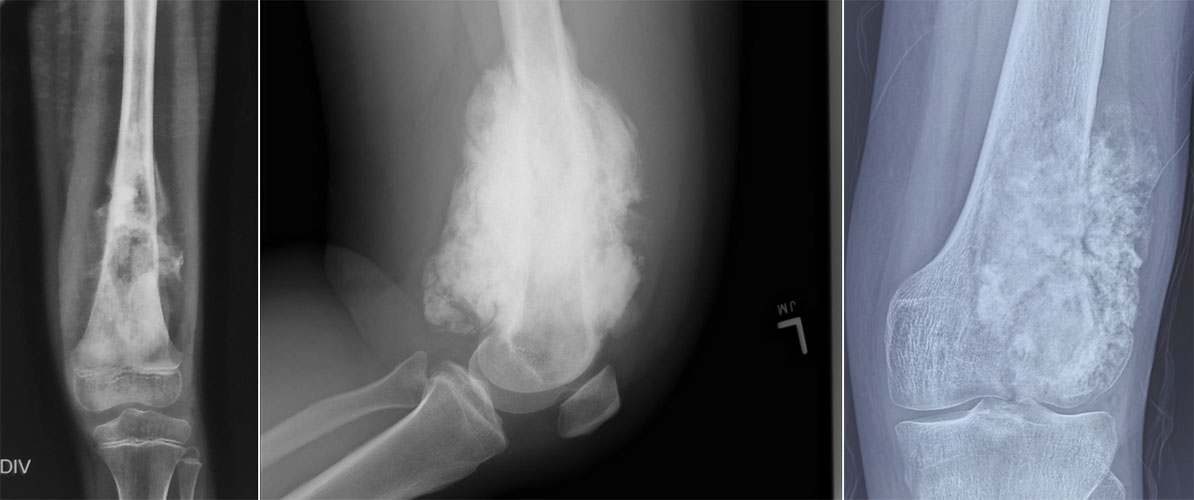
Figure 11. X-ray appearance of osteosarcoma: X-ray demonstrates the destructive bone lesion and abnormal bone formation commonly seen in osteosarcoma.
Step 2: MRI – Defining the Extent of the Tumor
Once a bone tumor is suspected, MRI (Magnetic Resonance Imaging) is the most important imaging study for local assessment.
MRI helps determine:
- The exact size of the tumor
- Involvement of the bone marrow
- Extension into surrounding muscles
- Relationship to nearby nerves and blood vessels
- Joint involvement
- Skip lesions (additional tumor deposits within the same bone)
This information is essential for planning surgery and determining whether limb salvage is feasible.
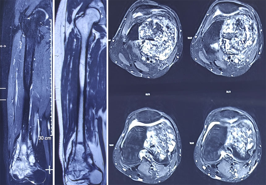
Figure 12. MRI evaluation of osteosarcoma: MRI helps determine the local extent of the tumor and its relationship with nearby muscles and other soft tissues.
Internal Link: → MRI for Bone Tumors
Clinical Pearl
MRI should ideally be performed before a biopsy. This helps guide biopsy planning and ensures the imaging accurately reflects the tumor before any procedure alters the surrounding tissues.
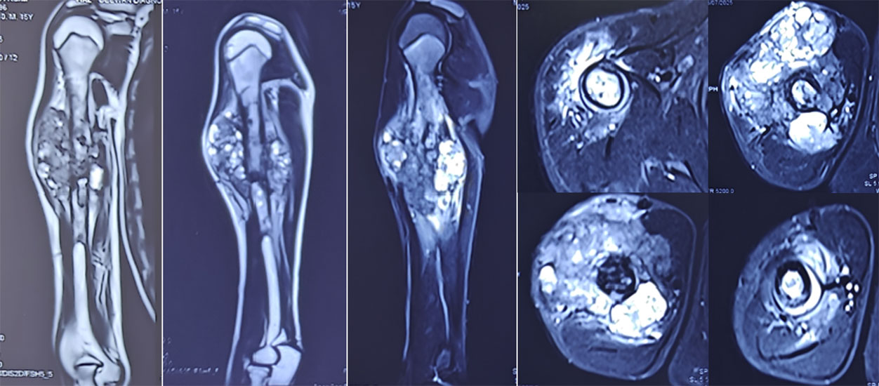
Figure 13. MRI assessment of osteosarcoma: Multiple MRI views help define the extent of the tumor within the bone and surrounding soft tissues before treatment.
Step 3: Biopsy – Confirming the Diagnosis
A biopsy involves obtaining a small sample of the tumor for examination under a microscope. It is the only way to confirm the diagnosis and determine the exact subtype of osteosarcoma.
Biopsies are commonly performed using a core needle, guided by imaging. In selected situations, an open biopsy may be recommended.
The biopsy should be carefully planned because the biopsy tract is usually removed during definitive surgery.
Internal Link: → Bone Tumor Biopsy
Is a Biopsy Safe?
Many patients worry that a biopsy could cause the cancer to spread.
When performed by an experienced musculoskeletal oncology team, a biopsy is a safe and essential procedure. Careful planning minimizes risks and provides the information needed to select the most appropriate treatment.
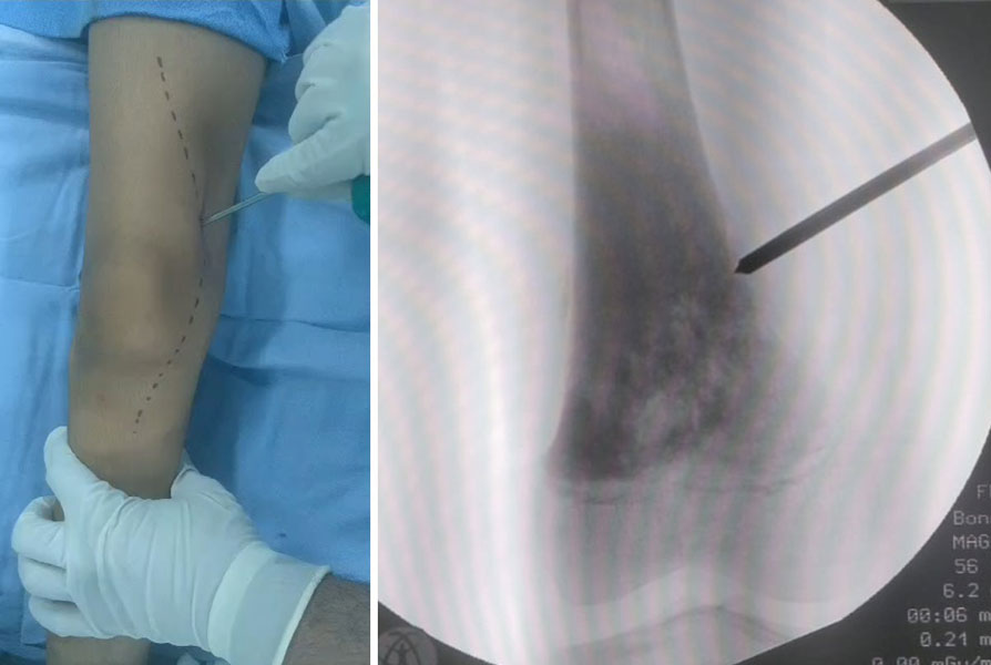
Figure 14. Image-guided biopsy for suspected osteosarcoma: A biopsy needle is accurately placed into the bone lesion to obtain tissue for diagnosis.
Step 4: Staging Investigations
Once osteosarcoma has been confirmed, additional investigations determine whether the disease has spread.
These commonly include:
CT Scan of the Chest
The lungs are the most common site of metastasis in osteosarcoma. A CT scan of the chest is therefore routinely performed before treatment begins.
Bone Scan or PET-CT
Depending on the individual situation, a bone scan or PET-CT may be used to identify additional bone lesions or distant metastases.
Not every patient requires every imaging study; investigations are tailored to the clinical scenario.
Internal Links:
- CT Scan for Bone Tumors
- PET-CT in Bone Tumors
Step 5: Laboratory Tests
Routine blood tests cannot diagnose osteosarcoma but help assess overall health before treatment.
Common investigations include:
- Complete blood count
- Kidney function
- Liver function
- Alkaline phosphatase (ALP)
- Lactate dehydrogenase (LDH)
Some markers, such as ALP and LDH, may be elevated in certain patients, but they are not specific for osteosarcoma.
Doctor Explains
Patients often ask, “Why do I need so many tests?”
Each investigation answers a different question:
- X-ray: Could this be a bone tumor?
- MRI: How far has it spread within the limb?
- Biopsy: What type of tumor is it?
- CT chest: Has it spread to the lungs?
- PET-CT or bone scan: Has it spread elsewhere?
Only after combining all of this information can the healthcare team develop the most appropriate treatment plan.
Patient Tip
Bring all your imaging studies—not just the reports—to your consultation. Reviewing the actual X-rays, MRI scans, and CT images is essential for accurate assessment and treatment planning.
Video
- How Osteosarcoma Is Diagnosed: From X-ray to Biopsy
Staging and Classification of Osteosarcoma
Once osteosarcoma has been confirmed, the next step is to determine how advanced the disease is. This process is called staging.
Staging describes:
- The size and extent of the primary tumor
- Whether the cancer has spread to nearby tissues
- Whether it has spread (metastasized) to distant organs, most commonly the lungs
- The biological aggressiveness (grade) of the tumor
Accurate staging helps the multidisciplinary team recommend the most appropriate treatment and estimate prognosis.
Why Is Staging Important?
Not every patient with osteosarcoma has the same disease.
Two patients may both have osteosarcoma but require different treatment depending on:
- Tumor size
- Tumor location
- Response to chemotherapy
- Presence or absence of metastases
- Ability to achieve complete surgical removal
Staging ensures that treatment is individualized rather than one-size-fits-all.
Staging helps answer four important questions:
- How large is the tumor?
- Has it spread beyond the bone?
- Has it spread to the lungs or other organs?
- What treatment offers the best chance of cure?
Tumor Grade
Tumor grade describes how abnormal the cancer cells appear under the microscope and how aggressively they are expected to behave.
Most osteosarcomas are high-grade tumors, meaning they grow rapidly and require treatment with both chemotherapy and surgery.
Low-grade osteosarcomas, such as parosteal osteosarcoma, are much less common and are managed differently.
Staging
The American Joint Committee on Cancer (AJCC) staging system is widely used internationally.
It considers:
- T (Tumor): Size and extent of the primary tumor
- N (Nodes): Whether nearby lymph nodes are involved (rare in osteosarcoma)
- M (Metastasis): Whether the cancer has spread to distant organs
- G (Grade): How aggressive the tumor appears under the microscope
Enneking Surgical Staging
Orthopaedic oncologists commonly use the Enneking staging system, which combines:
- Tumor grade
- Local extent of the tumor
- Presence of metastases
These factors are combined to determine the overall stage of the disease.
For most patients, the most important distinction is whether the disease is localized or metastatic.
Localized vs Metastatic Osteosarcoma
Localized Osteosarcoma
The cancer is confined to the bone and surrounding tissues without evidence of spread to distant organs.
Most patients are diagnosed at this stage.
Treatment usually involves:
- Neoadjuvant chemotherapy (before surgery)
- Surgical removal of the tumor
- Adjuvant chemotherapy (after surgery)
Metastatic Osteosarcoma
The cancer has spread beyond the primary bone.
The lungs are the most common site of metastasis, although spread to other bones or, rarely, other organs can occur.
Even when metastases are present, treatment may still aim for long-term disease control or cure in selected patients, depending on the extent and location of the disease.
Does a Higher Stage Mean There Is No Hope?
No.
Although staging helps doctors estimate prognosis and plan treatment, it does not predict the outcome for an individual patient.
Many factors influence recovery, including:
- Response to chemotherapy
- Complete removal of the tumor
- Overall health
- Advances in modern treatment
- Ongoing follow-up and rehabilitation
Treatment decisions are based on the whole clinical picture, not on the stage alone.
Patient Tip
Ask your treating team to explain:
- Your stage of disease
- Whether the cancer is localized or metastatic
- Whether chemotherapy has reduced the tumor
- What your pathology report shows after surgery
Understanding these terms can help you participate more confidently in treatment decisions.
The Typical Osteosarcoma Treatment Pathway
Although every patient is different, treatment for localized high-grade osteosarcoma usually follows a predictable sequence.
Step 1: Neoadjuvant Chemotherapy
Treatment usually begins with chemotherapy before surgery.
This is called neoadjuvant chemotherapy, meaning chemotherapy given before an operation.
Often patients ask, “Why is chemotherapy given before surgery?” “Why can’t the tumor simply be removed first?”
Chemotherapy before surgery is necessary:
- To destroy microscopic cancer cells that cannot yet be detected on scans
- To reduce the biological activity of the tumor
- To allow doctors to assess how well the tumor responds to treatment
- To improve the chances of successful surgery
Chemotherapy for Osteosarcoma
Step 2: Surgery
After completing the planned preoperative chemotherapy cycles, repeat imaging is performed to assess the tumor before surgery.
The multidisciplinary team then reviews:
- Response to chemotherapy
- Local extent of the tumor
- Surgical margins required
- Reconstruction options
- Overall fitness for surgery
The timing of surgery is carefully coordinated to achieve the best possible outcome.
Depending on the individual situation, surgery may involve:
- Limb salvage surgery
- Biological reconstruction
- Tumor prosthesis (mega prosthesis)
- Amputation (only when necessary)
The surgical approach is individualized and aims to completely remove the tumor while preserving as much function as possible.
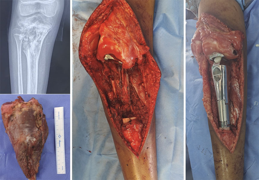
Figure 15A. Limb-salvage surgery for osteosarcoma around the knee: After chemotherapy, the tumor was removed with wide resection and the knee was reconstructed using an endoprosthetic replacement.
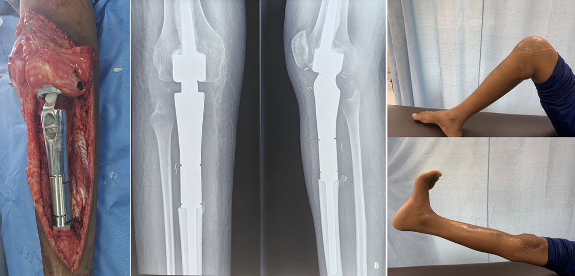
Figure 15B. Good knee function after limb-salvage surgery: The patient demonstrates good knee movement following osteosarcoma surgery and endoprosthetic reconstruction.
Step 3: Histopathology
After surgery, the removed tumor is examined under a microscope.
This helps determine:
- Whether the tumor has been completely removed
- The percentage of tumor necrosis (response to chemotherapy)
- Whether additional treatment recommendations need to be modified
Step 4: Adjuvant Chemotherapy
Following recovery from surgery, patients usually receive additional chemotherapy.
This is called adjuvant chemotherapy, meaning chemotherapy given after surgery.
The aim is to eliminate any remaining microscopic cancer cells and reduce the risk of recurrence.
Step 5: Rehabilitation
Recovery begins immediately after surgery.
A structured rehabilitation program focuses on:
- Regaining joint movement
- Improving muscle strength
- Restoring walking ability
- Returning to school, work, and daily activities
Internal Link: → Rehabilitation After Bone Tumor Surgery
Step 6: Long-Term Follow-up
Even after treatment has finished, regular follow-up remains essential.
Follow-up appointments help monitor:
- Recovery
- Implant function
- Local recurrence
- Lung metastases
- Long-term effects of treatment
Doctor Explains
The treatment sequence is carefully coordinated. Each stage builds on the previous one:
- Chemotherapy treats cancer throughout the body.
- Surgery removes the remaining tumor.
- Pathology evaluates the response.
- Additional chemotherapy helps reduce the risk of recurrence.
- Rehabilitation restores function and independence.
Each step has a specific purpose and contributes to the overall treatment plan.
Clinical Pearl
Successful osteosarcoma treatment depends not only on excellent surgery but also on appropriate chemotherapy, careful pathological assessment, structured rehabilitation, and regular follow-up. Each component is essential.
Did You Know?
💡 Modern treatment has made limb salvage surgery possible for many patients who, decades ago, might have required amputation. Advances in chemotherapy, imaging, and reconstruction have played a major role in this progress.
What Should I Expect After Diagnosis?
It is common to feel overwhelmed by the number of appointments and investigations that follow a diagnosis of osteosarcoma.
Remember:
- Treatment usually takes several months, not just a few days.
- Each step has a specific purpose.
- Your healthcare team will guide you through the process.
- You will have opportunities to ask questions before every major decision.
- Recovery continues long after surgery, and rehabilitation is an important part of returning to everyday life.
Taking the journey one step at a time often makes the process feel more manageable.
Histological Response to Chemotherapy
One of the most important factors influencing prognosis in osteosarcoma is how well the tumor responds to chemotherapy before surgery.
After the tumor is removed, a pathologist examines the specimen to determine the percentage of tumor necrosis, meaning the proportion of cancer cells that have been destroyed by chemotherapy.
A higher percentage of tumor necrosis generally indicates a better response to treatment.
Although this information helps estimate prognosis, it is only one of several factors considered when planning further management.
Doctor Explains
Patients often ask:
“If the chemotherapy has already worked, why do I still need surgery?”
Chemotherapy treats cancer cells throughout the body, including those that may be too small to detect on scans. However, the primary tumor usually still contains living cancer cells. Surgery removes the remaining tumor completely, providing the best opportunity for long-term disease control.
Clinical Pearl
The percentage of tumor necrosis is one of the strongest prognostic indicators in localized osteosarcoma, but it should always be interpreted alongside surgical margins, tumor stage, and the overall clinical picture.
Did You Know?
💡 In osteosarcoma, the final pathology report after surgery provides information that cannot be determined before treatment, including the tumor’s response to chemotherapy. This helps guide follow-up and provides valuable prognostic information.
Which Chemotherapy Drugs Are Used for Osteosarcoma?
Treatment protocols vary between institutions and countries. The exact medicines are selected by the medical oncology team based on factors such as age, overall health, and the treatment protocol being followed.
Commonly used drugs include:
- High-dose Methotrexate
- Doxorubicin (Adriamycin)
- Cisplatin
These medicines form the backbone of treatment for many children, adolescents, and young adults with localized osteosarcoma.
In selected situations, additional medicines may be considered depending on the individual clinical scenario.
Common Side Effects
Not every patient experiences the same side effects, and many can be prevented or managed effectively.
Possible side effects include:
- Fatigue
- Nausea and vomiting
- Hair loss
- Reduced appetite
- Mouth ulcers
- Increased susceptibility to infections due to low white blood cell counts
- Temporary reduction in red blood cells or platelets
Your healthcare team will monitor you closely throughout treatment and provide supportive medications to reduce these effects whenever possible.
Monitoring During Chemotherapy
Regular monitoring helps ensure that treatment remains both safe and effective.
This typically includes:
- Blood tests before each cycle
- Kidney and liver function tests
- Heart assessment when indicated
- Hearing assessment for selected medications
- Imaging to monitor treatment response
Occasionally, chemotherapy schedules may need to be adjusted to allow the body time to recover.
Doctor Explains
Patients often ask:
“Will chemotherapy make the tumor disappear?”
The goal of chemotherapy is not always to shrink the tumor dramatically. In osteosarcoma, chemotherapy is highly valuable because it destroys microscopic cancer cells and reduces the viability of the primary tumor. The true response is assessed after surgery by examining the tumor under the microscope and measuring the percentage of tumor necrosis.
Clinical Pearl
The effectiveness of chemotherapy in osteosarcoma is measured not only by imaging but also by the pathological response after surgery. A good histological response is associated with a more favourable prognosis.
Patient Tip
Before starting chemotherapy, discuss practical issues with your healthcare team, including:
- School or work planning
- Fertility preservation, when appropriate
- Vaccinations
- Dental care
- Nutrition
- Physical activity during treatment
Planning ahead can make the treatment journey smoother.
Did You Know?
💡 Many patients are able to continue aspects of their education or work during chemotherapy with appropriate adjustments. Your healthcare team can advise what is realistic and safe based on your treatment schedule and overall health.
Is Chemotherapy Always Difficult?
Many people feel anxious when they hear the word “chemotherapy.”
While chemotherapy can cause side effects, today’s supportive treatments have greatly improved patients’ ability to complete treatment safely and comfortably. Medications to control nausea, careful monitoring, infection prevention, and experienced oncology teams all play an important role in supporting patients throughout treatment.
Every person’s experience is different. Some patients experience relatively mild side effects, while others require additional support. Your healthcare team will work closely with you to manage any problems that arise.
Surgical Treatment of Osteosarcoma
After the initial phase of chemotherapy, the tumor is reassessed using MRI and other imaging studies.
If surgery is appropriate, the aim is to:
- Remove the entire tumor with a clear margin of healthy tissue.
- Preserve the limb whenever it is oncologically safe.
- Restore stability and function using appropriate reconstruction techniques.
Today, limb salvage surgery is possible for many patients, avoiding amputation while maintaining excellent function.
Reconstruction After Osteosarcoma Removal
Removing an osteosarcoma often leaves a significant bone defect that must be reconstructed.
Common reconstruction options include:
- Mega prosthesis (most common around major joints)
- Biological reconstruction using bone grafts
- Expandable prosthesis in growing children
- Rotationplasty in selected paediatric patients
The choice depends on the patient’s age, tumor location, expected activity level, and remaining bone and soft tissues.
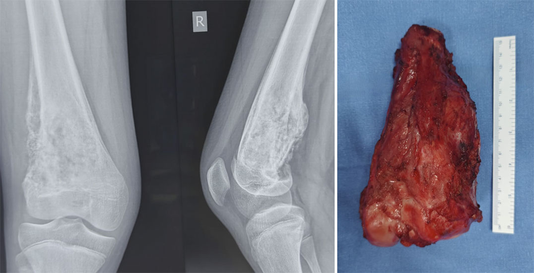
Figure 16A. Osteosarcoma treated with limb-salvage surgery: Wide resection of the tumor around the knee followed by reconstruction with a custom endoprosthesis.
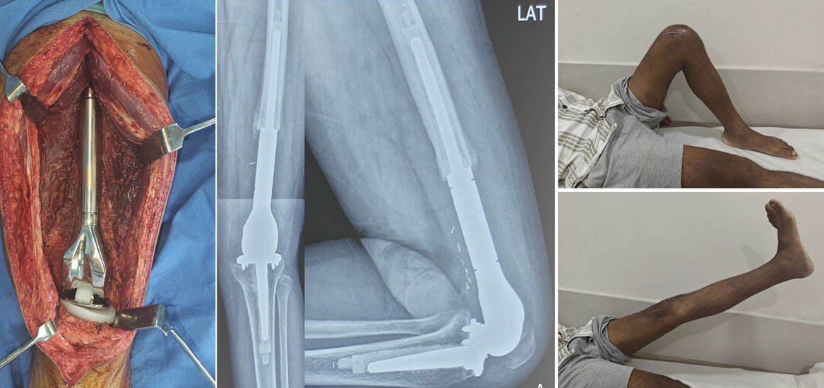
Figure 16B. Functional recovery after osteosarcoma surgery: Good knee movement demonstrating successful functional rehabilitation after limb-salvage surgery.
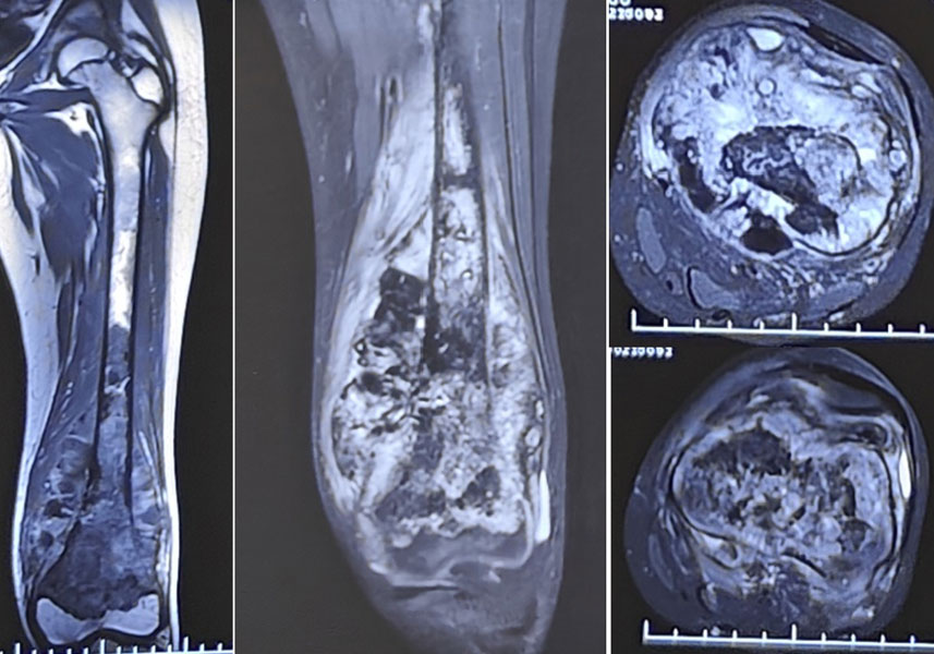
Figure 17A. Osteosarcoma involving the growth plate: Imaging shows a tumor extending to and involving the growth plate in a young patient.
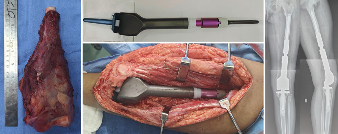
Figure 17B. Limb-salvage surgery with an expandable prosthesis: Wide resection of osteosarcoma followed by reconstruction using an expandable endoprosthesis to accommodate future growth.
-shown-on-X-ray-and-MRI-before-treatment.jpg "Osteosarcoma of the tibia (shin bone) shown on X-ray and MRI before treatment")
Figure 18A. Osteosarcoma of the tibia (shin bone) shown on X-ray and MRI before treatment
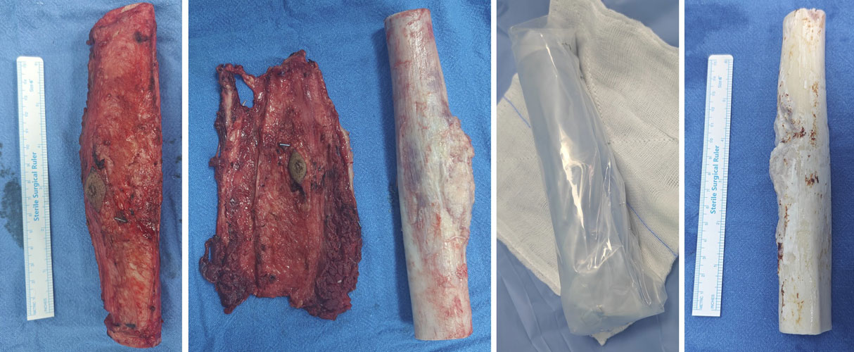
Figure 18B. Limb-salvage surgery for tibial osteosarcoma using wide resection, high-dose radiation and reimplantation of the patient’s own bone.
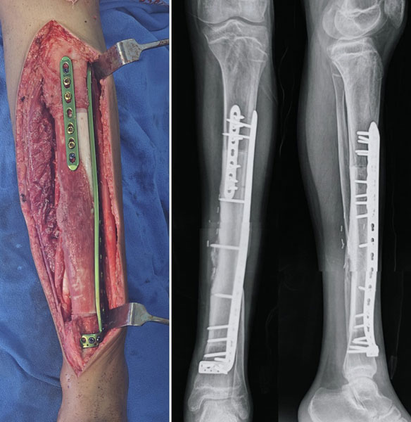
Figure 18C. Follow-up X-ray showing good bone union and incorporation after limb-salvage surgery for osteosarcoma.
-shown-on-X-ray-and-MRI.jpg "Osteosarcoma of the tibia (shin bone) shown on X-ray and MRI")
Figure 19A. Osteosarcoma of the tibia (shin bone) shown on X-ray and MRI.
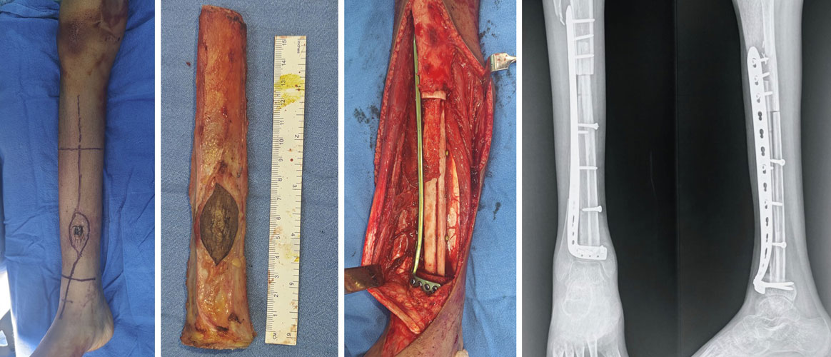
Figure 19B. Limb-salvage reconstruction of tibial osteosarcoma using the patient’s own fibula after wide tumor resection
- Mega Prosthesis Surgery
- Bone Reconstruction Surgery
- Rotationplasty
Clinical Pearl
Successful limb salvage depends on complete tumor removal. Preserving the limb should never compromise cancer control. The safest operation is the one that achieves both adequate oncological clearance and the best possible long-term function.
What We Look For During Surgery
Although every operation is individualized, the surgical team aims to achieve several important goals:
- Complete removal of the tumor with an adequate margin.
- Protection of nearby nerves and blood vessels whenever possible.
- Preservation of muscles and joints when oncologically safe.
- Stable reconstruction of the bone.
- Planning for long-term function and rehabilitation.
These decisions are made before surgery using detailed imaging, biopsy findings, and multidisciplinary discussion.
Is Surgery Necessary for Every Patient?
For high-grade osteosarcoma, surgery is almost always an essential part of treatment.
Although chemotherapy destroys many cancer cells, it cannot reliably eliminate the primary bone tumor on its own. Removing the remaining tumor surgically remains the standard of care.
- Limb Salvage Surgery
- Amputation for Bone Tumors
Doctor Insight
Unlike many routine orthopaedic procedures, bone tumor surgery begins long before entering the operating room. Every incision, biopsy tract, bone cut, and reconstruction is planned in advance to achieve complete tumor removal while preserving function whenever oncologically safe.
Clinical Pearl
The first operation offers the best opportunity for cure. Every step—from biopsy planning to definitive surgery—is performed with long-term cancer control and functional recovery in mind.
Doctor Explains
Patients often ask:
“Can you tell during surgery if all the cancer has been removed?”
The surgeon removes the tumor according to the preoperative plan and the planned surgical margins. The final confirmation comes from the pathology examination, where the entire specimen is carefully assessed under the microscope to ensure that the margins are free of tumor.
Is Amputation Ever Necessary?
Although much less common today, amputation remains the safest option in selected situations, such as:
- Extensive involvement of major nerves or blood vessels
- Very large recurrent tumors
- Severe infection after previous reconstruction
- Non-functional limb despite salvage options
The decision is individualized and based on achieving the best long-term oncological and functional outcome.
Internal Link: → Amputation for Bone Tumors
Doctor Insight
The goal is not simply to save the limb—it is to save a useful limb while completely removing the cancer.
Clinical Pearl
Limb preservation should never compromise complete tumor removal. Oncological safety always comes first.
Rehabilitation After Surgery
Rehabilitation begins soon after surgery and is an essential part of treatment.
The programme focuses on:
- Pain control
- Wound healing
- Joint movement
- Muscle strengthening
- Walking training
- Functional independence
Recovery varies depending on the type of surgery, reconstruction, and individual patient factors.
Internal Link: → Rehabilitation After Bone Tumor Surgery
Patient Tip
Recovery is gradual. Consistent physiotherapy and following your rehabilitation programme are among the most important factors influencing long-term function.
Prognosis
The outlook for osteosarcoma depends on several factors, including:
- Whether the disease is localized or metastatic
- Complete surgical removal
- Response to chemotherapy
- Tumor subtype
- Long-term follow-up
Patients with localized osteosarcoma generally have a more favourable prognosis than those with metastatic disease. Your treating team can provide individualized information based on your specific clinical situation.
Reassurance Box
Statistics describe outcomes for groups of patients—not individuals. Many factors influence prognosis, and every patient’s journey is unique.
Follow-up After Treatment
Regular follow-up is essential to:
- Detect recurrence early
- Monitor lung metastases
- Assess implant function
- Identify late effects of chemotherapy
- Support long-term recovery
Follow-up is usually more frequent during the first few years after treatment and becomes less frequent over time.
| Time After Treatment |
Typical Follow-up* |
| Years 1–2 |
Every 3–4 months |
| Years 3–5 |
Every 4–6 months |
| After 5 years |
Annually (individualized) |
*Follow-up schedules may vary depending on institutional protocols and individual clinical circumstances.
Survivorship After Osteosarcoma
Completing treatment is an important milestone, but survivorship extends well beyond the end of chemotherapy or surgery.
Long-term care focuses on:
- Returning to education or work
- Physical activity
- Emotional well-being
- Monitoring implant durability
- Screening for late treatment effects
- Maintaining overall health
Most patients gradually resume many of their normal daily activities, although recovery timelines vary.
Did You Know?
Many osteosarcoma survivors go on to complete their education, build careers, participate in sports, and lead fulfilling lives after treatment.
Frequently Asked Questions (FAQs)
1. What is osteosarcoma?
Osteosarcoma is the most common primary malignant bone tumor, meaning it is a cancer that starts within the bone rather than spreading from another organ. It most commonly affects children, teenagers, and young adults, although it can occur at any age.
2. Is osteosarcoma curable?
Many patients with localized osteosarcoma can be successfully treated with modern chemotherapy and surgery. The outcome depends on several factors, including the stage of the disease, response to chemotherapy, and complete surgical removal of the tumor.
3. What are the earliest symptoms of osteosarcoma?
The earliest symptom is usually persistent, localized bone pain. Swelling, limping, or reduced joint movement may develop later. Pain that continues despite rest or treatment should be evaluated.
4. Does osteosarcoma always cause severe pain?
Not always. Some patients experience mild discomfort initially, while others develop more significant pain as the tumor grows. Persistent pain is more important than pain intensity.
5. Why is osteosarcoma often mistaken for a sports injury?
Many patients are active adolescents, and the early symptoms—pain during activity or after exercise—can resemble common sports injuries. Symptoms that persist or worsen should be investigated further.
6. Why do I need both an MRI and a biopsy?
MRI shows where the tumor is and how far it extends, while a biopsy confirms exactly what type of tumor it is. Both are essential before treatment begins.
7. Why is chemotherapy given before surgery?
Chemotherapy treats microscopic cancer cells throughout the body and helps assess how the tumor responds to treatment before surgery.
8. Will I definitely need chemotherapy?
Most patients with high-grade osteosarcoma require chemotherapy. However, treatment is individualized based on the tumor subtype, stage, and overall health.
9. Will I lose my arm or leg?
Not necessarily. Advances in limb salvage surgery mean that many patients can keep their limb while still achieving effective cancer treatment. The safest option depends on the location and extent of the tumor.
10. Can osteosarcoma spread?
Yes. Osteosarcoma can spread to other parts of the body, most commonly the lungs. This is why CT scans of the chest are routinely performed before treatment.
11. What is the survival rate for osteosarcoma?
Survival varies depending on the stage of the disease, response to chemotherapy, and complete removal of the tumor. Your treating team can provide information specific to your individual situation.
12. Can osteosarcoma come back after treatment?
Yes. Although many patients remain disease-free, recurrence can occur. Regular follow-up appointments are important for early detection and timely management.
13. Is osteosarcoma hereditary?
Most cases are not inherited. Rare genetic syndromes can increase the risk, but these account for only a small proportion of patients.
14. Can osteosarcoma be prevented?
At present, there is no proven way to prevent osteosarcoma. Most cases occur without an identifiable cause.
15. Will treatment affect fertility?
Some chemotherapy medicines may affect fertility. Fertility preservation options should be discussed with your oncology team before treatment begins whenever appropriate.
16. Can children return to school after treatment?
Yes. Most children gradually return to school after recovering from surgery and chemotherapy, although additional support or a phased return may be helpful.
17. Will I be able to play sports again?
Many patients return to recreational activities after rehabilitation. The type of surgery and reconstruction influences which sports are recommended.
18. How long will follow-up continue?
Follow-up usually continues for several years after treatment to monitor for recurrence, lung metastases, implant function, and long-term effects of therapy.
19. Should I seek a second opinion?
Yes. Seeking a second opinion is entirely appropriate, particularly before major surgery or if you have questions about your diagnosis or treatment options.
20. What can I do to improve my recovery?
Following your chemotherapy schedule, participating in physiotherapy, attending follow-up appointments, maintaining good nutrition, and communicating openly with your healthcare team all contribute to the best possible recovery.
Myths and Facts
| Myth |
Fact |
| Osteosarcoma is caused by a fall or sports injury. |
Trauma does not cause osteosarcoma, although an injury may draw attention to an existing tumor. |
| Every patient with osteosarcoma needs amputation. |
Many patients are successfully treated with limb salvage surgery. |
| A biopsy causes cancer to spread. |
A properly planned biopsy is safe and essential for accurate diagnosis. |
| MRI alone can diagnose osteosarcoma. |
MRI is crucial, but a biopsy is required to confirm the diagnosis. |
| Chemotherapy is only needed if the cancer has spread. |
Chemotherapy is a standard part of treatment for most high-grade osteosarcomas, even when the disease appears localized. |
| Osteosarcoma only affects children. |
It is most common in young people but can also occur in adults. |
| Persistent bone pain always means cancer. |
Most bone pain is caused by non-cancerous conditions, but persistent symptoms deserve evaluation. |
| Once treatment is completed, follow-up is no longer necessary. |
Long-term follow-up is an essential part of osteosarcoma care. |
References
The recommendations and information presented on this page are based on current evidence and internationally accepted guidelines, including:
- National Comprehensive Cancer Network (NCCN). Clinical Practice Guidelines in Oncology: Bone Cancer. Latest available version.
- European Society for Medical Oncology (ESMO). Bone Sarcomas: ESMO Clinical Practice Guidelines.
- World Health Organization (WHO). WHO Classification of Soft Tissue and Bone Tumours, 5th Edition.
- Musculoskeletal Tumor Society (MSTS). Clinical recommendations and educational resources.
- International Society of Limb Salvage (ISOLS). Educational statements and consensus guidance.
- Children’s Oncology Group (COG). Osteosarcoma treatment protocols and supportive care recommendations.
- Strauss SJ, Frezza AM, Abecassis N, et al. Bone sarcomas: ESMO–EURACAN–GENTURIS Clinical Practice Guideline. Annals of Oncology.
- Isakoff MS, Bielack SS, Meltzer P, Gorlick R. Osteosarcoma: Current Treatment and a Collaborative Pathway to Success. Journal of Clinical Oncology.
- Ritter J, Bielack SS. Osteosarcoma. Annals of Oncology.
- National Cancer Institute (NCI). Osteosarcoma and Undifferentiated Pleomorphic Sarcoma of Bone (PDQ®) – Health Professional and Patient Versions.
Note: This page is reviewed periodically to reflect evolving evidence. Treatment recommendations should always be individualized according to the patient’s clinical condition and current international guidelines.
Medical Disclaimer
The information on this page is intended for educational purposes only and should not replace consultation with a qualified healthcare professional. Every patient with osteosarcoma is unique, and treatment decisions depend on the tumor’s characteristics, stage, overall health, and individual circumstances.
If you have persistent bone pain, swelling, or have been diagnosed with osteosarcoma, consult an orthopaedic oncologist or a multidisciplinary bone tumor team for a comprehensive evaluation. Do not delay seeking medical advice based solely on information found online.
 +91 93558 33005