Quick Summary
Bone tumors around the knee are among the most common bone tumors seen by orthopaedic oncologists. Fortunately, most are not cancerous (benign), and many can be treated successfully while preserving the leg and knee function. Early diagnosis, accurate imaging, proper biopsy, and treatment by an experienced orthopaedic oncology team are the key to achieving the best possible outcome.
In this guide, you will learn:
- Which bone tumors commonly occur around the knee
- Symptoms that should never be ignored
- How knee bone tumors are diagnosed
- Treatment options, including limb salvage surgery
- Recovery and rehabilitation
- What to expect in the long term
Understanding Bone Tumors Around the Knee
Hearing the words “bone tumor” can be frightening. Many patients immediately worry about cancer, losing their leg, or needing extensive surgery. In reality, most bone tumors around the knee are benign (non-cancerous), and even many malignant (cancerous) tumors can now be treated with limb salvage surgery, allowing the leg to be preserved.
A bone tumor is an abnormal growth of cells within the bone. Some tumors grow slowly and never spread to other parts of the body, while others are aggressive and require specialized treatment. The treatment depends on the type of tumor, its location, its size, and whether it has spread beyond the bone.
The knee is one of the most common sites for bone tumors because it contains areas of rapid bone growth, particularly near the ends of the femur (thigh bone) and tibia (shin bone). These regions, known as the growth plates in children and adolescents, are where many primary bone tumors develop.
Understanding exactly where the tumor is located helps surgeons choose the safest treatment while preserving as much healthy bone, joint function, and mobility as possible.
Did You Know?
The area around the knee—especially the distal femur (lower end of the thigh bone) and proximal tibia (upper end of the shin bone)—is the most common location for osteosarcoma, the most common primary bone cancer in children, adolescents, and young adults.
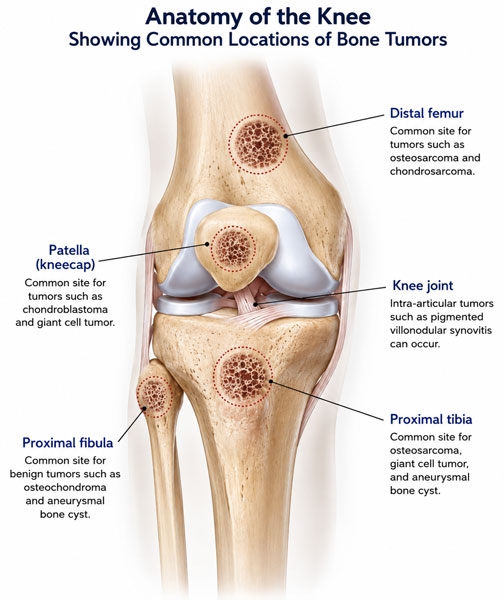
Figure 1. Anatomy of the knee and common locations of bone tumors
The distal femur, proximal tibia, patella and proximal fibula are common locations for different types of bone tumors around the knee.
Understanding the Bones Around the Knee
The knee is formed by the meeting of three bones—the distal femur (lower end of the thigh bone), the proximal tibia (upper end of the shin bone), and the patella (kneecap). Just to the outside of the knee lies the proximal fibula, a smaller bone that provides attachment for important ligaments and muscles.
Most primary bone tumors around the knee arise from the distal femur or proximal tibia, making this the most common location for bone cancers such as osteosarcoma and many benign tumors such as giant cell tumor of bone. These bones are covered by smooth articular cartilage, which allows painless movement, while strong muscles, ligaments, and tendons provide stability and help the knee bend and straighten.
Because the knee is a major weight-bearing joint, treatment aims not only to remove the tumor safely but also to preserve stability, movement, and long-term function.
Why Is the Knee the Most Common Location for Bone Tumors?
The knee is more than a joint—it is where the long bones of the leg meet and grow. During childhood and adolescence, the ends of these bones undergo rapid growth, making them more susceptible to certain bone tumors.
Several common bone tumors—including osteosarcoma, giant cell tumor of bone, chondroblastoma, and aneurysmal bone cyst—frequently arise around the knee.
The close relationship between the bone, cartilage, ligaments, muscles, nerves, and blood vessels also makes surgery in this region particularly demanding. The surgeon must remove the tumor completely while preserving as much normal anatomy as possible to maintain walking ability and knee function.
Modern advances such as high-resolution MRI, computer navigation, and patient-specific reconstruction have significantly improved the precision of surgery in selected patients.
Table. Bones Around the Knee and Common Bone Tumors
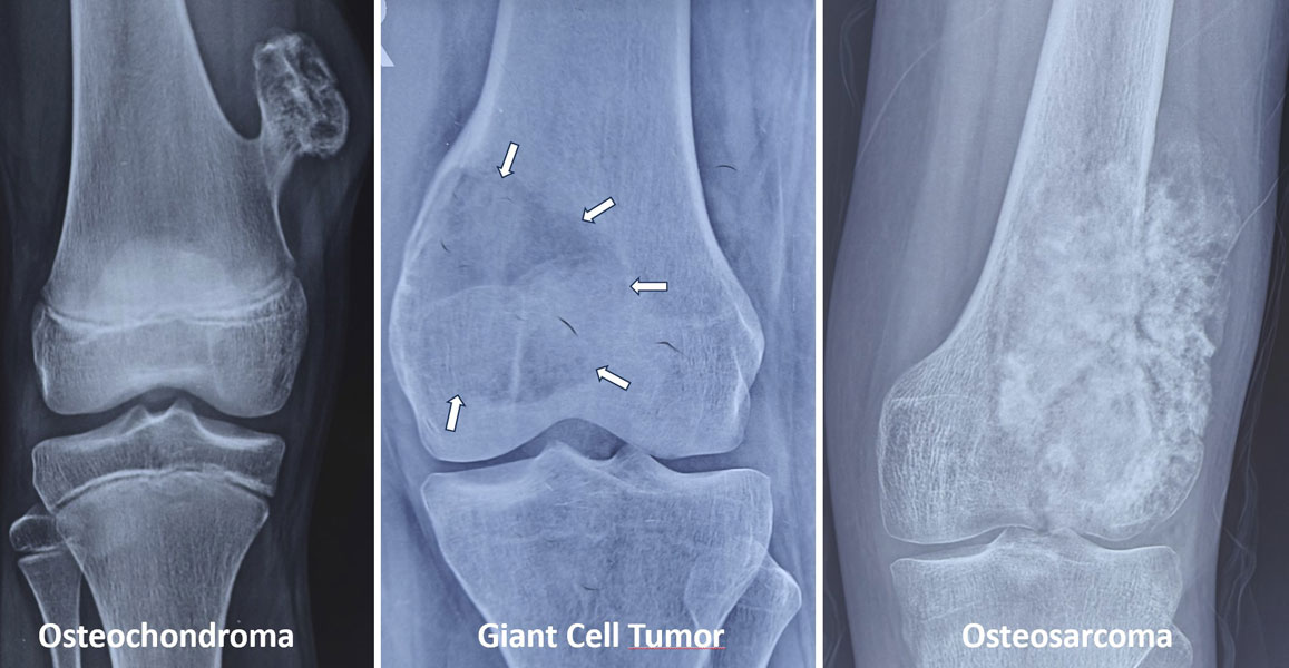
Figure 2. Common bone tumors involving the lower femur
X-rays showing a range of bone tumors that can occur around the lower end of the femur, including osteochondroma, giant cell tumor and osteosarcoma.
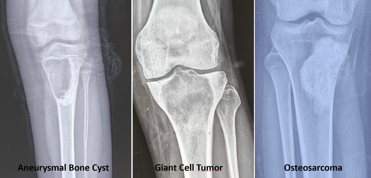
Figure 3. Common bone tumors involving the upper tibia
X-rays demonstrating different bone tumors around the upper tibia, including aneurysmal bone cyst, giant cell tumor and osteosarcoma.
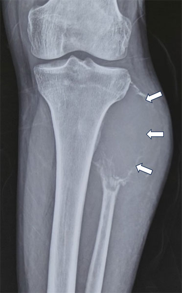
Figure 4. Bone tumor involving the upper fibula
X-ray showing a bone tumor involving the proximal fibula, an important but less common location for tumors around the knee.
Clinical Pearl
The location of a bone tumor often provides important clues about its diagnosis. Around the knee, certain tumors have characteristic locations that help guide further investigations, although a biopsy is usually required for a definitive diagnosis.
Symptoms of a Knee Bone Tumor
The symptoms of a bone tumor depend on its type, size, and location. Some tumors are discovered incidentally during an X-ray performed for an unrelated injury, while others cause progressive symptoms over weeks or months.
The most common symptom is persistent pain around the knee. Unlike pain caused by a minor injury, tumor-related pain often does not improve with rest and may gradually worsen over time. Some patients notice that the pain is more severe at night or becomes increasingly frequent.
As the tumor enlarges, additional symptoms may develop, including swelling, stiffness, reduced joint movement, or difficulty walking. Occasionally, the first sign is a pathological fracture—a break occurring in bone weakened by the tumor after minimal trauma.
Common Symptoms
- Persistent knee pain
- Swelling around the knee
- A lump or visible enlargement
- Reduced knee movement
- Difficulty walking or limping
- Night pain
- Pain that progressively worsens
- Bone fracture after a minor injury
Children may complain of persistent knee pain that is initially mistaken for a sports injury or “growing pains.” Adults may attribute symptoms to arthritis or overuse, leading to delays in diagnosis.
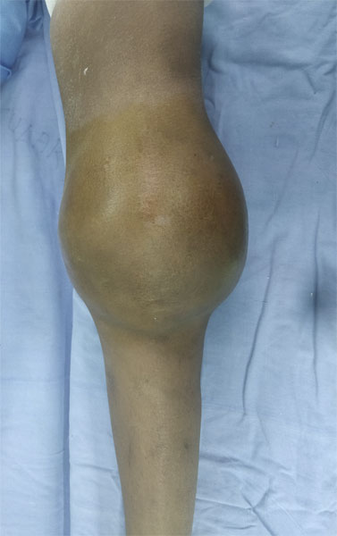
Figure 5. Bone tumor around the knee presenting as swelling
Clinical photograph showing a visible swelling around the knee caused by a bone tumor.
Red Flag Symptoms That Should Never Be Ignored
🚩 Seek prompt medical evaluation if you experience:
- Knee pain lasting more than 4–6 weeks
- Pain that wakes you from sleep
- Progressive swelling around the knee
- A hard lump near the bone
- Pain that does not improve despite rest or medication
- A fracture after a trivial injury
- Persistent pain in a child or teenager without a clear cause
These symptoms do not necessarily mean cancer, but they warrant further assessment with imaging and specialist evaluation.
Patient Tip
If your knee pain continues despite rest, physiotherapy, or pain medication—or if you notice swelling or a lump—request an X-ray and consult an orthopaedic specialist. Early diagnosis often leads to simpler treatment and better outcomes.
Could My Knee Pain Be Cancer?
Probably not.
The vast majority of people with knee pain have common conditions such as ligament injuries, tendon problems, arthritis, or overuse injuries—not a bone tumor. Bone tumors are uncommon, and primary bone cancers are rare.
However, certain features make a bone tumor more likely and should prompt further evaluation. These include persistent pain without a clear injury, pain that worsens over time, swelling over the bone, or an abnormal X-ray.
Because many benign and malignant bone tumors can initially resemble routine knee problems, persistent or unexplained symptoms should never be ignored.
Table 2. Bone Tumor vs Common Causes of Knee Pain
Reassurance Box
Most knee pain is not caused by a bone tumor. Nevertheless, persistent pain, swelling, or an abnormal X-ray should always be evaluated by an experienced specialist. Seeking medical advice early does not mean you have cancer—it simply helps ensure the correct diagnosis and timely treatment if needed.
How Are Knee Bone Tumors Diagnosed?
Accurate diagnosis is the foundation of successful treatment. Before any surgery is planned, your doctor must determine the exact type of bone tumor, its size, location, and whether it has spread beyond the bone. This ensures that treatment is tailored to your condition while maximizing the chances of preserving both your limb and knee function.
Diagnosis follows a carefully planned sequence. Performing surgery or a biopsy before completing the appropriate investigations can complicate future treatment.
The Diagnostic Process
Most patients undergo the following steps:
- Clinical Evaluation – Your doctor will assess your symptoms, examine the knee, and review your medical history.
- Imaging Studies – An X-ray is usually the first investigation, followed by an MRI, which is the most important scan for defining the extent of the tumor and planning surgery. Depending on the suspected diagnosis, CT, PET-CT, or a bone scan may also be required.
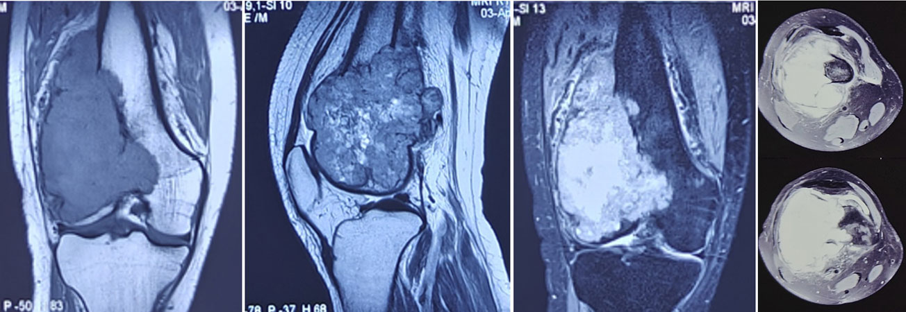
Figure 6. MRI evaluation of a bone tumor around the knee
MRI scans help define the size and extent of a bone tumor and its relationship with the knee joint and surrounding soft tissues.
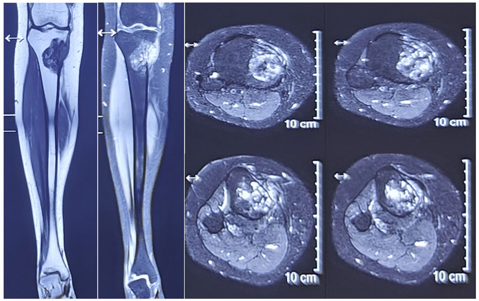
Figure 7. MRI assessment of a bone tumor around the knee
Detailed MRI images showing the extent of a bone tumor and its involvement of the surrounding tissues.
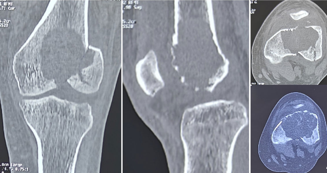
Figure 8. CT scan evaluation of a bone tumor around the knee
CT imaging provides detailed information about the bone involvement, cortical destruction and extent of a tumor around the knee.
- Biopsy – A small tissue sample is obtained to establish the exact diagnosis. In most cases, a core needle biopsy is sufficient. The biopsy should ideally be planned and performed by the same specialist team that will undertake the definitive surgery.
- Multidisciplinary Team (MDT) Review – Orthopaedic oncologists, radiologists, pathologists, medical oncologists, and other specialists review all findings together to develop an individualized treatment plan.
Clinical Pearl
Treat the diagnosis before treating the tumor. A well-planned biopsy is the first step toward successful limb salvage surgery.
🚩 Red Flag
Never undergo a biopsy or surgical removal of a suspected bone tumor before consultation with an orthopaedic oncologist. An unplanned procedure can affect future treatment options.
Reassurance Box
Waiting for investigations and biopsy results can be stressful. However, a careful diagnosis is essential to choosing the safest and most effective treatment. Taking the time to establish the correct diagnosis often improves the chances of successful limb salvage and long-term function.
Treatment of Knee Bone Tumors
Treatment depends on the type of tumor, whether it is benign or malignant, its size, location, and your age and activity level. There is no single operation suitable for every patient. The treatment plan is individualized after careful evaluation by a multidisciplinary bone tumor team.
The goals of treatment are to:
- Completely remove the tumor while preserving healthy tissues whenever possible.
- Restore a stable, painless, and functional knee.
- Minimize the risk of local recurrence.
- Allow patients to return to their normal daily activities as safely as possible.
Treatment may involve observation, curettage, limb salvage surgery, chemotherapy, radiotherapy, or, in selected situations, amputation.
Curettage for Benign Bone Tumors Around the Knee
Many benign but locally aggressive tumors around the knee can be treated without removing the entire bone.
The most common example is a Giant Cell Tumor of Bone (GCT), which frequently occurs in the distal femur and proximal tibia of young adults.
(Giant Cell Tumor of Bone)
The standard treatment is extended curettage, in which the tumor is carefully scraped out from inside the bone cavity. To reduce the chance of recurrence, surgeons usually combine curettage with additional local treatments (known as adjuvants) such as a high-speed burr, chemical agents, thermal techniques, or argon beam coagulation. The resulting cavity is then reconstructed using bone cement (PMMA), bone graft, or a combination of both, depending on the location and size of the defect.
This approach preserves the patient’s own knee joint while providing excellent function in appropriately selected patients.
Other benign tumors such as chondroblastoma and selected aneurysmal bone cysts may also be treated using curettage.
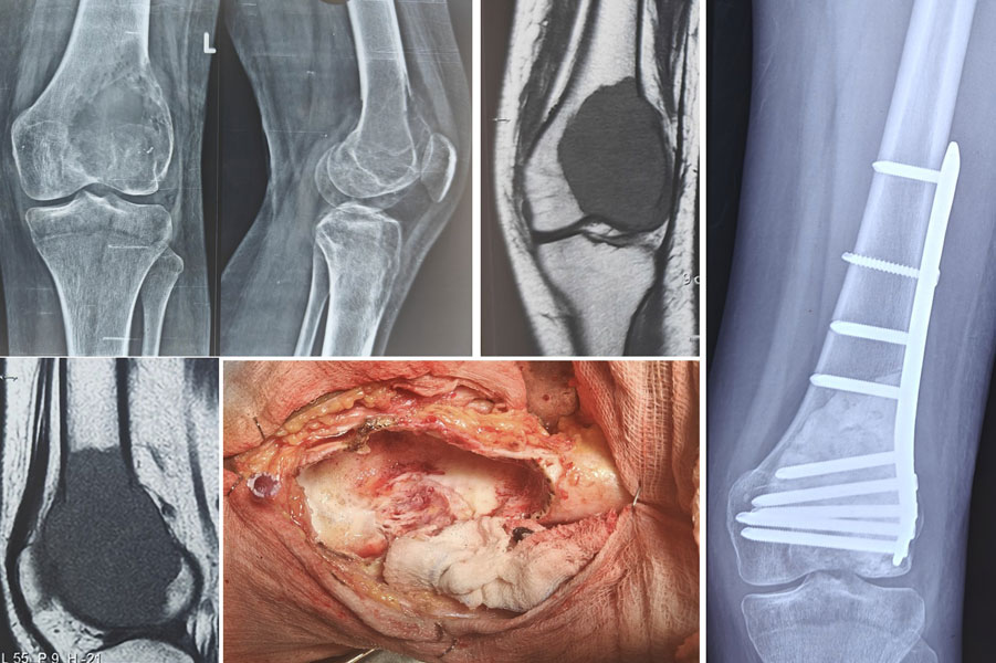
Figure 9. Extended curettage for a benign bone tumor around the knee
Surgical treatment of a giant cell tumor using extended curettage to remove the tumor while preserving the surrounding bone and knee joint.
Clinical Pearl
Joint-preserving curettage is preferred whenever oncologically safe, particularly in young patients with benign tumors around the knee.
Limb Salvage Surgery for Malignant Bone Tumors
For most primary bone cancers around the knee, the preferred treatment today is limb salvage surgery, in which the tumor is removed with an adequate margin of healthy tissue while preserving the limb.
(Limb Salvage Surgery for Bone Tumors)
Following tumor removal, the resulting bone and joint defect must be reconstructed to restore stability and allow walking.
The two most common sites requiring reconstruction are the distal femur and proximal tibia.
Distal Femur Tumor Resection and Megaprosthetic Reconstruction
Tumors involving the distal femur (the lower end of the thigh bone) are most commonly reconstructed using a distal femoral megaprosthesis, also known as a tumor prosthesis.
(Tumor Prosthesis (Megaprosthesis): A Complete Patient Guide)
A megaprosthesis is a specially designed metallic implant that replaces the portion of bone removed with the tumor, together with the knee joint surface when necessary. Modern modular systems allow surgeons to customize the implant according to the amount of bone resected.
Most patients are able to begin standing and walking with physiotherapy within a few days after surgery, making megaprosthetic reconstruction one of the most reliable options for restoring early mobility.
Fixed-Hinge vs Rotating-Hinge Prosthesis
Modern tumor prostheses are available in two principal designs:
- Fixed-hinge prostheses have a rigid connection between the femoral and tibial components. Although they provide excellent stability, they transmit greater rotational forces to the implant-bone interface and are now used less commonly.
- Rotating-hinge prostheses allow controlled rotational movement in addition to flexion and extension, better reproducing normal knee mechanics. By reducing torsional stress, they may decrease mechanical loosening and have become the preferred design in most contemporary limb salvage procedures.
The choice of implant depends on factors such as bone loss, ligament preservation, patient age, and surgeon preference.
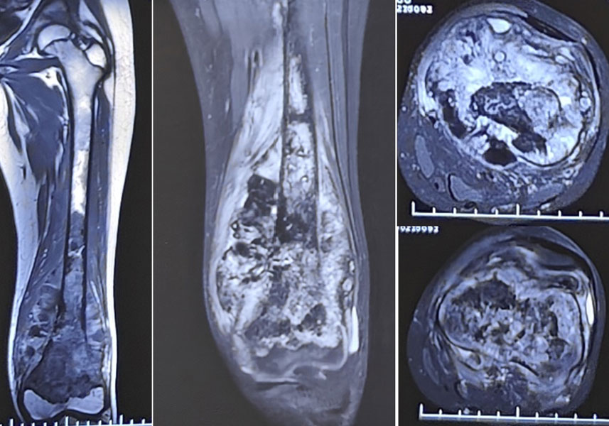
Figure 10A. Osteosarcoma of the lower femur in a 12-year-old boy
X-ray and MRI showing osteosarcoma involving the lower end of the femur in a young patient.
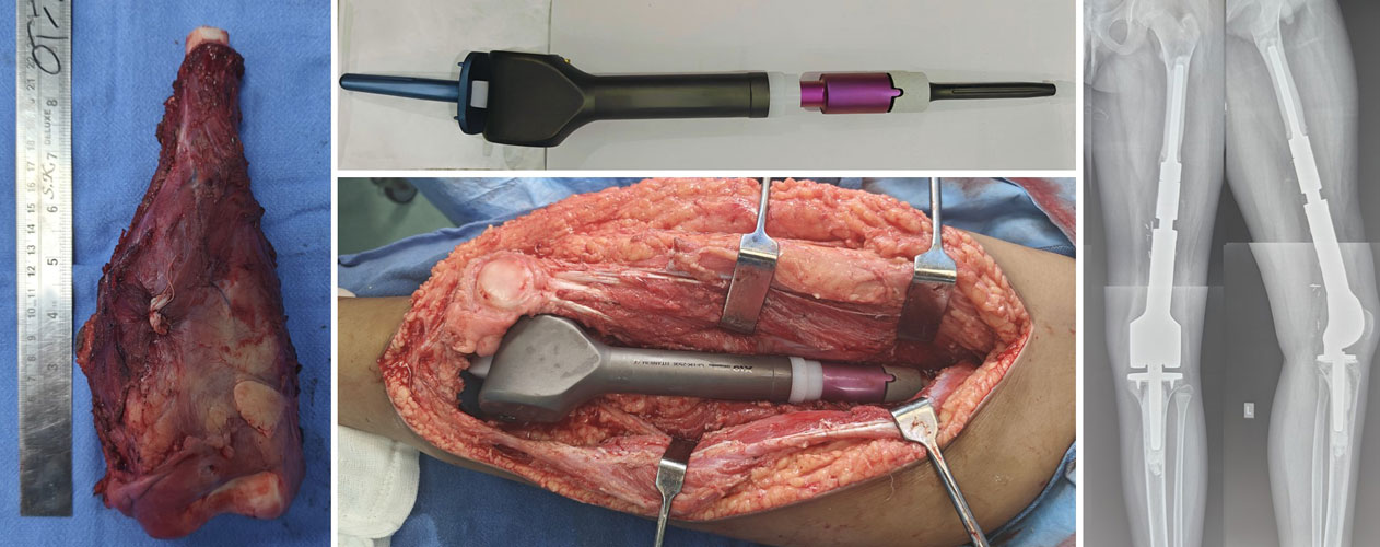
Figure 10B. Distal femur reconstruction after wide resection of osteosarcoma
After wide removal of the tumor, the knee was reconstructed using a distal femur endoprosthesis to preserve limb function.
Proximal Tibia Tumor Resection and Reconstruction
Reconstruction after proximal tibia tumor resection is technically more demanding because the tumor often involves the attachment of the patellar tendon, which forms part of the extensor mechanism—the system of muscles and tendons that allows the knee to straighten.
Most patients undergo reconstruction using a proximal tibial megaprosthesis.
( Tumor Prosthesis (Megaprosthesis): A Complete Patient Guide)
Extensor Mechanism Reconstruction
Restoring the extensor mechanism is one of the most important aspects of proximal tibial reconstruction. Without it, patients may have difficulty actively straightening the knee or walking confidently.
Several techniques are available, including:
- Reattachment of the patellar tendon to the prosthesis using dedicated fixation systems.
- Reinforcement with synthetic materials or biological grafts in selected patients.
- Coverage with a medial gastrocnemius muscle flap, which improves soft tissue coverage, protects the prosthesis, and enhances healing.
Postoperative rehabilitation is carefully supervised because the reconstructed tendon requires time to heal before full knee function can be regained.
Doctor Insight
Although proximal tibial reconstruction is more complex than distal femoral reconstruction, meticulous extensor mechanism reconstruction and structured physiotherapy allow most patients to regain independent walking and satisfactory knee function.
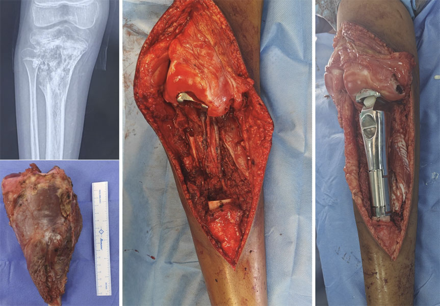
Figure 11A. Osteosarcoma of the upper tibia treated with wide resection
Imaging and surgical photographs showing wide removal of an osteosarcoma involving the upper end of the tibia.
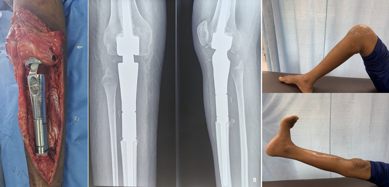
Figure 11B. Proximal tibia reconstruction after bone tumor surgery
The knee was reconstructed with a proximal tibia endoprosthesis following wide resection, allowing useful knee movement and limb function.
Management of Proximal Fibula Tumors
The proximal fibula is an uncommon site for bone tumors but presents unique surgical challenges because of its close relationship to the common peroneal nerve, which controls ankle and toe movement.
Benign tumors such as osteochondroma are often treated by en bloc excision, particularly when they cause pain, nerve compression, or progressive enlargement.
For malignant tumors, wide excision of the proximal fibula is usually required to achieve adequate oncological clearance while protecting the nearby neurovascular structures whenever possible.
Although removal of the proximal fibula generally has little effect on weight-bearing, careful reconstruction of the lateral collateral ligament and associated soft tissues may be required to maintain knee stability in selected patients.
Patients should also be counselled regarding the small but important risk of temporary or permanent weakness of ankle dorsiflexion due to involvement of the common peroneal nerve.
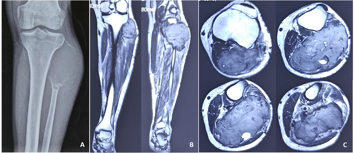
Figure 12A. Giant cell tumor of the upper fibula
X-ray showing a giant cell tumor involving the proximal fibula near the knee.
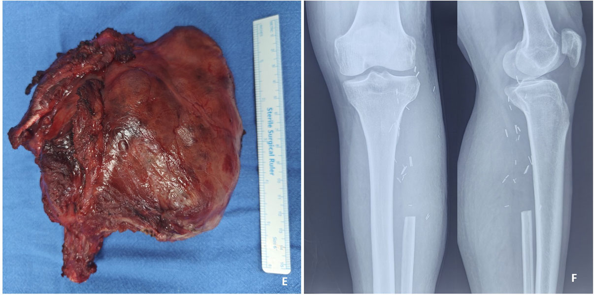
Figure 12B. Surgical resection of a giant cell tumor of the upper fibula
The tumor was treated with surgical resection of the involved proximal fibula while preserving the surrounding structures.
Table. Surgical Treatment Options for Knee Bone Tumors
Reassurance Box
Advances in surgical techniques and implant technology mean that most patients with malignant tumors around the knee can now undergo limb salvage surgery instead of amputation. Although every operation is individualized, the primary goals remain the same—to remove the tumor safely, restore function, and help patients return to an active and independent life.
Recovery and Rehabilitation After Knee Bone Tumor Surgery
Recovery after knee bone tumor surgery is a gradual process that continues for several months. Although the exact timeline varies depending on the type of tumor, the surgical procedure performed, and whether chemotherapy is required, most patients steadily regain independence through structured rehabilitation.
Recovery begins immediately after surgery. A multidisciplinary rehabilitation team—including physiotherapists, occupational therapists, rehabilitation physicians, nursing staff, and your orthopaedic oncologist—works together to restore mobility while protecting the surgical reconstruction.
The rehabilitation programme is individualized according to the type of reconstruction performed. Patients who undergo joint-preserving curettage for benign tumors generally recover much faster than those requiring megaprosthetic reconstruction after limb salvage surgery.
Walking After Surgery
One of the most common questions patients ask is:
“When will I be able to walk again?”
The answer depends on the operation performed.
Following distal femoral megaprosthetic reconstruction, many patients begin standing and walking with a walker or crutches within the first few days after surgery under the supervision of a physiotherapist.
Patients undergoing proximal tibial reconstruction often require a more gradual rehabilitation programme because the reconstructed extensor mechanism needs time to heal. A knee brace may be used during the early weeks to protect the repair while allowing controlled rehabilitation.
Patients treated with curettage for benign tumors usually recover more quickly and often regain normal knee function sooner.
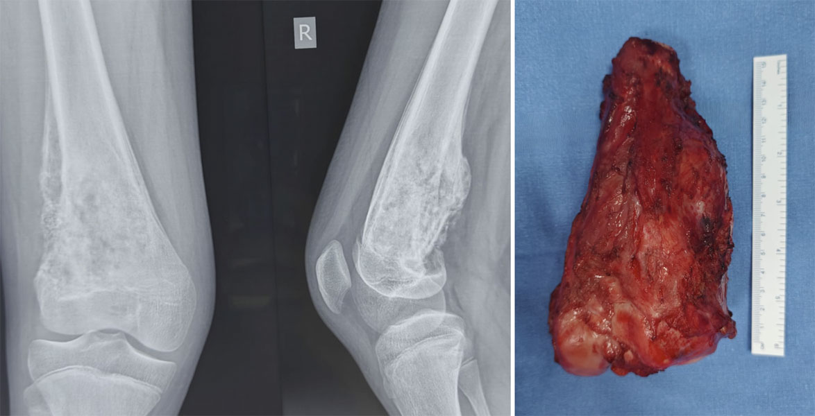
Figure 13A. Osteosarcoma of the lower femur before wide resection
X-rays showing an osteosarcoma involving the lower end of the femur around the knee before limb-sparing surgery.
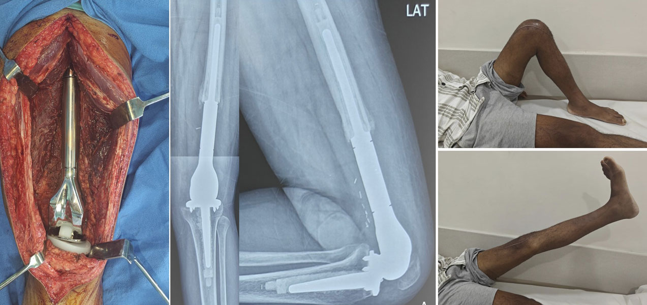
Figure 13B. Regaining knee movement after osteosarcoma surgery
After distal femur reconstruction with an endoprosthesis, structured physiotherapy helped restore good knee movement and function.
Physiotherapy
Physiotherapy is an essential part of treatment and begins soon after surgery.
The goals include:
- Restoring knee movement
- Improving muscle strength
- Regaining balance and coordination
- Returning to independent walking
- Preventing joint stiffness
- Building confidence during daily activities
Exercises are progressed gradually according to bone healing, soft tissue recovery, and implant stability.
Table. General Recovery Timeline After Knee Bone Tumor Surgery
Recovery varies depending on the type of surgery, reconstruction, chemotherapy, and individual healing.
Returning to Daily Life
Most patients gradually resume everyday activities over the months following surgery.
The timeline for returning to work, school, driving, or recreational activities depends on several factors, including:
- Type of surgery
- Functional recovery
- Occupation
- Ongoing chemotherapy, if required
- Overall health
High-impact activities such as running or contact sports are generally discouraged after megaprosthetic reconstruction to maximize implant longevity. However, many patients return to an active lifestyle that includes walking, cycling, swimming, travelling, and low-impact recreational activities.
Patient Tip
Recovery is not a race. Consistent physiotherapy, good nutrition, and regular follow-up appointments are often more important than trying to regain function too quickly.
Doctor Insight
The success of limb salvage surgery depends not only on removing the tumor but also on committed rehabilitation. Patients who actively participate in physiotherapy generally achieve better mobility, greater confidence, and improved long-term function.
Reassurance Box
Recovery after bone tumor surgery requires patience, but most improvements occur gradually over many months. Every patient’s journey is unique, and progress should be measured against your own recovery rather than someone else’s. With expert surgical care, dedicated rehabilitation, and regular follow-up, many patients regain independence and enjoy an excellent quality of life.
Long-Term Outlook
A diagnosis of a bone tumor naturally raises many questions about the future. Patients often wonder whether the tumor will return, how long the implant will last, whether they will walk normally again, or if lifelong follow-up is necessary.
The outlook depends on several factors, including the type of tumor, stage of disease, treatment received, and response to therapy. While every patient’s journey is unique, advances in orthopaedic oncology have significantly improved both survival and functional outcomes, allowing many patients to return to independent and productive lives.
Regular follow-up remains an essential part of treatment. Follow-up appointments allow your medical team to monitor recovery, detect any signs of recurrence at an early stage, evaluate implant function, and address any new symptoms promptly.
Patients treated for malignant bone tumors usually require follow-up for several years, while those with benign tumors may need periodic monitoring depending on the diagnosis and risk of recurrence.
For detailed information about surveillance schedules and recurrence, visit Follow-up After Bone Tumor Treatment .
For a cornerstone page like “Knee Bone Tumor Surgery: Symptoms, Diagnosis & Treatment”, the FAQs should target Google’s People Also Ask (PAA), Featured Snippets, and AI Overviews. They should answer the highest-intent patient questions while naturally incorporating relevant keywords. The answers should be concise (80–150 words), medically accurate, and encourage internal navigation where appropriate.
Frequently Asked Questions
1. What are the first signs of a bone tumor around the knee?
The earliest symptom of a bone tumor around the knee is usually persistent pain that does not improve with rest or routine treatment. Some patients notice swelling, a gradually enlarging lump, stiffness, or difficulty walking. Pain that worsens at night or continues for several weeks without a clear injury should be evaluated by a doctor. Although most knee pain is caused by common conditions such as arthritis or sports injuries, persistent symptoms warrant an X-ray and, if necessary, further imaging with MRI.
Related page: Bone Tumor Symptoms: When Should You Worry?
2. Is every bone tumor around the knee cancer?
No. Most bone tumors around the knee are benign (non-cancerous). Common benign tumors include osteochondroma, giant cell tumor of bone, chondroblastoma, and aneurysmal bone cyst. Malignant tumors such as osteosarcoma, Ewing sarcoma, and chondrosarcoma are much less common. Imaging studies and a biopsy are often required to determine the exact diagnosis and guide treatment.
Related pages: Giant Cell Tumor of Bone, Osteosarcoma, Ewing Sarcoma
3. How is a knee bone tumor diagnosed?
Diagnosis begins with a detailed medical history, physical examination, and X-ray, followed by MRI to determine the extent of the tumor. Additional tests such as CT, PET-CT, or a bone scan may be recommended in selected patients. A biopsy is usually required to confirm the diagnosis before treatment is planned.
4. Do all knee bone tumors require surgery?
No. Treatment depends on the type of tumor. Some small, asymptomatic benign tumors may only require observation with regular follow-up. Others, such as giant cell tumors or malignant bone cancers, usually require surgery. Your orthopaedic oncologist will recommend the most appropriate treatment based on the diagnosis, symptoms, and risk of complications.
5. Can a bone tumor around the knee be treated without amputation?
Yes. Most patients with primary bone tumors around the knee can now undergo limb salvage surgery, where the tumor is removed while preserving the leg. Amputation is reserved for selected situations in which limb preservation would not provide safe cancer control or satisfactory function.
Related page: Limb Salvage Surgery for Bone Tumors
6. What is a tumor prosthesis (megaprosthesis)?
A tumor prosthesis, also called a megaprosthesis, is a specially designed artificial implant used to replace bone and joint removed during tumor surgery. Around the knee, it is commonly used after distal femur or proximal tibia tumor resection. Modern modular prostheses restore limb length, provide immediate stability, and allow many patients to begin walking soon after surgery.
Related page: Tumor Prosthesis (Megaprosthesis): A Complete Patient Guide
7. Will I be able to walk normally after knee bone tumor surgery?
Most patients regain independent walking after successful surgery and rehabilitation. Recovery depends on the type of operation, muscle strength, physiotherapy, and overall health. Patients treated with joint-preserving surgery often recover faster, while those undergoing megaprosthetic reconstruction require a structured rehabilitation programme over several months.
8. How long does recovery take after knee bone tumor surgery?
Initial recovery usually takes several weeks, while functional improvement continues for 6 to 12 months. The timeline varies according to the type of tumor, the reconstruction performed, and whether chemotherapy is required. Regular physiotherapy and follow-up appointments are essential for achieving the best possible outcome.
9. Can a bone tumor come back after surgery?
Some bone tumors can recur after treatment. Giant Cell Tumor of Bone has a recognized risk of local recurrence even after successful curettage, while malignant tumors require long-term surveillance to detect recurrence or spread at an early stage. Regular follow-up imaging is an important part of ongoing care.
Related pages: Giant Cell Tumor of Bone
10. Should a biopsy always be performed before surgery?
In most cases, yes. A biopsy confirms the exact diagnosis and helps determine the most appropriate treatment. The biopsy should ideally be planned and performed by the same specialist team that will carry out the definitive surgery, as an improperly performed biopsy can complicate future treatment.
Related page: Biopsy in Bone & Soft Tissue Tumor
11. When should I seek a second opinion for a knee bone tumor?
A second opinion is recommended before undergoing a biopsy or major surgery, particularly if you have been diagnosed with a malignant bone tumor or advised to undergo amputation. Because bone tumors are uncommon, evaluation by an experienced orthopaedic oncologist and a multidisciplinary sarcoma team can help confirm the diagnosis and ensure that all treatment options have been considered.
12. Which specialist should treat a bone tumor around the knee?
Bone tumors should be treated by an orthopaedic oncologist—an orthopaedic surgeon with specialized training in the diagnosis and treatment of bone and soft tissue tumors. These patients benefit from care at dedicated bone tumor or sarcoma centres, where orthopaedic oncologists work closely with radiologists, pathologists, medical oncologists, radiation oncologists, plastic surgeons, and rehabilitation specialists to provide comprehensive, evidence-based treatment.
Medical Disclaimer
Disclaimer: The information provided on this page is intended for educational purposes only and should not be used as a substitute for professional medical advice, diagnosis, or treatment. Knee bone tumors include a wide range of benign and malignant conditions, and management varies depending on the diagnosis, stage, and individual patient factors. If you have persistent knee pain, swelling, a bone lesion on imaging, or have been diagnosed with a bone tumor, consult an experienced orthopaedic oncologist for a comprehensive evaluation and personalized treatment plan.
References
- Jentzsch T, Erschbamer M, Seeli F, et al. Extensor function after medial gastrocnemius flap reconstruction of the proximal tibia. Clin Orthop Relat Res. 2013;471:2333-2339.
- Myers GJ, Abudu AT, Carter SR, Tillman RM, Grimer RJ. The long-term results of endoprosthetic replacement of the proximal tibia for bone tumours. J Bone Joint Surg Br. 2007;89(12):1632-1637.
- Kapoor L, Banjara R, Khan SA, Majeed A. Extensor apparatus reconstruction using prolene mesh tube and medial gastrocnemius flap following proximal tibial endoprosthetic reconstruction. Tech Orthop. 2022;37(1):65-69.
- Myers GJ, Abudu AT, Carter SR, et al. Endoprosthetic replacement of the distal femur for bone tumors: long-term results. J Bone Joint Surg Br. 2007;89:521-526.
- Sharma S, Turcotte RE, Isler MH, et al. Cemented rotating-hinge endoprosthesis for limb salvage of distal femur tumors. Clin Orthop Relat Res. 2006;450:28-32.
 +91 93558 33005