Quick Summary
Hand and wrist bone tumors are uncommon, but they should never be ignored. Early diagnosis allows appropriate treatment while preserving hand function.
Key Points
- Most hand and wrist bone tumors are benign (non-cancerous).
- Persistent pain, swelling, or an unexplained bone lesion should be evaluated by an orthopaedic oncologist.
- X-rays are usually the first investigation, followed by MRI when needed.
- A biopsy (removing a small tissue sample for microscopic examination) is often required before treatment.
- Many tumors can be treated with curettage (surgical removal of the tumor from within the bone) or limb-sparing surgery, avoiding amputation.
- Early diagnosis improves treatment options and long-term function.
Reassurance
Finding out that you have a bone tumor can be overwhelming. Fortunately, the majority of hand and wrist bone tumors are benign, and even many malignant tumors can be treated successfully while preserving the hand and wrist using modern limb-salvage techniques.
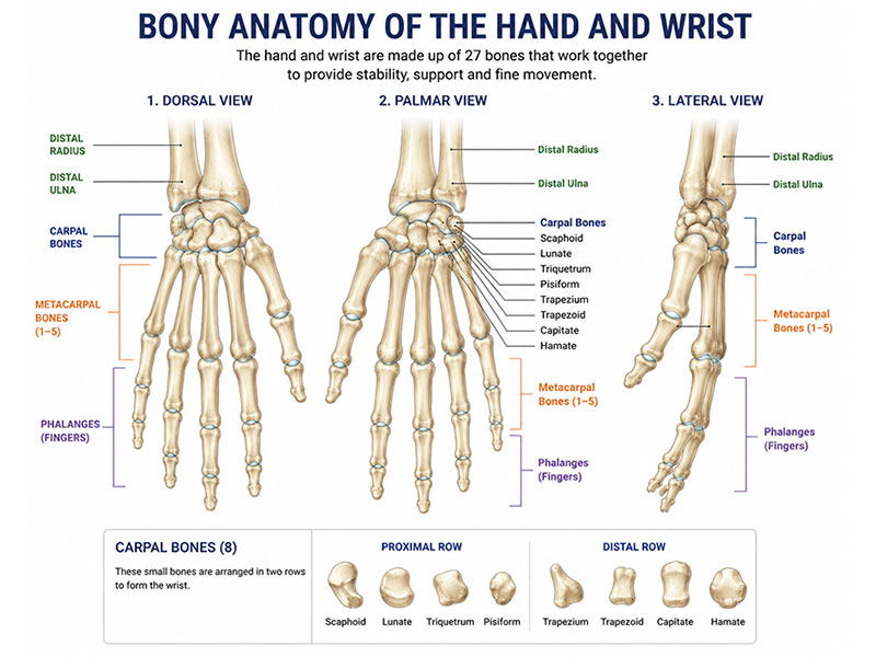
Figure 1. Bony anatomy of the hand and wrist
Understanding the bones of the hand and wrist is the first step in planning safe and precise bone tumor surgery.
Understanding the Anatomy of the Hand & Wrist
Understanding where a tumor develops helps explain the symptoms, treatment options, and expected recovery. The hand and wrist consist of 29 bones, multiple joints, tendons, nerves, blood vessels, and ligaments that work together to provide strength, mobility, and fine motor control.
Unlike tumors around larger joints such as the hip or knee, even small lesions in the hand may significantly affect grip, finger movement, or dexterity.
Distal Radius
The distal radius is the lower end of the radius, the larger of the two forearm bones. It forms most of the wrist joint and plays a crucial role in wrist motion and load transmission.
Tumors in this region may cause:
- Wrist pain
- Swelling
- Reduced wrist movement
- Weak grip
- Pathological fracture (a fracture occurring through weakened bone)
Certain tumors, particularly giant cell tumors, commonly occur around the distal radius and may require specialized reconstructive surgery.
Distal Ulna
The distal ulna forms the inner side of the wrist and contributes to forearm rotation and stability of the distal radioulnar joint (DRUJ)—the joint that allows the forearm to rotate during activities such as turning a key or using a screwdriver.
Although tumors of the distal ulna are relatively uncommon, treatment aims to preserve both wrist stability and forearm rotation whenever possible.
Carpal Bones
The carpal bones are eight small bones arranged in two rows that form the wrist.
Primary bone tumors of the carpal bones are rare but may present with persistent wrist pain that resembles a ligament injury or arthritis. Because of their small size and complex anatomy, these tumors often require advanced imaging for accurate diagnosis.
Metacarpals
The metacarpals are the five long bones that form the palm.
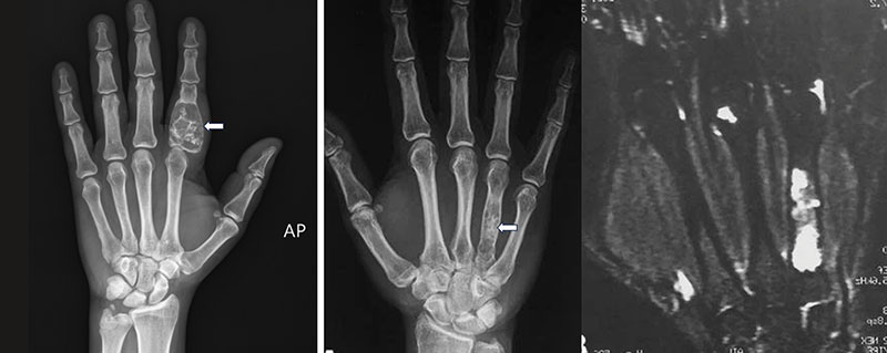
They are among the most frequently affected bones in the hand, particularly by enchondromas, which are benign cartilage tumors. Although many enchondromas are discovered incidentally, larger tumors may weaken the bone and increase the risk of fracture.
Phalanges
The phalanges are the bones of the fingers and thumb.
Tumors in the phalanges may present as:
- Finger swelling
- Persistent pain
- Progressive deformity
- Reduced finger motion
- Pathological fracture
Because the fingers are essential for fine motor tasks, treatment focuses on removing the tumor while preserving finger function whenever possible.
Doctor Explains
The location of a bone tumor is just as important as its diagnosis. The same type of tumor may require different treatments depending on whether it occurs in the distal radius, a carpal bone, a metacarpal, or a finger bone because each area has unique functional demands.
Clinical Pearl
Preserving hand function is the primary goal of treatment. Surgical planning considers not only tumor removal but also grip strength, finger motion, wrist stability, and long-term quality of life.
Did You Know?
The metacarpals and phalanges are the most common sites of bone tumors in the hand, while tumors arising in the carpal bones are relatively rare.
What Are Hand & Wrist Bone Tumors?
A bone tumor is an abnormal growth of cells arising within the bone. Bone tumors may be benign (non-cancerous), locally aggressive, or malignant (cancerous).
Benign tumors usually grow slowly and do not spread to other parts of the body, although some can weaken the bone or damage nearby joints if left untreated. Malignant tumors have the potential to invade surrounding tissues and spread to distant organs, most commonly the lungs.
Fortunately, malignant tumors of the hand and wrist are uncommon. Most lesions encountered in these regions are benign cartilage tumors, cystic lesions, or other non-cancerous conditions.
It is important to understand that not every abnormality seen on an X-ray represents cancer. Some lesions require only observation, while others may need surgery depending on their size, symptoms, and risk of fracture.
Doctor Insight
A diagnosis of a bone tumor should not automatically be equated with cancer. Careful imaging, biopsy, and expert evaluation are essential before deciding on treatment.
Reassurance
Many patients referred with a “bone tumor” ultimately have a benign condition that can be treated successfully with excellent long-term function.
Types of Hand & Wrist Bone Tumors
Different tumors behave differently. Some require only observation, while others need surgery or multidisciplinary cancer treatment.
Common Benign Tumors
The most frequently encountered benign tumors include:
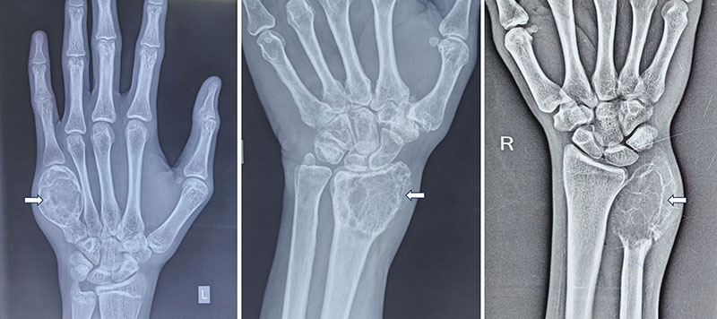
Figure 3. Giant cell tumor of the hand and wrist
Imaging showing a giant cell tumor (GCT) involving a bone of the hand and wrist.
Primary Malignant Tumors
Malignant tumors of the hand and wrist are rare but include:
- Chondrosarcoma – a cancer arising from cartilage-producing cells, more common in adults.
- Osteosarcoma – a malignant bone-forming tumor, uncommon in the hand but important to recognize.
- Ewing Sarcoma – a highly aggressive tumor that usually affects children and young adults.
Clinical Pearl
Enchondroma is the most common bone tumor of the hand, whereas giant cell tumor most frequently affects the distal radius. Recognizing these characteristic patterns helps guide diagnosis and treatment.
Symptoms of Hand & Wrist Bone Tumors
The symptoms of hand and wrist bone tumors depend on the type, size, and location of the tumor. While some tumors cause persistent pain and swelling, others are discovered incidentally on an X-ray after a minor injury. Early evaluation is important because prompt diagnosis often leads to simpler treatment and better preservation of hand function.
Pain
Pain is the most common symptom and may develop gradually. It can occur during activity, while gripping objects, or even at rest. Some tumors, such as osteoid osteoma (a small benign bone tumor), characteristically cause night pain that improves with anti-inflammatory medications. In contrast, enchondromas (benign cartilage tumors) are often painless until the bone weakens or fractures.
Swelling or a Lump
A firm swelling over the wrist, palm, or finger may indicate a bone tumor. It is usually slow-growing and may initially be painless. Larger tumors can cause tenderness, visible deformity, or difficulty wearing rings or using the hand comfortably.
Reduced Hand or Wrist Function
As the tumor enlarges, it may affect normal movement and strength. Patients may experience:
- Weak grip strength
- Wrist or finger stiffness
- Difficulty lifting objects
- Reduced range of motion
- Problems with fine tasks such as writing or buttoning clothes
Pathological Fracture
A pathological fracture is a break that occurs because the bone has been weakened by a tumor rather than significant trauma. Many enchondromas of the metacarpals and phalanges are first detected after such a fracture.
Symptoms According to Tumor Location
Doctor Explains
Bone tumors of the hand and wrist often mimic common conditions such as arthritis, tendon injuries, or ganglion cysts. Persistent symptoms that do not improve should be evaluated rather than repeatedly treated with pain medications.
When Should You See an Orthopaedic Oncologist?
Consult a specialist if you have persistent pain lasting more than 4–6 weeks, a hard bony swelling, an unexplained lesion on X-ray, a pathological fracture, worsening symptoms despite treatment, or a previously diagnosed bone tumor requiring follow-up.
Red Flag
Never ignore a persistent bony swelling or unexplained bone lesion. Proper imaging should always be performed before any biopsy or surgery.
Patient Tip
Bring all previous X-rays, MRI scans, and reports to your appointment. Comparing earlier imaging often helps determine whether a lesion is stable or growing.
Reassurance
Most patients with hand or wrist pain do not have cancer. Even when a bone tumor is diagnosed, the majority are benign and can often be treated successfully while preserving excellent hand function.
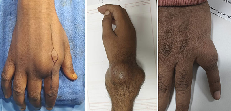
Figure 4. Swelling caused by a bone tumor of the hand and wrist
Visible swelling may be an important warning sign of a bone tumor in the hand or wrist.
Diagnosis
Accurate diagnosis is essential before treatment. The goals are to determine whether the tumor is benign or malignant, identify its type, assess its extent, and plan the safest treatment.
Clinical Assessment
Evaluation begins with a detailed history and physical examination. Your orthopaedic oncologist will assess pain, swelling, duration of symptoms, previous fractures, and any history of cancer. The examination also evaluates hand function, grip strength, joint movement, nerve function, and circulation.
Doctor Explains
A thorough examination helps determine not only the diagnosis but also which joints, tendons, and nerves can be preserved during surgery.
X-rays
An X-ray is the first imaging test and provides valuable information about the location, size, and appearance of the bone lesion. Although it often suggests the diagnosis, additional tests are usually required.
Magnetic Resonance Imaging (MRI)
MRI (Magnetic Resonance Imaging) uses magnetic fields to produce detailed images of bones and surrounding soft tissues. It is the most important investigation for assessing tumor extent and planning surgery.
Computed Tomography (CT)
A CT scan (Computed Tomography) provides detailed images of bone and is especially useful for evaluating cortical destruction, small carpal bone lesions, and complex wrist anatomy. A CT scan of the chest may also be performed in malignant tumors to check for spread to the lungs.
PET-CT and Bone Scan
PET-CT and bone scans are reserved for selected patients with suspected malignant tumors to determine whether the disease has spread or to assess treatment response.
Why Is a Biopsy Necessary?
A biopsy is the removal of a small tissue sample for microscopic examination and is the only way to confirm the diagnosis of most bone tumors. A core needle biopsy is commonly performed, while an open biopsy is reserved for selected situations. Biopsy should always follow complete imaging and be planned by the surgical team whenever possible.
Clinical Pearl
The biopsy should ideally be performed by the same musculoskeletal oncology team that will carry out the definitive surgery.
Red Flag
Never undergo an unplanned biopsy or excision of a suspected bone tumor, as it may complicate future treatment and reduce the chances of successful limb-sparing surgery.
Treatment Planning
Once the diagnosis has been confirmed, treatment is individualized for each patient. There is no single treatment that is suitable for every hand or wrist bone tumor. The recommended approach depends on the tumor type, its location, symptoms, and the expected impact on hand function.
The primary goals of treatment are to:
- Completely treat or remove the tumor
- Preserve hand and wrist function whenever possible
- Maintain grip strength and finger movement
- Prevent recurrence
- Achieve the best long-term quality of life
Many benign tumors require only observation, while others may need surgery to relieve pain, prevent fractures, or preserve joint function. Malignant tumors often require a combination of surgery and, in selected cases, chemotherapy or radiotherapy.
The Multidisciplinary Team
Treatment is best planned by a Multidisciplinary Team (MDT)—a group of specialists who work together to determine the safest and most effective treatment plan.
Depending on the diagnosis, the MDT may include:
- Orthopaedic oncologist
- Musculoskeletal radiologist
- Pathologist
- Medical oncologist
- Radiation oncologist
- Plastic and reconstructive surgeon
- Hand therapist and physiotherapist
Collaboration between these specialists ensures that both tumor control and hand function are carefully considered before treatment begins.
Factors That Influence Treatment
Several factors determine the most appropriate treatment strategy:
- Whether the tumor is benign, locally aggressive, or malignant
- Its location (distal radius, distal ulna, carpal bones, metacarpals, or phalanges)
- Size of the tumor and the amount of bone involved
- Involvement of nearby joints, tendons, nerves, or blood vessels
- Presence of a pathological fracture
- Age, occupation, hand dominance, and activity level of the patient
For example, a small, painless enchondroma may simply be monitored, whereas a giant cell tumor of the distal radius often requires surgical removal and reconstruction. Similarly, treatment for a manual laborer may differ from that for someone with lower functional demands.
Doctor Insight
Treatment planning is not focused solely on removing the tumor. Equal importance is given to preserving wrist stability, finger movement, grip strength, and the ability to return to daily activities.
Patient Tip
Ask your surgeon to explain the treatment options, expected hand function after surgery, possible complications, and the rehabilitation process. Understanding the plan helps you participate confidently in decision-making.
Reassurance
Modern treatment aims to preserve the hand whenever it is safe to do so. Advances in limb-salvage surgery and reconstruction mean that amputation is required only in exceptional situations.
Non-Surgical Treatment
Not every hand or wrist bone tumor requires an operation. Some benign tumors remain stable for years without causing symptoms, while others respond well to medical treatments used alongside surgery.
Observation
Small, asymptomatic benign tumors may only require regular follow-up with clinical examination and periodic X-rays to ensure they are not increasing in size or weakening the bone.
Medications
Pain caused by certain benign tumors, particularly osteoid osteoma, may improve with anti-inflammatory medications. In selected giant cell tumors, medications such as denosumab may be used to shrink or control the tumor before surgery.
Chemotherapy
Chemotherapy uses medicines to destroy cancer cells and is recommended for certain malignant tumors, particularly osteosarcoma and Ewing sarcoma. It is usually given both before and after surgery to improve outcomes.
Clinical Pearl
Observation does not mean ignoring the tumor. Regular follow-up is essential to detect growth, increasing bone destruction, or a rising risk of fracture before complications develop.
Surgical Treatment
Surgery is recommended when a tumor causes pain, weakens the bone, threatens joint function, continues to grow, or is confirmed to be malignant. The objective is to completely treat the tumor while preserving as much normal bone, joint, and hand function as possible.
The type of surgery depends on the location of the tumor rather than adopting a single approach for all hand bones.
Curettage
Curettage is a procedure in which the tumor is carefully scraped out from within the bone while preserving the surrounding healthy bone. It is commonly performed for benign tumors such as enchondromas and selected giant cell tumors.
After curettage, the cavity may be filled with:
- Bone graft (bone taken from the patient or a donor)
- Synthetic bone substitute
- Bone cement in selected cases
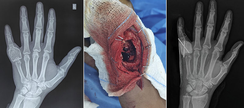
Figure 6. Curettage of a giant cell tumor of the hand
Careful curettage removes the tumor while preserving as much healthy bone and function as possible.
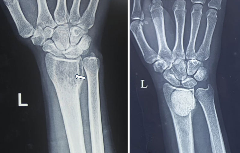
Figure 7. Reconstruction following curettage of a giant cell tumor
After tumor removal, the bone is reconstructed to restore strength and preserve hand function.
Wide Resection
A wide resection involves removing the tumor together with a margin of healthy tissue. This procedure is generally required for aggressive or malignant tumors to reduce the risk of recurrence.
The amount of bone removed depends on the tumor’s size and extent.
Surgery According to Tumor Location
Distal Radius Tumors
Tumors of the distal radius, particularly giant cell tumors and malignant lesions, often require removal of the affected bone followed by reconstruction to restore wrist stability and function.
Distal Ulna Tumors
Because the distal ulna contributes to forearm rotation and wrist stability, surgery aims to remove the tumor while preserving the function of the distal radioulnar joint whenever feasible.
Carpal Bone Tumors
Carpal bone tumors are uncommon. Depending on the affected bone, surgery may involve curettage, partial excision, or reconstruction while preserving wrist movement.
Metacarpal and Phalangeal Tumors
Most tumors of the metacarpals and phalanges are benign and can be treated with curettage and bone grafting. Larger or malignant tumors may require segmental resection followed by biological reconstruction to maintain finger length and function.
Doctor Explains
The same diagnosis does not always require the same operation. Surgical planning depends on the exact bone involved, the size of the tumor, and the importance of preserving hand function.
Clinical Pearl
Whenever it is oncologically safe, limb-salvage surgery is preferred over amputation. Modern reconstructive techniques allow preservation of function in the vast majority of patients.
Reassurance
Many patients worry that surgery will result in the loss of a finger or hand. Fortunately, with current surgical techniques, most hand and wrist bone tumors can be treated while preserving both appearance and function.
Reconstruction After Hand & Wrist Bone Tumor Surgery
Following removal of a bone tumor, the surgeon may need to reconstruct the resulting bone defect to restore the strength, stability, alignment, and function of the hand or wrist. The type of reconstruction depends on the location of the tumor, the amount of bone removed, whether the joint is involved, and the patient’s age, occupation, and functional demands.
Small defects after curettage can often be managed with bone grafting alone. However, larger resections—particularly around the distal radius—require more complex reconstruction to restore wrist function and maintain hand strength.
The goals of reconstruction are to:
- Restore skeletal stability
- Preserve hand and wrist function
- Maintain grip strength
- Achieve durable long-term outcomes
- Minimize the risk of complications and recurrence
Biological Reconstruction
Biological reconstruction uses living bone to replace the bone removed during tumor surgery. The graft may come from the patient’s own body (autograft) or from a donated bone (allograft).
Common biological options include:
- Cancellous bone grafting for small defects
- Structural iliac crest grafts
- Non-vascularized fibular strut grafts
- Vascularized fibular grafts for larger defects
- Structural allografts in selected patients
These techniques allow the graft to gradually unite with the patient’s own bone and may provide long-lasting reconstruction, particularly in younger individuals.
Bone Grafting
Bone grafting is commonly performed after curettage of benign tumors such as enchondromas or aneurysmal bone cysts.
The cavity may be filled with:
- Autograft (bone taken from the patient)
- Allograft (donor bone)
- Synthetic bone substitutes
Bone grafting restores structural support, encourages bone healing, and reduces the risk of future fracture.
Distal Radius Reconstruction
Reconstruction of the distal radius is one of the most challenging procedures in orthopaedic oncology because this bone forms the major load-bearing part of the wrist joint. The reconstructive strategy depends largely on whether the wrist joint can be preserved.
Wrist Arthrodesis (Wrist Fusion)
Wrist arthrodesis means permanently joining the wrist bones to create a stable, pain-free wrist. Although wrist motion is sacrificed, finger movement and forearm rotation are largely preserved.
Several biological reconstruction options are available:
- Ipsilateral distal ulna translocation (using the patient’s own ulna to reconstruct the radius)
- Non-vascularized fibular strut graft
- Structural iliac crest graft
- Structural allograft
Advantages
- Durable long-term reconstruction
- Excellent stability
- Lower risk of implant wear
- Suitable for younger and physically active patients
Limitations
- Permanent loss of wrist movement
- Longer time required for bone healing
- Risk of graft non-union or fracture
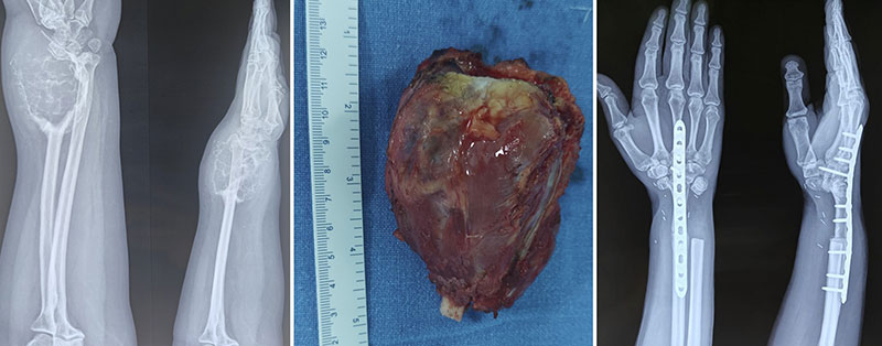
Figure 8. Distal radius tumor resection with ulna translocation and wrist fusion
For an extensive distal radius tumor, wide resection followed by ulna translocation and wrist fusion can provide a stable, functional reconstruction.
Wrist Arthroplasty (Motion-Preserving Reconstruction)
In selected patients, the wrist joint can be reconstructed to preserve movement.
Options include:
- Custom-made endoprosthesis (patient-specific metallic implant)
- Proximal fibular autograft, where the upper end of the fibula is used to recreate the wrist joint
Advantages
- Preserves some wrist motion
- Better functional range for selected daily activities
- Faster early rehabilitation with prosthetic reconstruction
Limitations
- Higher technical complexity
- Potential implant loosening or wear over time
- Biological grafts may develop joint degeneration in the long term
- Some procedures may require revision surgery years later
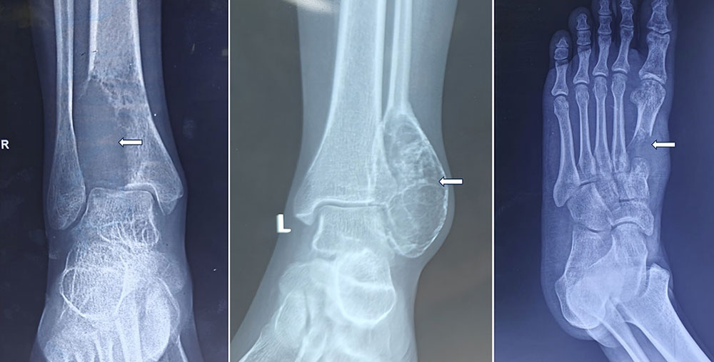
Figure 9. Distal radius tumor resection with proximal fibula wrist reconstruction
After removal of a giant cell tumor of the distal radius, the wrist is reconstructed using a proximal fibula graft to preserve useful hand function.
There is no universally superior reconstruction. The choice is individualized after considering tumor extent, patient age, expected activity level, and surgeon experience.
Doctor Explains
Some patients benefit more from a stable, pain-free fused wrist, while others may be better candidates for a motion-preserving reconstruction. The decision is based on balancing durability, function, and long-term outcomes rather than simply preserving movement.
Distal Ulna Reconstruction
The distal ulna contributes to forearm rotation and stability of the distal radioulnar joint (DRUJ). Depending on the extent of tumor removal, reconstruction may not always be necessary.
When reconstruction is indicated, options include:
- Soft tissue stabilization after distal ulna excision
- Structural bone graft reconstruction
- Custom distal ulna prosthesis in selected patients
The objective is to maintain forearm rotation, improve grip strength, and prevent instability of the wrist.
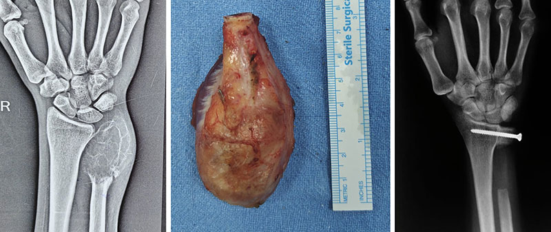
Figure 10. Giant cell tumor of the distal ulna treated with tumor resection
Wide resection of a distal ulna giant cell tumor with reconstruction designed to maintain wrist and forearm function.
Carpal Bone Reconstruction
Tumors involving the carpal bones are uncommon and reconstruction depends on the specific bone affected.
Possible options include:
- Cancellous bone grafting after curettage
- Partial carpal excision
- Intercarpal fusion for stability
- Patient-specific 3D-printed custom implants in selected complex defects
Because the wrist contains multiple small joints working together, preserving alignment and stability is often more important than replacing every individual bone.
Metacarpal and Phalangeal Reconstruction
Following resection of tumors involving the metacarpals or phalanges, reconstruction aims to preserve finger length, alignment, and hand function.
Common reconstructive techniques include:
- Cancellous or structural autografts
- Non-vascularized fibular grafts for larger defects
- Vascularized toe or fibular grafts in selected situations
- Osteoarticular grafts when joint preservation is possible
- Custom 3D-printed implants, increasingly used for complex segmental defects where conventional reconstruction is difficult
Advances in 3D printing technology now allow implants to be designed specifically for an individual patient’s anatomy, improving fit and potentially restoring finger alignment more accurately in carefully selected cases.
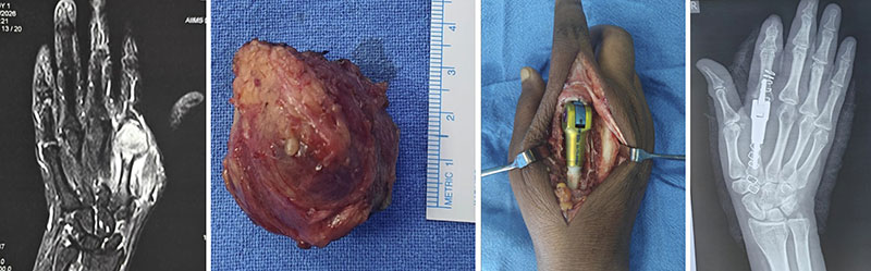
Figure 11. Metacarpal tumor resection with custom 3D-printed reconstruction
Patient-specific 3D-printed reconstruction can restore the shape and function of the hand after metacarpal bone tumor resection.
Joint Preservation
Whenever it is oncologically safe, surgeons aim to preserve the native joint.
Potential benefits include:
- Better wrist movement
- Improved grip strength
- More natural hand mechanics
- Easier performance of everyday activities
However, if the tumor extensively involves the joint surface, complete removal of the joint may be necessary to achieve adequate tumor clearance and reduce the risk of recurrence.
Reconstruction Options at a Glance
Clinical Pearl
There is no single “best” reconstruction for every patient. Successful reconstruction depends on selecting the technique that offers the optimal balance between oncological safety, durability, preservation of function, and long-term quality of life.
Recovery and Rehabilitation
Recovery after hand and wrist bone tumor surgery is a gradual process that extends beyond wound healing. The final outcome depends not only on successful tumor removal but also on bone healing, structured rehabilitation, and restoration of hand function.
Recovery varies according to the type of surgery performed. Patients undergoing curettage with bone grafting generally recover faster than those requiring major reconstruction or joint reconstruction. The ultimate goal is to achieve a pain-free, stable, and functional hand that allows patients to return to their daily activities with confidence.
Hospital Stay
The length of hospital stay depends on the complexity of surgery.
- Curettage and bone grafting: Usually 1–2 days
- Complex reconstruction or joint replacement: Typically 3–7 days
During hospitalization, the healthcare team focuses on:
- Pain management
- Wound care
- Swelling control
- Early finger mobilization
- Splint or cast care
- Planning postoperative rehabilitation
Whenever possible, finger movement is encouraged early to minimize stiffness while protecting the reconstruction.
Hand Therapy and Rehabilitation
Hand therapy is a specialized rehabilitation program conducted by therapists trained in the treatment of hand and upper limb conditions. It is one of the most important factors influencing the final functional outcome.
Rehabilitation is tailored to the type of surgery performed and generally progresses through three phases:
Early Phase (0–6 Weeks)
The priorities are to:
- Protect the reconstruction
- Control pain and swelling
- Maintain finger mobility
- Prevent joint stiffness
- Preserve tendon gliding
Intermediate Phase (6–12 Weeks)
As healing progresses, therapy focuses on:
- Improving wrist and finger range of motion
- Restoring forearm rotation
- Beginning gentle strengthening exercises
- Improving coordination
Advanced Phase (After 3 Months)
The emphasis shifts to:
- Grip strengthening
- Fine motor skills
- Dexterity training
- Functional use of the hand
- Return to work, sports, and hobbies
Patients who participate actively in rehabilitation generally achieve better functional outcomes than those who discontinue therapy prematurely.
Return to Daily Activities
Recovery is individualized and depends on the diagnosis, surgical procedure, bone healing, and occupation.
*These timelines are approximate and vary between individuals.
Patients should return to activities only after approval from their treating surgeon.
Follow-Up After Surgery
Regular follow-up is an essential part of treatment. It allows the surgical team to monitor healing, assess function, and detect recurrence at an early stage.
Follow-up commonly includes:
- Clinical examination
- Serial X-rays to assess bone healing
- MRI when recurrence is suspected
- Functional assessment of grip strength and joint movement
Patients treated for malignant tumors may also undergo CT scans of the chest, as the lungs are the most common site for metastasis (spread of cancer).
The frequency of follow-up gradually decreases over time but may continue for several years depending on the tumor type.
Possible Complications
Most patients recover without major problems, but every surgical procedure carries some risks.
Potential complications include:
- Infection
- Delayed bone healing or non-union
- Fracture of the reconstructed bone or graft
- Implant loosening or failure
- Joint stiffness
- Reduced grip strength
- Local tumor recurrence
- Need for revision surgery
Fortunately, many complications can be successfully treated when recognized early through regular follow-up.
Doctor Explains
Recovery does not end when the operation is over. Surgery removes the tumor, but rehabilitation restores movement, strength, and confidence. A dedicated rehabilitation program is just as important as the surgery itself.
Clinical Pearl
Early controlled movement of the fingers helps prevent stiffness without compromising bone healing. The rehabilitation protocol should always be individualized according to the type of reconstruction performed.
Patient Tip
Do not compare your recovery with someone else’s. The time required to regain function depends on the tumor type, the extent of surgery, and the reconstruction performed. Following your therapist’s instructions consistently is the best way to achieve an optimal outcome.
Did You Know?
Patients who undergo hand therapy after surgery often regain significantly better movement, grip strength, and hand function than those who rely on natural recovery alone.
Reassurance
It is normal to experience temporary weakness, stiffness, and swelling after surgery. With appropriate rehabilitation and regular follow-up, most patients are able to return to independent daily activities, work, and many recreational pursuits while maintaining good hand function.
Frequently Asked Questions (FAQs)
1. Is every hand or wrist bone tumor cancer?
No. The majority of hand and wrist bone tumors are benign (non-cancerous). However, some benign tumors can still weaken the bone or damage nearby joints, while a small proportion are malignant and require specialized treatment. Proper imaging and, in many cases, a biopsy are necessary to establish the diagnosis.
2. What is the most common bone tumor of the hand?
Enchondroma, a benign cartilage tumor, is the most common bone tumor of the hand. It frequently affects the metacarpals and phalanges and is often discovered incidentally or after a pathological fracture.
3. Will I need surgery?
Not always. Small, asymptomatic benign tumors may only require observation. Surgery is recommended when the tumor causes pain, weakens the bone, continues to grow, affects function, or is malignant.
4. Why is a biopsy necessary?
A biopsy involves removing a small tissue sample for microscopic examination. It is the most reliable way to determine the exact type of tumor and helps ensure that the appropriate treatment is chosen.
5. Will I lose my hand or fingers?
In most cases, no. Modern limb-salvage techniques allow surgeons to remove the tumor while preserving the hand and fingers whenever it is oncologically safe. Amputation is required only in rare situations where adequate tumor removal cannot otherwise be achieved.
6. Can the wrist be reconstructed after tumor removal?
Yes. Depending on the extent of bone loss, reconstruction may involve bone grafts, wrist fusion (arthrodesis), motion-preserving procedures (arthroplasty), custom endoprostheses, or biological grafts such as the proximal fibula. The choice depends on the tumor location and the patient’s functional needs.
7. How long does recovery take?
Recovery varies according to the type of surgery. Many patients resume light activities within a few weeks, while complete recovery after complex reconstruction may take 6–12 months.
8. Can bone tumors come back after treatment?
Some tumors, particularly giant cell tumors, have a risk of local recurrence. Regular follow-up with clinical examinations and imaging helps detect recurrence early, when treatment is often more successful.
9. Will I regain normal hand function?
Many patients regain excellent function after treatment, although recovery depends on the tumor type, surgical procedure, rehabilitation, and individual healing. Consistent hand therapy plays a major role in achieving the best outcome.
10. When should I seek a second opinion?
A second opinion is particularly valuable when:
- A biopsy has not yet been performed.
- Major surgery or reconstruction has been recommended.
- The diagnosis is uncertain.
- You have a recurrent tumor.
- You have been advised that amputation may be necessary.
Consulting an orthopaedic oncologist before definitive treatment can help ensure that all appropriate treatment options have been considered.
Medical Disclaimer
The information provided on this page is intended for educational purposes only and should not be considered a substitute for professional medical advice, diagnosis, or treatment. Every patient and every bone tumor is unique, and treatment decisions should be based on a thorough clinical evaluation, appropriate imaging, and, when indicated, a biopsy performed by a qualified healthcare professional.
If you have persistent hand or wrist pain, swelling, an unexplained bone lesion, or have been diagnosed with a bone tumor, consult an orthopaedic oncologist for an accurate diagnosis and individualized treatment plan. Never delay or disregard professional medical advice based on information found online.
References
- Lazerges C, Degeorge B, Coulet B, Chammas M. Diagnosis and treatment of hand tumors. Orthop Traumatol Surg Res. 2022;108(1S):103153.
- Hsu CS, Hentz VR, Yao J. Tumours of the hand. Lancet Oncol. 2007;8(2):157-166.
- Liu W, Wang B, Zhang S, Li Y, Hu B, Shao Z. Wrist Reconstruction after En bloc Resection of Bone Tumors of the Distal Radius. Orthop Surg. 2021;13(2):376-383.
- Kapoor L, Kumar VS, Ansari MT, Khan SA. Ulnar buttress arthroplasty without ulnar stump stabilization for giant cell tumor of distal ulna. J Wrist Surg. 2023;13(3):215-221.
- Qu H, Guo W, Li D, et al. Functional results of wrist arthrodesis versus arthroplasty with proximal fibula following giant cell tumour excision of the distal radius. J Hand Surg Eur Vol. 2019;44(4):394-401.
 +91 93558 33005