Quick Summary
- Most shoulder bone tumors are benign, not cancerous.
- Persistent shoulder pain should not automatically be attributed to a rotator cuff injury.
- MRI and a carefully planned biopsy are essential before treatment.
- Most malignant shoulder bone tumors can now be treated with limb salvage surgery rather than amputation.
- Treatment is individualized based on the tumor type, location, age, and functional requirements.
Reassurance Box
Being told that you have a bone tumor can be frightening. However, most shoulder bone tumors are not cancer, and even when a malignant tumor is diagnosed, modern surgical techniques often allow the arm to be preserved while achieving excellent cancer control. An accurate diagnosis and treatment by a specialist bone tumor team offer the best chance of a successful outcome.
Understanding the Shoulder Anatomy
The shoulder is the most mobile joint in the human body, allowing us to reach overhead, lift objects, throw, push, pull, and position the hand in almost any direction. This remarkable range of movement is possible because several bones, muscles, tendons, and ligaments work together as a coordinated unit.
The three bones that form the shoulder are:
- Proximal humerus – the upper end of the arm bone that forms the ball of the shoulder joint.
- Scapula (shoulder blade) – a flat triangular bone on the back of the chest that forms the socket (glenoid) of the shoulder joint.
- Clavicle (collarbone) – the bone connecting the shoulder to the breastbone, helping stabilize the shoulder girdle.
Most primary bone tumors around the shoulder arise in the proximal humerus, making it the second most common location for many primary bone sarcomas after the region around the knee. Tumors may also occur in the scapula or clavicle, although these are considerably less common.
Unlike the lower limb, where the primary goal after surgery is restoring weight-bearing, reconstruction around the shoulder focuses on preserving hand and elbow function while providing a stable, painless upper limb. Even if shoulder movement cannot be completely restored, retaining a functional arm allows patients to perform most daily activities independently.
Understanding which bone is affected helps explain why treatment varies from one patient to another. For example, a tumor involving the proximal humerus may require reconstruction with a tumor megaprosthesis or reverse shoulder replacement, whereas tumors of the scapula or clavicle often require different surgical approaches.
Did You Know?
The proximal humerus is one of the most common locations for osteosarcoma, chondrosarcoma, and several benign bone tumors, making the shoulder an important site in orthopaedic oncology.
Clinical Pearl
The shoulder is a non-weight-bearing joint, allowing surgeons greater flexibility in reconstruction than around the hip or knee. As a result, many patients retain excellent hand and elbow function even after extensive tumor surgery.
Symptoms of Shoulder Bone Tumors
The symptoms of a shoulder bone tumor depend on its size, location, growth rate, and whether it is benign or malignant. In the early stages, many tumors produce only mild discomfort and may be mistaken for common shoulder problems such as tendinitis, frozen shoulder, or a rotator cuff injury.
The most common symptom is persistent shoulder pain. Unlike pain caused by muscle strain, this discomfort often develops gradually, becomes progressively worse, and may not improve with rest, pain medication, or physiotherapy.
Other symptoms may include:
- Swelling around the shoulder or upper arm
- A slowly enlarging lump
- Reduced shoulder movement
- Pain during routine activities or at rest
- Night pain that disturbs sleep
- Weakness while lifting the arm
- A pathological fracture (a bone breaking after minimal injury because it has been weakened by a tumor)
Large tumors involving the scapula may present as a painless swelling over the back of the shoulder, while tumors of the clavicle may appear as a firm lump above the collarbone.
Because many shoulder tumors develop close to important nerves, very large lesions may occasionally cause numbness, tingling, or weakness in the arm or hand. These symptoms are uncommon but require prompt specialist evaluation.
It is important to remember that shoulder pain is extremely common, whereas bone tumors are rare. Most patients with shoulder pain have conditions such as rotator cuff disease, arthritis, bursitis, or frozen shoulder rather than a bone tumor. However, pain that persists despite appropriate treatment deserves further investigation.
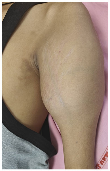
Figure 1. Swelling around the shoulder may be a sign of an underlying bone tumor.
Red Flag Box
Seek medical evaluation if you experience:
- Persistent shoulder pain lasting more than 4–6 weeks
- Pain that worsens at night
- An unexplained swelling or lump
- Progressive limitation of shoulder movement
- A fracture after a minor injury
- An abnormal bone lesion detected on an X-ray or MRI
Patient Tip
If your shoulder pain continues despite physiotherapy, injections, or routine treatment, ask whether an X-ray is needed before pursuing further interventions. Although bone tumors are uncommon, persistent symptoms should always be explained.
Doctor Explains
One of the commonest reasons for delayed diagnosis is that shoulder tumors initially resemble much more common conditions such as rotator cuff disease or frozen shoulder. A simple X-ray often provides the first clue that a bone tumor may be present, allowing timely referral for specialist assessment.
Reassurance Box
Most people with shoulder pain do not have a bone tumor. Nevertheless, persistent or unexplained symptoms should never be ignored. Early diagnosis allows appropriate treatment before complications such as pathological fractures or extensive bone destruction develop.
Types of Bone Tumors Around the Shoulder
Bone tumors around the shoulder can arise in the proximal humerus (upper arm bone), scapula (shoulder blade), or clavicle (collarbone). They are broadly classified as benign (non-cancerous) or malignant (cancerous). Knowing the exact type of tumor is essential because treatment, prognosis, and follow-up vary considerably.
Fortunately, most bone tumors around the shoulder are benign. However, some benign tumors can grow aggressively, weaken the bone, or damage the shoulder joint, making treatment necessary.
Benign Bone Tumors
Benign tumors do not spread to other parts of the body, but some can enlarge over time, cause pain, weaken the bone, or increase the risk of fracture.
Common benign tumors around the shoulder include:
- Osteochondroma – the most common benign bone tumor, often presenting as a painless bony prominence near the shoulder. Surgery is considered if it causes pain, restricts movement, compresses nearby nerves, or continues to enlarge after skeletal maturity. (Osteochondroma)
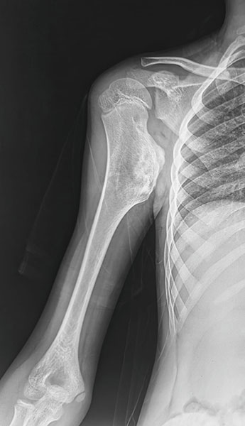
Figure 2. X-rays showing an osteochondroma of the proximal humerus, a common benign bone tumor near the shoulder.
- Giant Cell Tumor of Bone (GCT) – a locally aggressive tumor that usually develops after skeletal maturity, most commonly around the ends of long bones, including the proximal humerus. Although classified as benign, it can destroy bone and occasionally recur after treatment. (Giant Cell Tumor of Bone)
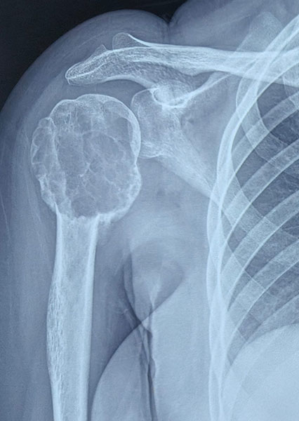
Figure 3. X-rays showing a giant cell tumor of the proximal humerus involving the shoulder region.
- Chondroblastoma – a rare cartilage-forming tumor typically affecting adolescents and young adults. It commonly arises near the shoulder joint and often presents with pain and restricted movement. (Chondroblastoma)
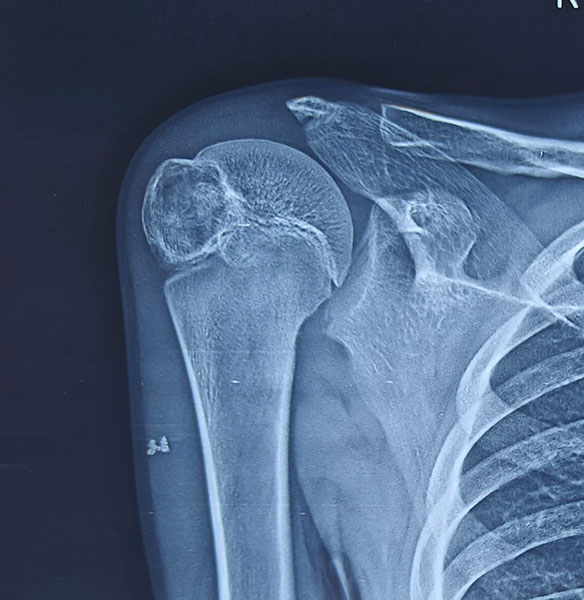
Figure 4. X-rays showing a chondroblastoma of the proximal humerus near the shoulder joint.
- Aneurysmal Bone Cyst (ABC) – a blood-filled benign lesion that expands the bone and may present with pain, swelling, or a pathological fracture. (Internal link: Aneurysmal Bone Cyst)
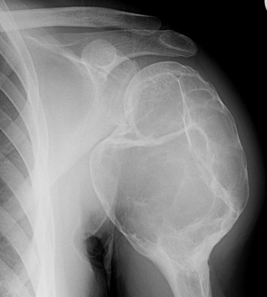
Figure 5. X-rays showing an aneurysmal bone cyst of the proximal humerus.
Many of these tumors can be treated successfully using joint-preserving surgery, such as curettage (careful removal of the tumor from within the bone), followed by bone grafting or bone cement where appropriate.
Malignant Bone Tumors
Malignant tumors are cancers that can invade surrounding tissues and, in some cases, spread to other organs. Although uncommon, they require prompt diagnosis and treatment by a specialized multidisciplinary sarcoma team.
The most important primary malignant bone tumors around the shoulder are:
- Osteosarcoma – the most common primary bone cancer in children, adolescents, and young adults. It frequently involves the proximal humerus and is usually treated with chemotherapy combined with limb salvage surgery. (Osteosarcoma)
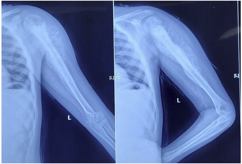
Figure 6. X-rays showing an osteosarcoma of the proximal humerus, a malignant bone tumor involving the shoulder region.
- Chondrosarcoma – a cartilage-producing cancer that occurs mainly in adults. It commonly affects the proximal humerus, scapula, and shoulder girdle. Unlike osteosarcoma, treatment is primarily surgical because most conventional chondrosarcomas respond poorly to chemotherapy and radiotherapy. ( Chondrosarcoma)
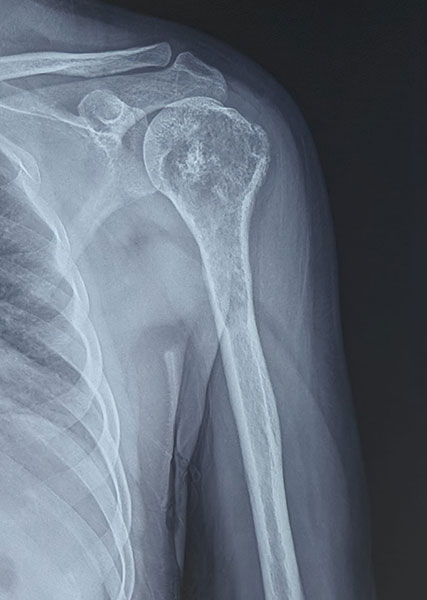
Figure 7. X-rays showing a chondrosarcoma of the proximal humerus, a malignant cartilage-forming bone tumor.
- Ewing Sarcoma – a highly aggressive tumor affecting children and young adults. It often requires a combination of chemotherapy, surgery, and occasionally radiotherapy. (Ewing Sarcoma)
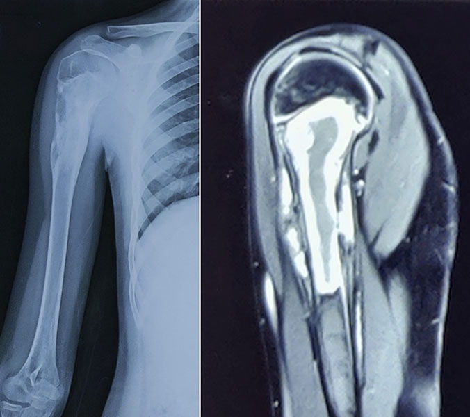
Figure 8. X-ray and MRI showing Ewing sarcoma of the proximal humerus and the extent of the tumor.
Metastatic Bone Disease
Not every cancer involving the shoulder originates in the bone itself.
In adults over 40 years of age, a destructive bone lesion in the shoulder is more likely to represent metastatic disease—cancer that has spread from another organ—than a primary bone sarcoma. Common primary cancers that spread to bone include breast, lung, kidney, thyroid, and prostate cancer.
Treatment focuses on relieving pain, preventing fractures, preserving function, and controlling the underlying cancer. Depending on the situation, management may involve surgery, radiotherapy, systemic therapy, or a combination of these treatments.
(Bone Metastasis)
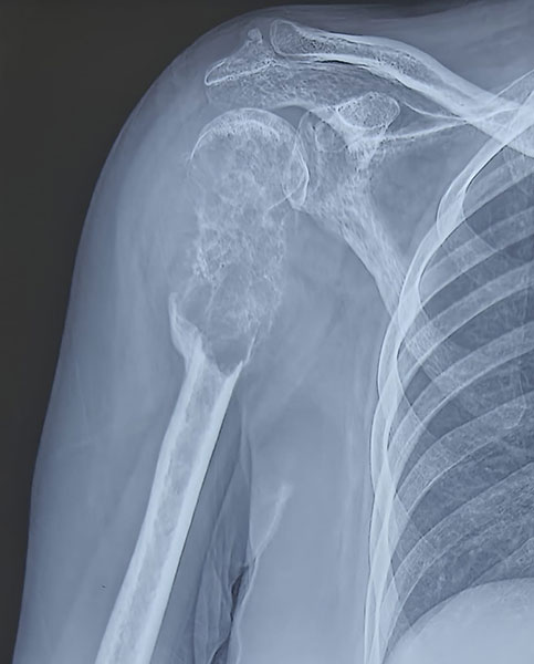
Figure 9. X-ray showing a metastatic bone lesion in the proximal humerus from breast cancer.
Table. Common Bone Tumors Around the Shoulder
Doctor Explains
Two tumors may appear very similar on an X-ray but require completely different treatments. For example, a giant cell tumor may be treated with joint-preserving curettage, whereas an osteosarcoma usually requires chemotherapy and wide surgical resection. This is why treatment should never be planned based on imaging alone.
Clinical Pearl
The patient’s age is one of the most valuable clues in narrowing the diagnosis. A bone lesion in a teenager has a very different list of possible causes than a similar lesion in a 65-year-old patient.
Reassurance Box
Hearing the word “tumor” does not necessarily mean cancer. Many shoulder bone tumors are benign and can be treated successfully while preserving the patient’s own shoulder joint or arm.
Diagnosis of Shoulder Bone Tumors
Accurate diagnosis is the foundation of successful treatment. Because many shoulder tumors resemble common conditions such as rotator cuff disease or arthritis, diagnosis requires a careful stepwise evaluation rather than relying on a single test.
The process usually begins with a detailed medical history and physical examination, followed by imaging studies to define the location and extent of the lesion. If a bone tumor is suspected, a biopsy is often required to confirm the diagnosis before any definitive treatment is planned.
A multidisciplinary team—including an orthopaedic oncologist, musculoskeletal radiologist, pathologist, medical oncologist, and radiation oncologist—reviews all clinical, imaging, and biopsy findings before recommending treatment.
Clinical Evaluation
Your doctor will ask about:
- Duration and progression of pain
- Night pain or pain at rest
- Previous injuries
- Swelling or enlarging mass
- Loss of shoulder function
- History of cancer elsewhere in the body
- General symptoms such as weight loss or fever
A thorough examination helps assess shoulder movement, muscle strength, nerve function, and the presence of any swelling or tenderness.
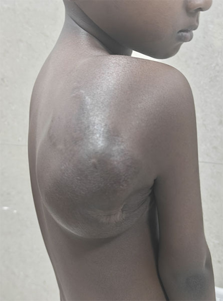
Figure 10. Clinical photograph showing a visible swelling caused by a scapular bone tumor.
Imaging Studies
Most patients undergo a sequence of investigations that may include:
- X-ray – usually the first imaging test and often the most important initial investigation.
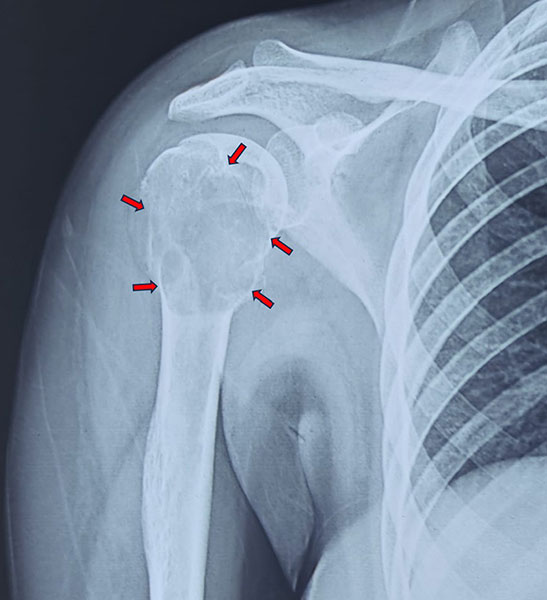
Figure 11. X-ray showing a tumor involving the proximal humerus near the shoulder joint.
- MRI (Magnetic Resonance Imaging) – defines the extent of the tumor within the bone and surrounding soft tissues. (MRI in Bone Tumors)
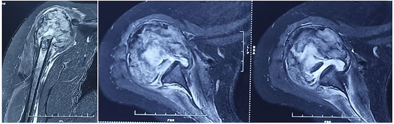
Figure 12. X-ray and MRI showing osteosarcoma of the proximal humerus. MRI helps assess the tumor within the bone, surrounding soft tissues, nearby nerves and blood vessels, and the shoulder joint.
- CT (Computed Tomography) – provides detailed information about bone destruction and is particularly useful for tumors of the scapula and clavicle. CT Scan for Bone Tumors)
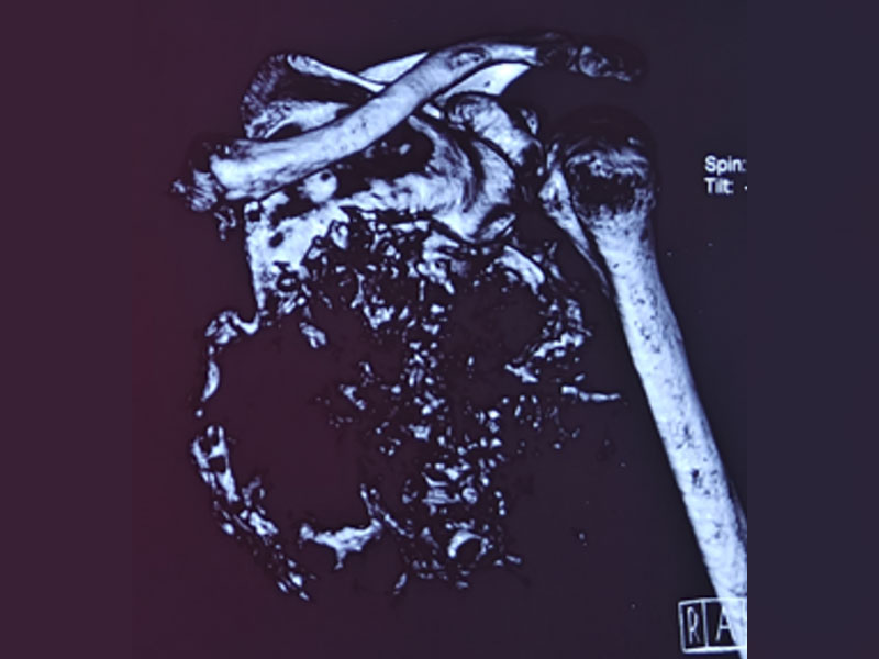
Figure 13. CT scan showing the extent of a scapular bone tumor, helping guide surgical planning
- PET-CT or Bone Scan – used selectively for staging and detecting disease elsewhere in the body. (Bone Tumor Staging)
Biopsy
A biopsy involves removing a small sample of tissue so that it can be examined under a microscope. It is the only reliable way to establish the exact diagnosis before definitive surgery.
Whenever possible, the biopsy should be performed by the same specialist team that will carry out the final treatment, as the biopsy approach can influence subsequent surgery.
(Biopsy in Bone & Soft Tissue Tumors)
Clinical Pearl
MRI should usually be performed before biopsy, as the biopsy tract must be carefully planned to avoid compromising future limb salvage surgery.
Red Flag Box
Do not undergo an unplanned biopsy or excision of a suspected bone tumor. An improperly performed biopsy can complicate definitive surgery and may reduce limb salvage options.
Reassurance Box
Modern imaging and biopsy techniques allow most shoulder bone tumors to be diagnosed accurately before treatment begins. This careful planning enables surgeons to select the safest procedure while preserving as much normal function as possible.
Treatment of Shoulder Bone Tumors
Treatment depends on several factors, including the type of tumor, whether it is benign or malignant, its size, location, relationship to nearby nerves and blood vessels, and the patient’s age and activity level. There is no single operation that is suitable for every patient.
The primary goals of treatment are to:
- Completely remove or control the tumor.
- Preserve as much normal shoulder function as possible.
- Maintain a stable, painless upper limb.
- Reduce the risk of recurrence.
- Allow patients to return to independent daily activities whenever feasible.
Every patient should be evaluated by a multidisciplinary sarcoma team, where surgeons, radiologists, pathologists, medical oncologists, and radiation oncologists jointly determine the most appropriate treatment plan.
Treatment Planning
Before surgery, the multidisciplinary team carefully reviews:
- Clinical examination findings
- X-rays, MRI, and CT scans
- Biopsy results
- Tumor stage
- Expected shoulder function after reconstruction
- The patient’s occupation, lifestyle, and overall health
This individualized approach ensures that treatment balances cancer control with long-term function.
Doctor Insight
The “best” operation is not always the most complex one. The ideal procedure is the one that completely removes the tumor while providing the safest reconstruction and the best expected function for that individual patient.
Curettage for Benign Bone Tumors
Many benign but locally aggressive tumors can be treated without removing an entire segment of bone.
Curettage is a procedure in which the surgeon carefully removes the tumor from inside the bone while preserving the surrounding healthy bone and the shoulder joint.
After the tumor is removed, the cavity may be treated with local adjuvants—additional treatments applied directly inside the bone to reduce the chance of recurrence. These may include high-speed burring, chemical agents, or thermal techniques. The resulting cavity is then filled with bone graft (living or donated bone used to promote healing) or bone cement (a special orthopedic cement that provides immediate structural support).
This approach is commonly used for:
Whenever oncologically safe, preserving the patient’s own shoulder joint remains the preferred option.
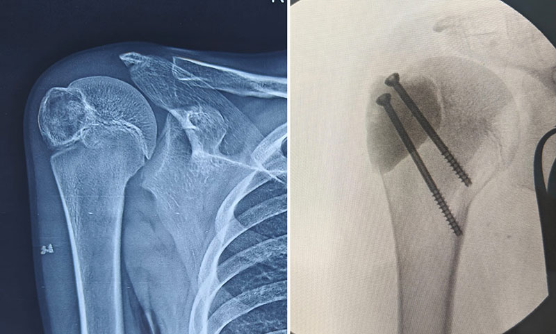
Figure 14. Chondroblastoma of the proximal humerus treated with extended curettage and bone cement reconstruction
Table. Treatment of Common Benign Shoulder Bone Tumors
Clinical Pearl
For many benign shoulder tumors, preserving the native shoulder joint provides better long-term function than replacing it.
Limb Salvage Surgery
For most malignant shoulder bone tumors, the standard treatment today is limb salvage surgery.
Limb salvage surgery involves removing the tumor with a margin of healthy tissue while preserving the arm. The removed bone is then reconstructed using an artificial implant, biological graft, or a combination of both.
Several decades ago, amputation or forequarter amputation was commonly required for aggressive shoulder tumors. Today, improvements in imaging, chemotherapy, surgical techniques, and reconstruction allow limb preservation in the majority of patients without compromising cancer control.
( Limb Salvage Surgery for Bone Tumors)
Reassurance Box
Most patients with a malignant shoulder bone tumor do not require amputation. Advances in modern orthopaedic oncology have made limb salvage the preferred treatment whenever it can be performed safely.
Does the Tumor Involve the Shoulder Joint?
One of the most important questions before planning surgery is whether the tumor has extended into the shoulder joint.
The shoulder joint is formed where the head of the humerus (ball) meets the glenoid of the scapula (socket). If imaging shows that the tumor is confined to the bone and the joint remains uninvolved, surgeons may be able to preserve much of the normal shoulder anatomy during reconstruction.
However, when the tumor extends into the joint or destroys the joint surfaces, a more extensive operation may be required. This often influences:
- The amount of bone that must be removed.
- Whether the shoulder joint can be preserved.
- The type of reconstruction performed.
- Expected shoulder movement after surgery.
Determining joint involvement requires careful evaluation of MRI, and in selected cases CT scans, before any operation is planned. This information helps the surgical team choose the reconstruction that offers the best balance between complete tumor removal and long-term function.
Patients frequently ask whether preserving the shoulder joint guarantees normal movement. Unfortunately, the answer is not always yes. Shoulder function also depends on preservation of the rotator cuff, deltoid muscle, and axillary nerve, all of which are essential for lifting the arm.
How Much Shoulder Bone Needs to Be Removed?
Proximal Humerus Reconstruction
The proximal humerus is the most frequent location for bone tumors around the shoulder. After removing the tumor, the surgeon must reconstruct the shoulder to restore stability and maximize function.
The choice of reconstruction depends largely on:
- How much bone must be removed
- Whether the shoulder socket is involved
- The condition of the deltoid muscle
- The rotator cuff tendons
- The axillary nerve
- The patient’s age and functional demands
No single reconstruction is ideal for every patient.
Tumor Megaprosthesis
A tumor megaprosthesis is a large modular metallic implant designed to replace the portion of bone removed during tumor surgery.
( Tumor Prosthesis)
Its advantages include:
- Immediate structural stability
- Early rehabilitation
- No need for bone healing before movement begins
- Reliable reconstruction for large bone defects
Modern modular designs allow the implant to be customized to the amount of bone removed during surgery.
Although shoulder movement may not return to normal, most patients regain excellent elbow and hand function and are able to perform many routine daily activities independently.
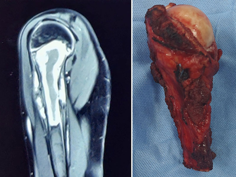
Figure 15. Ewing sarcoma of the proximal humerus treated with wide tumor resection and limb-salvage surgery.
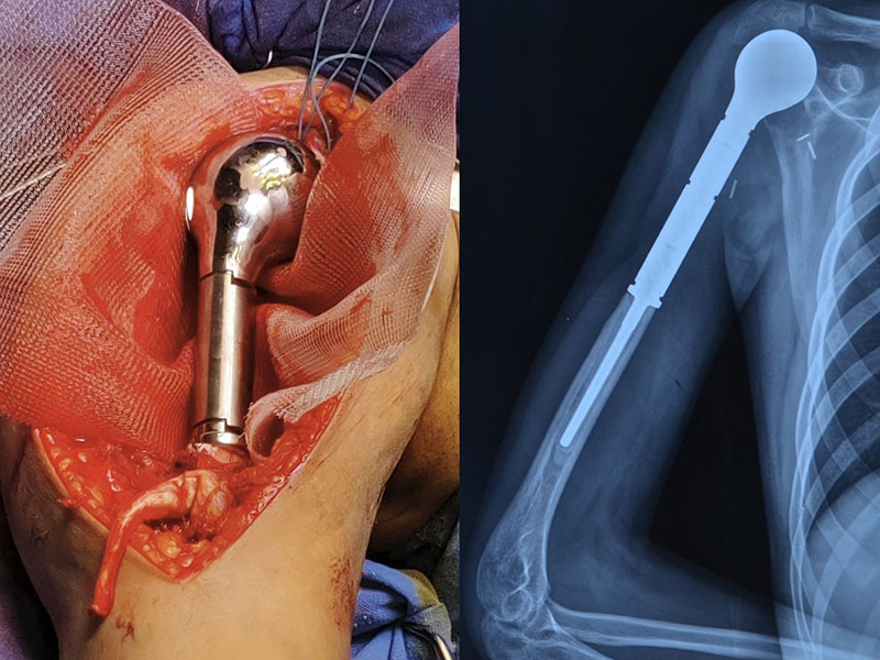
Figure 16. Endoprosthetic reconstruction of the proximal humerus after tumor removal as part of limb-salvage surgery.
Reverse Shoulder Arthroplasty
A reverse shoulder arthroplasty (reverse shoulder replacement) is a specialized shoulder replacement in which the normal ball-and-socket anatomy is reversed. This design allows the deltoid muscle, rather than the damaged rotator cuff, to lift the arm.
Reverse shoulder arthroplasty may be appropriate when:
- The rotator cuff cannot be preserved
- The deltoid muscle and axillary nerve remain functional
- The glenoid (shoulder socket) can safely support the implant
Compared with conventional shoulder replacement, reverse shoulder arthroplasty often provides better overhead function in carefully selected patients after tumor resection.
However, it is not suitable for every patient, and careful preoperative planning is essential.
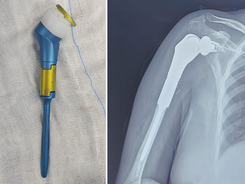
Figure 17. Reverse shoulder tumor prosthesis used for reconstruction after removal of a shoulder bone tumor.
Biological Reconstruction
In selected patients—particularly younger individuals—surgeons may reconstruct the bone using living or donated biological tissue rather than a metallic implant.
Options include:
- Allograft-Prosthetic Composite (APC)—a combination of donated bone (allograft) and a prosthesis that restores bone stock while providing mechanical stability.
(Internal link: Allograft-Prosthetic Composite Reconstruction)
- Free Vascularized Fibular Graft—a segment of the fibula (the smaller bone in the leg) transferred with its blood supply to reconstruct the shoulder. Because the graft remains alive, it can gradually unite with the patient’s own bone and remodel over time.
(Internal link: Vascularized Fibular Graft Reconstruction)
These procedures are technically demanding and generally reserved for carefully selected patients.
Cement Spacer Reconstruction
In some patients, a conventional tumor megaprosthesis may not be the safest reconstruction. This is particularly true when the tumor has required extensive removal of muscles and soft tissues, leaving insufficient coverage for a large metallic implant or increasing the risk of wound complications and infection.
In these situations, the surgeon may choose a cement spacer reinforced with an intramedullary nail or plate as a definitive reconstruction. The internal metal implant provides structural support, while the surrounding bone cement restores limb length and creates a stable construct.
Although this technique does not recreate a normal shoulder joint, it can provide:
- A stable, painless upper limb
- Preservation of arm length
- Reliable soft tissue coverage
- Lower implant-related complexity in selected patients
- Good elbow and hand function for everyday activities
Because shoulder movement depends largely on the surrounding muscles, active shoulder motion is usually limited. Nevertheless, many patients achieve a comfortable and functional arm for daily activities.
Doctor Insight
Sometimes, the safest reconstruction is not the most complex one. When extensive muscles have been removed with the tumor, a cement-and-nail reconstruction may provide a more durable and reliable solution than a megaprosthesis, particularly when soft tissue coverage is limited.
Clinical Pearl
Successful shoulder reconstruction depends on adequate soft tissue coverage as much as on the implant itself. A well-covered, stable reconstruction generally performs better than a complex prosthesis placed beneath compromised soft tissues.
Doctor Explains
There is no “best” reconstruction after shoulder tumor surgery. The ideal option depends on the tumor, the remaining muscles and nerves, the patient’s age, expected lifespan, activity level, and personal goals.
Table. Reconstruction Options After Proximal Humerus Tumor Resection
Clinical Pearl
The quality of the remaining muscles and nerves often has a greater influence on shoulder function than the type of implant itself.
Patient Tip
Before surgery, ask your surgeon not only how the tumor will be removed, but also what level of shoulder function you can realistically expect afterward. Understanding these expectations helps guide rehabilitation and recovery.
Doctor Insight
The primary aim of reconstruction is not to restore a perfectly normal shoulder. It is to provide a stable, pain-free, functional upper limb that allows patients to remain independent in their daily lives.
Scapular Tumors
Tumors involving the scapula (shoulder blade) are relatively uncommon but often present unique reconstructive challenges because the scapula serves as the foundation for shoulder movement.
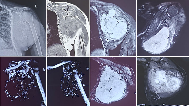
Figure 20. X-rays and MRI showing an extensive chondrosarcoma of the scapula.
Depending on the tumor, surgery may involve:
- Partial scapulectomy, where only the involved portion of the scapula is removed.
- Total scapulectomy, where the entire scapula is removed.
- Reconstruction using soft tissue techniques or custom implants in selected cases.
(Internal link: Scapulectomy for Bone Tumors)
Although shoulder movement may be reduced after extensive scapular surgery, preservation of the arm, elbow, and hand often allows patients to remain functionally independent.
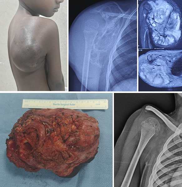
Figure 21. X-rays and MRI showing a scapular sarcoma. The tumor was treated with total scapulectomy as part of limb-salvage surgery.
Clavicle Tumors
Tumors of the clavicle (collarbone) are rare.
Treatment usually involves partial or total claviculectomy, meaning removal of part or all of the clavicle while preserving nearby muscles, nerves, and blood vessels whenever possible.
Unlike many other bones, the clavicle does not always require reconstruction after removal. Many patients adapt remarkably well and regain excellent arm function following rehabilitation.
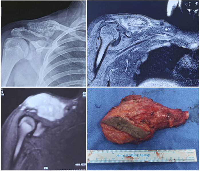
Figure 22. X-rays and MRI showing Ewing sarcoma of the clavicle, treated with wide surgical resection.
Recovery, Rehabilitation & Long-Term Outlook
Recovery after shoulder bone tumor surgery is a gradual process that depends on the type of tumor, the operation performed, and the patient’s overall health. Some people recover after relatively minor procedures such as curettage, while others require rehabilitation following complex limb salvage surgery and reconstruction.
The goals of rehabilitation are to:
- Control pain
- Protect the reconstruction while it heals
- Restore shoulder movement safely
- Improve strength and function
- Help patients return to daily activities as independently as possible
A structured physiotherapy programme is an essential part of treatment and often continues for several months after surgery.
What Happens After Surgery?
Immediately after surgery, your arm may be supported in a sling to protect the reconstruction. Pain control, wound care, and gentle exercises begin during the hospital stay under the supervision of your surgical and rehabilitation teams.
The exact rehabilitation programme depends on the type of reconstruction performed. Patients treated with curettage usually regain function more quickly than those undergoing major bone resection and reconstruction.
Early rehabilitation focuses on:
- Controlling swelling and pain
- Maintaining movement of the elbow, wrist, and hand
- Protecting soft tissue repair
- Preventing joint stiffness
As healing progresses, supervised shoulder exercises are gradually introduced to improve strength and mobility.
Expected Shoulder Function
One of the most common questions patients ask is, “Will I be able to use my arm normally again?”
The answer depends on several factors, including:
- The amount of bone removed
- Preservation of the deltoid muscle and rotator cuff
- Function of the axillary nerve
- Type of reconstruction performed
- Commitment to physiotherapy
Many patients regain excellent function of the hand and elbow and are able to perform most activities of daily living independently. Although overhead movement and heavy lifting may remain limited after extensive reconstruction, preserving a painless, functional upper limb is considered a successful outcome.
The ultimate goal is not to create a perfectly normal shoulder but to restore a stable, useful arm that allows patients to work, drive, write, eat, dress independently, and enjoy a good quality of life.
Table. Typical Recovery Timeline
Recovery varies depending on the diagnosis, reconstruction, and individual healing.
Patient Tip
Recovery after shoulder reconstruction is a marathon rather than a sprint. Small improvements over many months often lead to significant long-term gains.
Doctor Insight
Successful outcomes depend not only on the operation but also on dedicated rehabilitation. Consistent physiotherapy and realistic expectations are just as important as the surgery itself.
Clinical Pearl
Preservation of the deltoid muscle and axillary nerve often has a greater influence on postoperative shoulder function than the specific implant used for reconstruction.
Long-Term Follow-up
Regular follow-up visits allow your healthcare team to monitor:
- Healing of the reconstruction
- Implant function (if a prosthesis has been used)
- Bone healing after biological reconstruction
- Tumor recurrence
- Possible spread of disease in malignant tumors
The frequency of follow-up depends on the diagnosis and stage of the tumor. Patients treated for malignant bone tumors usually require long-term surveillance with clinical examination and imaging.
Reassurance Box
Modern limb salvage surgery allows many patients to return to independent living, employment, education, and recreational activities. While some limitations in shoulder movement may persist, preserving a functional arm provides excellent quality of life for most patients.
Frequently Asked Questions
Can shoulder pain be caused by a bone tumor?
Yes, but bone tumors are an uncommon cause of shoulder pain. Persistent pain that worsens over time, occurs at night, or is associated with swelling should be evaluated.
Is every shoulder bone tumor cancer?
No. Most shoulder bone tumors are benign, although some benign tumors may still require treatment because they can weaken the bone or damage the joint.
Will I lose my arm if I have bone cancer?
In most cases, no. Modern limb salvage surgery allows the arm to be preserved in the majority of patients while achieving appropriate cancer treatment.
What is a shoulder tumor megaprosthesis?
A tumor megaprosthesis is a specialized metallic implant used to replace bone removed during tumor surgery while preserving the arm.
(Tumor Megaprosthesis)
What is reverse shoulder replacement?
Reverse shoulder arthroplasty is a specialized shoulder replacement that allows the deltoid muscle to lift the arm when the rotator cuff cannot be preserved.
Will I regain full shoulder movement?
Not always. The amount of movement depends on the tumor, surgery performed, preserved muscles and nerves, and rehabilitation. Most patients regain useful function for daily activities.
Can shoulder bone tumors come back?
Some benign and malignant tumors can recur. Regular follow-up helps detect recurrence early and allows timely treatment.
Will I need chemotherapy?
Only certain malignant tumors, such as osteosarcoma and Ewing sarcoma, routinely require chemotherapy. Many benign tumors and chondrosarcomas are treated primarily with surgery.
Which doctor treats shoulder bone tumors?
Patients should be evaluated by an orthopaedic oncologist, a surgeon specializing in tumors of bone and soft tissues.
Should I seek a second opinion?
Because bone tumors are uncommon and treatment decisions can be complex, seeking a second opinion from a specialized bone tumor center is often valuable, particularly before biopsy or major surgery.
Medical Disclaimer
The information provided on this page is intended for educational purposes only and should not replace professional medical advice, diagnosis, or treatment. Bone tumors are uncommon and require individualized evaluation by an experienced multidisciplinary team. If you have persistent shoulder pain, an unexplained swelling, or have been diagnosed with a bone tumor, consult an orthopaedic oncologist or a specialized bone tumor center for appropriate assessment and management.
References
- D’Arienzo A, Ipponi E, Ruinato AD, et al. Proximal humerus reconstruction after tumor resection: an overview of surgical management. Adv Orthop. 2021;2021:5559377.
- Sirveaux F. Reconstruction techniques after proximal humerus tumour resection. Orthop Traumatol Surg Res. 2019;105(1):S153-S164.
- Kitagawa Y, Thai DM, Choong PF. Reconstructions of the shoulder following tumour resection. J Orthop Surg. 2007;15(2):201-206.
- Sehrawat S, Behera A, Kapoor L, et al. Endoprosthesis vs. nail-cement spacer application for reconstruction after oncologic proximal humeral resection: is there a difference in functional outcome? JSES Int. 2024;8(1):xx-xx
 +91 93558 33005