Quick Summary
What is Arm Bone Tumor Surgery?
Arm bone tumor surgery involves removing a tumor from the humerus, radius, or ulna while preserving as much normal bone, muscle, nerves, and joint function as possible. Depending on the type and location of the tumor, surgery may involve curettage (scraping out the tumor), segmental bone removal (resection), reconstruction with bone grafts or custom implants, or occasionally amputation when limb preservation is not safe.
Key Points
- Most arm bone tumors are benign (non-cancerous).
- Early diagnosis improves both cancer control and long-term arm function.
- MRI and a carefully planned biopsy are essential before surgery.
- Limb salvage surgery, which removes the tumor while preserving the arm, is possible in the majority of patients treated at specialist centers.
- Reconstruction is individualized and may involve bone grafts, metal implants, or biological techniques.
- Recovery requires a combination of surgery, physiotherapy, and regular follow-up.
Reassurance
Being told that you have a bone tumor can be overwhelming. It is important to remember that not every bone tumor is cancer, and even many malignant tumors can be successfully treated while preserving the arm. Modern multidisciplinary care has dramatically improved outcomes for both adults and children.
Understanding the Anatomy of the Arm
Understanding the basic anatomy of the arm helps explain why treatment differs depending on where the tumor develops. The upper limb is designed to provide both strength and remarkable flexibility, allowing us to lift, push, pull, rotate, and perform precise hand movements. A tumor affecting different parts of the arm can therefore have very different implications for movement and reconstruction.
The Humerus
The humerus is the long bone of the upper arm that extends from the shoulder to the elbow. It forms the shoulder joint at its upper end and the elbow joint at its lower end.
Tumors may occur in:
- Proximal humerus (near the shoulder)
- Humeral shaft (middle portion)
- Distal humerus (near the elbow)
Each location presents unique surgical challenges. For example, tumors near the shoulder may require reconstruction to restore shoulder stability, whereas tumors in the shaft often focus on preserving the major nerves and muscles that control the hand and elbow.
The Radius
The radius is one of the two forearm bones and lies on the thumb side. It is essential for:
- Rotating the forearm (turning the palm up and down)
- Wrist movement
- Grip strength
- Hand positioning
Tumors involving the distal radius (near the wrist) often require careful reconstruction to preserve wrist function.
The Ulna
The ulna runs alongside the radius on the little finger side of the forearm. It contributes to:
- Elbow stability
- Forearm strength
- Muscle attachment
- Coordination of forearm rotation
Unlike the radius, certain segments of the ulna can sometimes be removed with minimal loss of function, while tumors involving the upper end near the elbow often require more complex reconstruction.
Why Does Tumor Location Matter?
Two patients may both have a “bone tumor of the arm,” yet require completely different operations. Surgical planning depends on several factors, including:
- Which bone is involved
- Whether the tumor is close to the shoulder or elbow
- Involvement of the joint surface
- Relationship to major nerves and blood vessels
- Amount of bone that must be removed
- Whether reconstruction is necessary
For this reason, treatment is always individualized rather than based on the tumor name alone.
Doctor Explains
Orthopaedic oncologists do not simply remove the tumor—they plan surgery to achieve two equally important goals: complete tumor removal and the best possible long-term function of the arm.
Clinical Pearl
Preserving muscles, tendons, nerves, and joints is often just as important as preserving bone. These soft tissues play a major role in determining how well the arm functions after surgery.
Did You Know?
The humerus is the longest bone of the upper limb and provides attachment for more than 20 muscles that control shoulder, elbow, and arm movement.
Symptoms of Arm Bone Tumors
Bone tumors often develop gradually. In the early stages, symptoms may be subtle and easily mistaken for sports injuries, muscle strain, arthritis, or tendon problems. Because these conditions are far more common than bone tumors, diagnosis is sometimes delayed.
Recognizing persistent or unusual symptoms is the first step toward early treatment.
Persistent Bone Pain
Pain is the most common symptom of an arm bone tumor.
Unlike pain caused by a muscle injury, bone tumor pain often:
- Persists for several weeks or months
- Gradually becomes more severe
- Occurs even during rest
- May wake a person from sleep
- Does not improve with routine pain medication
Not every painful bone lesion is cancer, but persistent unexplained pain deserves medical evaluation.
Swelling or a Lump
As a tumor enlarges, it may produce:
- Visible swelling
- A firm lump
- Fullness around the shoulder, arm, elbow, or forearm
Some tumors remain hidden deep within the bone and may not produce any visible swelling until they become quite large.
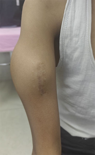
Figure 1. A swelling or lump in the arm can be a warning sign of an underlying bone or soft-tissue tumor and should be evaluated promptly.
Reduced Arm Function
Depending on its location, a tumor may interfere with normal movement by weakening bone or affecting nearby muscles and tendons.
Patients may notice:
- Difficulty lifting the arm
- Limited shoulder movement
- Reduced elbow motion
- Weak grip strength
- Difficulty rotating the forearm
- Trouble performing everyday activities such as dressing or lifting objects
Pathological Fracture
A pathological fracture is a broken bone that occurs because the bone has been weakened by disease rather than by a major injury.
In some patients, the first sign of a bone tumor is a fracture after a seemingly minor fall or even while performing routine daily activities.
A pathological fracture always requires careful evaluation because it may indicate an underlying bone tumor or another disease affecting bone strength.
Nerve-related Symptoms
Tumors located near major nerves may occasionally cause:
- Numbness
- Tingling
- Weakness of the hand
- Difficulty extending the wrist or fingers
- Loss of fine motor control
Fortunately, these symptoms are relatively uncommon and usually occur only when tumors become large or involve structures surrounding the bone.
Red Flag
Seek specialist evaluation if you experience:
- Persistent bone pain lasting more than six weeks
- Pain that worsens at night
- A growing swelling or lump
- A fracture after minimal trauma
- An unexplained bone lesion on an X-ray
Patient Tip
If your doctor identifies an unexplained bone lesion on an X-ray, avoid rushing into surgery. Additional imaging and a properly planned biopsy are often essential before treatment.
Reassurance
Most shoulder, arm, and forearm pain is caused by common conditions such as tendon injuries, arthritis, or muscle strain—not bone tumors. However, persistent or unusual symptoms should always be assessed by a healthcare professional.
Types of Arm Bone Tumors
The term bone tumor simply means an abnormal growth of cells within the bone. These tumors vary widely in behavior. Some remain harmless for life, while others require urgent treatment.
Understanding the broad categories helps patients appreciate why treatment recommendations differ from one individual to another.
Benign Bone Tumors
Benign tumors are non-cancerous growths that do not spread to other parts of the body. Many are discovered incidentally during imaging for unrelated injuries and may only require observation.
Examples include:
- Osteochondroma
- Enchondroma
- Unicameral bone cyst
- Fibrous dysplasia
Although benign, some tumors can weaken the bone, cause pain, or increase the risk of fracture, making surgery necessary in selected cases.
.jpg "Osteochondroma arising from the humerus (upper arm bone), a common benign bone tumor that may present as a painless lump")
Figure 2. Osteochondroma arising from the humerus (upper arm bone), a common benign bone tumor that may present as a painless lump.
Aggressive Benign Tumors
These tumors are not cancerous but behave more aggressively by expanding within the bone and damaging surrounding structures.
Common examples include:
- Giant cell tumor of bone
- Aneurysmal bone cyst
They often require surgery because of their tendency to recur or weaken the bone.
Malignant Bone Tumors
Malignant tumors are cancers that can grow into surrounding tissues and, in some cases, spread to distant organs such as the lungs.
Primary malignant bone tumors affecting the arm include:
- Osteosarcoma
- Chondrosarcoma
- Ewing sarcoma
Treatment often combines surgery with chemotherapy and, in selected situations, radiotherapy.
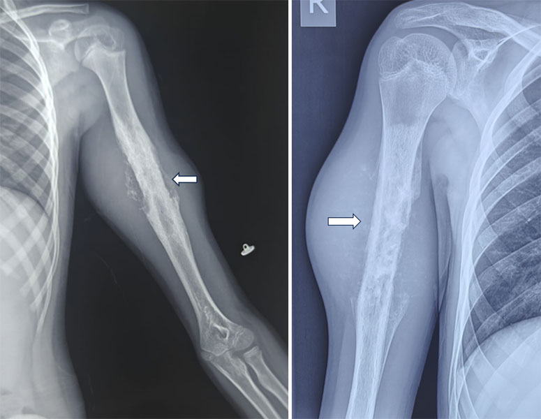
Figure 3. X-ray and MRI showing a malignant bone tumor of the humerus, demonstrating the importance of detailed imaging before arm bone tumor surgery.
Metastatic Bone Disease
Sometimes a tumor found in the humerus, radius, or ulna has spread from another cancer elsewhere in the body, most commonly the breast, lung, kidney, thyroid, or prostate.
Treatment focuses on relieving pain, preserving function, preventing fractures, and controlling the underlying cancer.
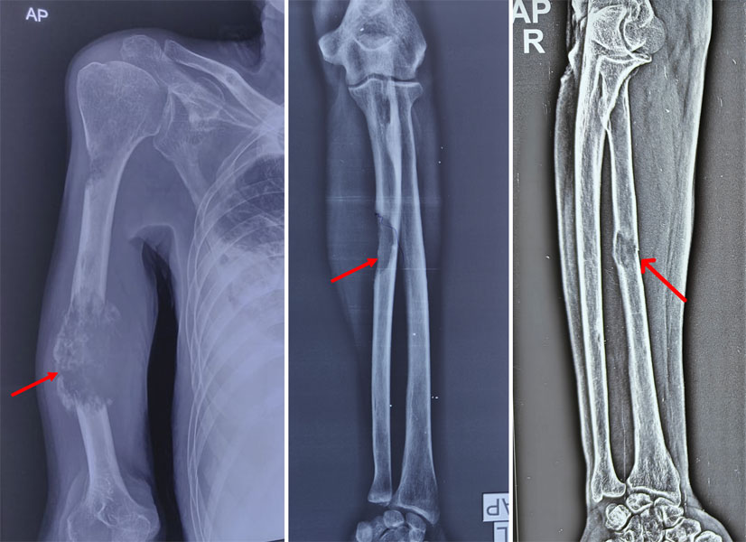
Figure 4. Metastatic bone lesions involving the humerus and forearm bones, showing how cancer from another organ can spread to the arm
Clinical Pearl
Imaging studies can strongly suggest the nature of a bone tumor, but a biopsy is often required to establish the definitive diagnosis before treatment is planned.
Reassurance
Hearing the word “tumor” does not necessarily mean cancer. Many arm bone tumors are benign, and even malignant tumors can often be treated successfully with modern limb-sparing techniques.
Diagnosis
Accurate diagnosis is the foundation of successful treatment. While imaging can strongly suggest the nature of a bone tumor, a definitive diagnosis requires careful evaluation and, in most cases, a biopsy. The goals are to determine whether the tumor is benign or malignant, identify its exact type, assess its extent within the bone and surrounding soft tissues, detect any spread, and plan treatment that preserves maximum arm function.
Clinical Assessment
Evaluation begins with a detailed medical history and physical examination. Your orthopaedic oncologist will assess symptoms such as persistent pain, night pain, swelling, previous fractures, and any history of cancer. The examination also evaluates the size and location of the lesion, joint movement, muscle strength, nerve function, blood circulation, and signs of an impending pathological fracture.
Doctor Explains: Clinical examination not only helps diagnose the tumor but also identifies which muscles, nerves, and joints can be safely preserved during surgery.
X-rays
An X-ray is usually the first investigation. It provides information about bone destruction, calcification, fracture risk, and the biological behavior of the lesion. Although X-rays help distinguish benign from aggressive tumors, they cannot confirm the diagnosis.
Magnetic Resonance Imaging (MRI)
MRI is the most important imaging study for surgical planning. It accurately defines the tumor’s size, bone marrow involvement, soft tissue extension, relationship to nerves and blood vessels, joint involvement, and any skip lesions, allowing precise planning of tumor removal while preserving normal tissues.
Computed Tomography (CT)
A CT scan provides detailed images of bone and is particularly useful for evaluating cortical destruction, complex joint anatomy, mineralization within tumors, planning reconstruction, and detecting lung metastases with a CT chest.
PET-CT and Bone Scan
In selected patients, PET-CT or a bone scan may be performed to determine whether the tumor has spread elsewhere or to assess the response to chemotherapy. These investigations are recommended based on the suspected diagnosis and multidisciplinary team discussion.
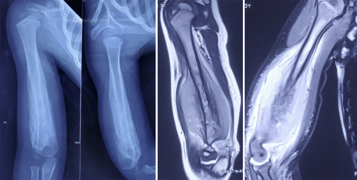
Figure 5. Radiological evaluation of an arm bone tumor using X-rays and MRI to assess the tumor’s location, extent and involvement of surrounding tissues
Why Is a Biopsy Necessary?
A biopsy—removal of a small tissue sample for microscopic examination—is essential for confirming the diagnosis. Most tumors are evaluated using a core needle biopsy, while an open biopsy is reserved for selected cases. Biopsy should always be performed after imaging and carefully planned so the biopsy tract can be removed during definitive surgery if required.
Red Flag: Never undergo an unplanned biopsy or excision of a suspected bone tumor. An incorrectly placed biopsy can compromise future surgery and reduce limb salvage options.
Clinical Pearl: Whenever possible, the biopsy should be planned or performed by the specialist musculoskeletal oncology team that will undertake the definitive surgery.
Treatment Planning
Every arm bone tumor is unique, and treatment is tailored to the individual patient. The choice of treatment depends on the tumor’s type, size, location, stage, and involvement of nearby joints, nerves, and blood vessels. To ensure the best outcome, treatment is planned by a Multidisciplinary Team (MDT) comprising specialists with expertise in musculoskeletal oncology.
The Multidisciplinary Team
Your MDT may include an orthopaedic oncologist, musculoskeletal radiologist, pathologist, medical and radiation oncologists, plastic and reconstructive surgeon, physiotherapist, occupational therapist, and specialist oncology nurses. Together, they develop a personalized treatment plan that aims to achieve both complete tumor control and optimal long-term arm function.
Factors That Influence Treatment
Treatment decisions are based on several factors, including:
- Whether the tumor is benign or malignant
- Histological grade (how aggressive the tumor appears under the microscope)
- Tumor size and location within the humerus, radius, or ulna
- Involvement of the shoulder, elbow, or wrist
- Relationship to major nerves and blood vessels
- Presence of a pathological fracture
- Whether the disease has spread
- The patient’s age, activity level, occupation, and overall health
Malignant tumors such as osteosarcoma and Ewing sarcoma often require chemotherapy before surgery (neoadjuvant chemotherapy) and sometimes additional chemotherapy or radiotherapy afterward. Many benign tumors can be treated with surgery alone.
Doctor Insight: The goal is not only to remove the tumor but also to preserve arm function and maintain the best possible quality of life.
Limb Salvage Surgery
One of the most common concerns is, “Will I lose my arm?” Fortunately, advances in imaging, chemotherapy, surgical techniques, and reconstruction mean that limb salvage surgery is possible for most patients.
What Is Limb Salvage Surgery?
Limb salvage surgery removes the tumor with a margin of healthy tissue while preserving the arm. The resulting bone defect is reconstructed using the most appropriate technique, with the aim of restoring stability and function.
When Is Limb Salvage Possible?
Limb salvage is usually feasible when the tumor can be completely removed with safe margins, major nerves and blood vessels can be preserved, and reconstruction is expected to provide a functional limb.
When Is Amputation Necessary?
Amputation is now uncommon and is considered only when safe tumor removal is not possible, critical nerves or blood vessels are extensively involved, reconstruction is unlikely to provide a useful limb, or severe infection or repeated reconstruction failure occurs.
Clinical Pearl:
Successful limb salvage is defined not simply by preserving the arm, but by preserving a pain-free, stable, and functional limb.
Reassurance:
Today, only a small proportion of patients with arm bone tumors require amputation. Most can be treated successfully with limb-preserving surgery at specialist musculoskeletal oncology centers.
Surgical Resection of Arm Bone Tumors
The extent of surgery depends on the location, size, and biological behavior of the tumor. The primary objective is to remove the tumor completely while preserving as much normal bone, muscle, nerves, and joint function as possible. Modern imaging and careful surgical planning allow most patients to undergo limb salvage surgery rather than amputation.
Humeral Shaft Tumor Resection
Tumors involving the shaft (middle portion) of the humerus are commonly treated by segmental (intercalary) resection, in which only the diseased portion of bone is removed while preserving the shoulder and elbow joints.
Whenever oncologically safe, important structures such as the radial nerve and surrounding muscles are carefully protected. After tumor removal, the bone defect is reconstructed using a biological graft, intercalary prosthesis, or other reconstructive technique depending on the size of the defect and patient factors.
Because both adjacent joints are preserved, many patients achieve excellent long-term arm function.
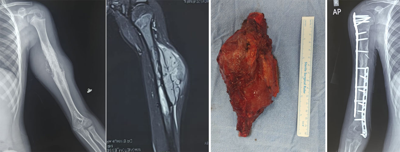
Figure 6. Ewing sarcoma of the humerus in an 8-year-old girl treated with chemotherapy and wide tumor resection. The resected tumor-bearing bone was treated with high-dose extracorporeal radiation therapy (ECRT) and reimplanted, allowing reconstruction using the patient’s own bone as a biological reconstruction.
Distal Humerus (Elbow) Tumor Resection
Tumors around the distal humerus are particularly challenging because they involve the elbow joint, one of the most complex joints in the upper limb.
Whenever possible, surgeons preserve the elbow joint by performing a partial bone resection. However, tumors extending into the joint often require en bloc resection (removal of the tumor in one piece) together with the involved portion of the distal humerus.
Depending on the extent of bone removal, reconstruction may include:
- Distal humeral endoprosthetic replacement
- Total elbow replacement following tumor resection
- Biological reconstruction using allograft or vascularized bone grafts in selected patients
- Soft tissue and ligament reconstruction to restore elbow stability
The choice of reconstruction depends on the patient’s age, activity level, tumor type, expected survival, and functional requirements.
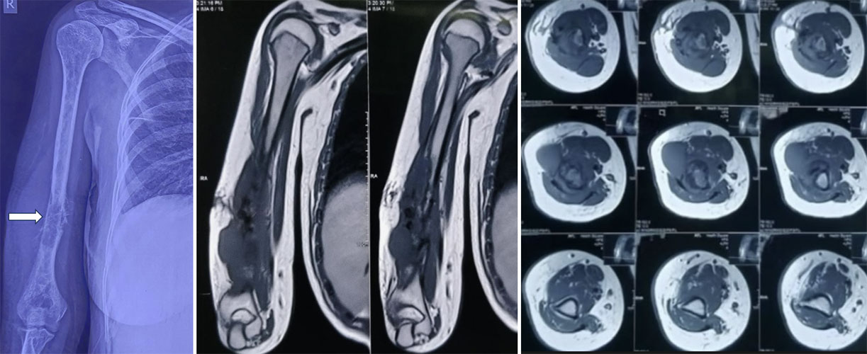
Figure 7A. Malignant bone tumor of the lower humerus extending to the elbow joint, requiring careful surgical planning to achieve tumor-free margins while preserving arm function.
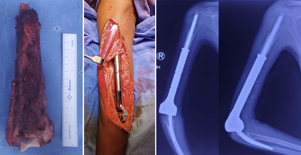
Figure 7B. Elbow reconstruction with an endoprosthesis after wide resection of the humeral tumor, designed to preserve elbow movement and arm function.
Radius Shaft Tumor Resection
Tumors involving the radius shaft are uncommon but require meticulous planning because the radius plays a critical role in forearm rotation and hand positioning.
Following tumor resection, reconstruction is usually recommended to restore forearm stability and maintain pronation and supination (rotation of the forearm). Depending on the defect, options include intercalary biological grafts or custom implants.
When adequate reconstruction is achieved, most patients regain useful hand and forearm function.
Ulna Tumor Resection
The ulna contributes to elbow stability and serves as an important attachment site for muscles controlling forearm movement.
Small or mid-shaft tumors can often be removed safely while preserving satisfactory function. Larger defects may require biological reconstruction or internal fixation, particularly when elbow stability is affected.
Compared with the radius, selected ulnar resections may require less extensive reconstruction because the radius bears a greater role in wrist function.
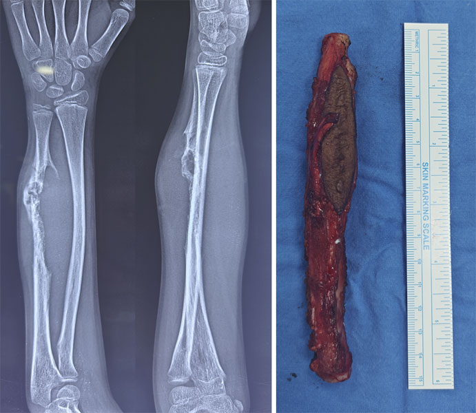
Figure 8. Malignant tumor of the ulna treated with wide resection, removing the tumor with a surrounding margin of healthy tissue.
Clinical Pearl
The surgical approach is determined not only by the bone involved, but also by the need to preserve elbow function, forearm rotation, nerve integrity, and long-term limb durability. A successful operation removes the tumor while maximizing the patient’s ability to perform everyday activities.
Excellent. Since this is a cornerstone procedure page, the next section should focus on reconstruction principles rather than describing every reconstructive technique in depth (those can have dedicated pages). This improves SEO, reduces duplication, and encourages internal linking.
Reconstruction After Arm Bone Tumor Surgery
Following removal of a bone tumor, the resulting defect must be reconstructed to restore the stability and function of the arm. The ideal reconstruction depends on the location and size of the defect, patient age, expected activity level, tumor biology, and the need for chemotherapy or radiotherapy.
The primary goals of reconstruction are to:
- Restore skeletal stability
- Preserve elbow and forearm function
- Enable early rehabilitation
- Provide a durable reconstruction with low complication rates
- Achieve the best possible long-term quality of life
Your orthopaedic oncologist will recommend the most suitable option based on your individual needs.
Biological Reconstruction
Biological reconstruction uses living bone to replace the segment removed during tumor surgery. It is generally preferred in younger patients because the graft has the potential to heal, remodel, and become incorporated into the patient’s own skeleton.
Common biological reconstruction techniques include:
- Autografts (bone taken from the patient’s own body)
- Allografts (donor bone)
- Vascularized fibular grafts
- Composite biological reconstructions
Although biological reconstruction offers excellent long-term durability, healing may take several months and requires close follow-up.
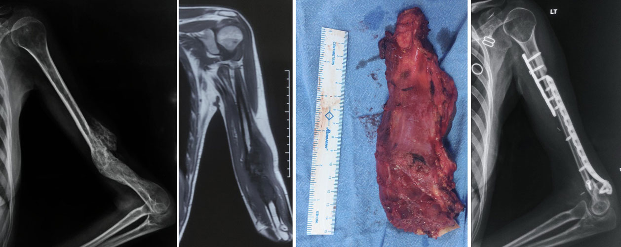
Figure 9. Osteosarcoma of the humerus treated with wide resection and reconstruction using a fibular bone graft to restore structural continuity.
Read more: Biological Reconstruction After Bone Tumor Surgery.
Endoprosthetic Reconstruction
When immediate structural stability is required, endoprosthetic reconstruction is an excellent option. Metal implants replace the resected segment of bone and allow earlier mobilization than biological grafts.
For tumors around the distal humerus, specialized tumor endoprostheses or total elbow replacement may be used to restore elbow function after wide tumor excision.
Advantages include:
- Immediate stability
- Early movement and rehabilitation
- Predictable functional recovery
- No need for bone graft healing
However, prostheses may require revision surgery over time because of wear, loosening, or mechanical failure, particularly in younger, active patients.
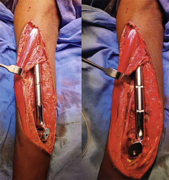
Figure 10. Endoprosthetic elbow reconstruction after tumor resection around the elbow, helping restore stability and useful elbow movement.
Intercalary Reconstruction
When both adjacent joints can be preserved—such as after humeral shaft, radial shaft, or ulnar shaft resections—the missing bone segment can be reconstructed without replacing the joint itself.
Options include:
- Intercalary allografts
- Vascularized fibular grafts
- Intercalary endoprostheses
- Composite biological-prosthetic reconstructions
Because the shoulder and elbow joints remain intact, patients often achieve excellent functional outcomes.
Elbow Reconstruction After Distal Humerus Resection
Reconstruction of the elbow is one of the most demanding procedures in musculoskeletal oncology. The elbow provides both stability and a wide range of motion required for daily activities such as eating, dressing, and personal hygiene.
Depending on the extent of resection, reconstruction may involve:
- Distal humeral tumor prosthesis
- Total elbow replacement
- Biological reconstruction using allografts or vascularized bone grafts
- Soft tissue and ligament reconstruction to restore elbow stability
The choice depends on the patient’s age, tumor type, expected longevity, functional demands, and surgeon experience.
Which Reconstruction Is Best?
There is no single reconstruction technique that is ideal for every patient. Each method has specific advantages, limitations, and potential complications.
The final decision is influenced by:
- Tumor location
- Length of bone removed
- Patient age
- Functional expectations
- Bone quality
- Previous treatments
- Risk of complications
- Need for additional cancer treatment
An individualized approach provides the best balance between durability, function, and oncological safety.
Doctor Explains
Successful reconstruction is not simply about replacing bone—it is about restoring a stable, pain-free, and functional arm that allows patients to return to work, recreation, and everyday life.
Reconstruction Options at a Glance
Clinical Pearl
Preserving a patient’s own elbow joint whenever oncologically safe usually provides better long-term function than replacing it. However, when the joint is involved by the tumor, modern elbow reconstruction techniques can still achieve excellent pain relief and meaningful upper-limb function.
Recovery and Rehabilitation After Arm Bone Tumor Surgery
Recovery after arm bone tumor surgery is a gradual process that involves wound healing, bone recovery, and rehabilitation. The duration varies depending on the type of tumor, extent of surgery, reconstruction performed, and the need for chemotherapy or radiotherapy.
Most patients begin gentle movement exercises soon after surgery under the guidance of a physiotherapist. Early rehabilitation helps prevent stiffness, restore muscle strength, and improve long-term arm function.
Recovery is not only about healing the bone—it is about helping you safely return to work, hobbies, and everyday activities.
Hospital Stay
The average hospital stay ranges from 3 to 7 days, although it may be longer after complex reconstructions or if additional medical treatment is required.
During your stay, the healthcare team will focus on:
- Pain control
- Wound care
- Early arm and hand exercises
- Prevention of complications
- Planning rehabilitation and discharge
Physiotherapy and Rehabilitation
Physiotherapy is an essential part of recovery. A personalized rehabilitation program is designed according to the type of reconstruction and the stability of the surgical repair.
Rehabilitation focuses on:
- Restoring elbow and forearm movement
- Improving muscle strength
- Reducing stiffness
- Regaining hand function
- Returning safely to daily activities
Patients undergoing elbow reconstruction may require a longer rehabilitation period to regain optimal range of motion.
Patient Tip: Follow your rehabilitation program consistently. Regular physiotherapy often has as much impact on the final outcome as the surgery itself.
Returning to Daily Activities
The timeline for returning to normal activities varies between individuals.
Most patients can expect to:
*Recovery timelines vary depending on the type of surgery and individual progress.
Follow-Up After Surgery
Regular follow-up is essential to monitor both recovery and tumor control.
During follow-up visits, your surgeon may assess:
- Wound healing
- Bone healing or implant stability
- Shoulder, elbow, wrist, and hand function
- Signs of local recurrence
- Complications such as infection, fracture, implant loosening, or non-union
- Surveillance imaging to detect recurrence or metastasis when appropriate
Follow-up is typically more frequent during the first 2–3 years, when the risk of recurrence is highest.
Possible Complications
Although most patients recover well, complications can occur after any major surgery.
Potential complications include:
- Infection
- Bleeding
- Delayed wound healing
- Nerve injury
- Joint stiffness
- Implant loosening or mechanical failure
- Non-union of biological grafts
- Local tumor recurrence
- Rarely, the need for additional surgery
Early recognition and prompt treatment of complications usually lead to better outcomes.
Clinical Pearl: Successful recovery depends on three equally important factors—complete tumor removal, appropriate reconstruction, and dedicated rehabilitation.
Reassurance: Most patients experience gradual improvement in pain, strength, and function over several months. With expert surgical care, structured rehabilitation, and regular follow-up, many are able to return to independent daily living and enjoy an excellent quality of life.
Frequently Asked Questions (FAQs)
1. Is every arm bone tumor cancer?
No. Most arm bone tumors are benign (non-cancerous). However, some may be malignant or aggressive benign tumors that require specialized treatment. Imaging and a biopsy are usually necessary to establish the correct diagnosis before any surgery is planned.
2. Will I lose my arm if I have a bone tumor?
In most cases, no. Advances in musculoskeletal oncology allow limb salvage surgery for the majority of patients. Amputation is now reserved for uncommon situations where the tumor cannot be safely removed while preserving a functional limb.
3. Is a biopsy always necessary?
Yes, in most cases. A biopsy is the only reliable way to confirm the exact type of bone tumor. It should always be carefully planned and ideally performed by the specialist team that will carry out the definitive surgery.
4. How long does recovery take after arm bone tumor surgery?
Recovery depends on the type of surgery, reconstruction performed, and whether chemotherapy is required. Most patients resume light daily activities within a few weeks, while complete recovery and rehabilitation may take several months.
5. Will I regain normal use of my arm?
Many patients regain excellent function after treatment. The outcome depends on the tumor location, the amount of bone removed, the reconstruction method, and commitment to physiotherapy. Preserving important nerves, muscles, and joints significantly improves long-term function.
6. Can bone tumors come back after surgery?
Yes. Both benign and malignant bone tumors can recur, although the risk varies depending on the tumor type and surgical margins. Regular follow-up with clinical examination and imaging is important for early detection of recurrence.
7. Will I need chemotherapy or radiotherapy?
Not every patient requires additional treatment. Benign tumors are often treated with surgery alone, whereas malignant tumors such as osteosarcoma or Ewing sarcoma commonly require chemotherapy. Radiotherapy is recommended only for selected tumor types.
8. Are metal implants safe inside the body?
Yes. Modern tumor prostheses are made from biocompatible materials designed for long-term use. Your surgeon will explain the expected lifespan of the implant and whether future revision surgery may be required.
9. Can I return to work and sports after surgery?
Most patients can return to work once healing and rehabilitation are complete. Office work is usually resumed earlier than physically demanding jobs. Participation in sports depends on the type of reconstruction and your surgeon’s recommendations.
10. When should I consult an orthopaedic oncologist?
You should seek specialist evaluation if you have persistent bone pain, an unexplained swelling, a bone lesion seen on imaging, or a suspected bone tumor. Early assessment improves diagnostic accuracy and increases the likelihood of successful limb-preserving treatment.
Medical Disclaimer
The information provided on this page is intended for educational purposes only and should not be considered a substitute for professional medical advice, diagnosis, or treatment. Every bone tumor is unique, and treatment recommendations vary depending on the type of tumor, its location, stage, and the patient’s overall health.
If you have persistent bone pain, swelling, or have been diagnosed with a bone tumor, consult a qualified orthopaedic oncologist for a comprehensive evaluation and individualized treatment plan.
References
- Biermann JS, Hirbe A, Ahlawat S, et al. Bone Cancer, Version 2.2025, NCCN Clinical Practice Guidelines in Oncology. Journal of the National Comprehensive Cancer Network. 2025;23(4):e250017. Available at:
- Casali PG, Bielack S, Abecassis N, et al. Bone sarcomas: ESMO–EURACAN–GENTURIS–ERN PaedCan Clinical Practice Guideline for diagnosis, treatment and follow-up. Annals of Oncology. 2021;32(12):1520–1536. Available at:
- National Comprehensive Cancer Network (NCCN). NCCN Guidelines for Patients®: Bone Cancer. 2026 Edition. Available at:
- American Academy of Orthopaedic Surgeons (AAOS). Orthopaedic oncology patient education resources. Available at:
- National Cancer Institute (NCI). Bone Cancer (Primary Bone Cancer) – Patient Information and Treatment Overview. Available at:
 +91 93558 33005