Quick Summary
A bone tumor is an abnormal growth of cells arising from bone. Bone tumors affecting the foot and ankle are uncommon, accounting for only a small proportion of all bone tumors. They may be benign (non-cancerous), aggressive benign (locally destructive but not spreading to other organs), or malignant (cancerous). Because the foot contains many small bones, joints, tendons, nerves, and weight-bearing structures packed into a limited space, treatment requires careful planning to remove the tumor while preserving walking ability and foot function.
The goals of surgery are to:
- Establish an accurate diagnosis.
- Remove the tumor completely and safely.
- Preserve as much normal bone, joints, muscles, and soft tissue as possible.
- Restore foot stability and function.
- Allow patients to return to everyday activities with the best possible quality of life.
Today, limb salvage surgery—an operation that removes the tumor while preserving the foot or ankle—is possible for many patients. Advances in imaging, surgical planning, reconstruction techniques, microsurgery, and rehabilitation have significantly reduced the need for amputation in appropriately selected patients.
Treatment, however, is never “one size fits all.” The best approach depends on several factors, including:
- The exact type of tumor
- Whether it is benign or malignant
- Its location within the foot or ankle
- The amount of bone and surrounding soft tissue involved
- The patient’s age, activity level, and overall health
A multidisciplinary team consisting of orthopaedic oncologists, radiologists, pathologists, medical oncologists, radiation oncologists, plastic surgeons, physiotherapists, and rehabilitation specialists often works together to achieve the best outcome.
Reassurance
Being told that you have a bone tumor in your foot or ankle can be overwhelming. Fortunately, many tumors are benign, and even when surgery is required, modern limb salvage techniques allow many patients to keep a functional foot. Your treatment plan will be tailored to your specific diagnosis rather than following a standard approach for every patient.
Understanding Foot & Ankle Anatomy
The foot and ankle form one of the most complex parts of the human body. Together they support the entire body weight, absorb impact during walking and running, and allow balance on uneven surfaces. Successful treatment of a bone tumor involves not only removing the disease but also preserving these important functions whenever possible.
The foot consists of 26 bones, more than 30 joints, and numerous muscles, tendons, ligaments, nerves, and blood vessels that work together to produce smooth and stable movement. Even removing a relatively small amount of bone can affect walking if the surrounding structures are not carefully preserved or reconstructed.
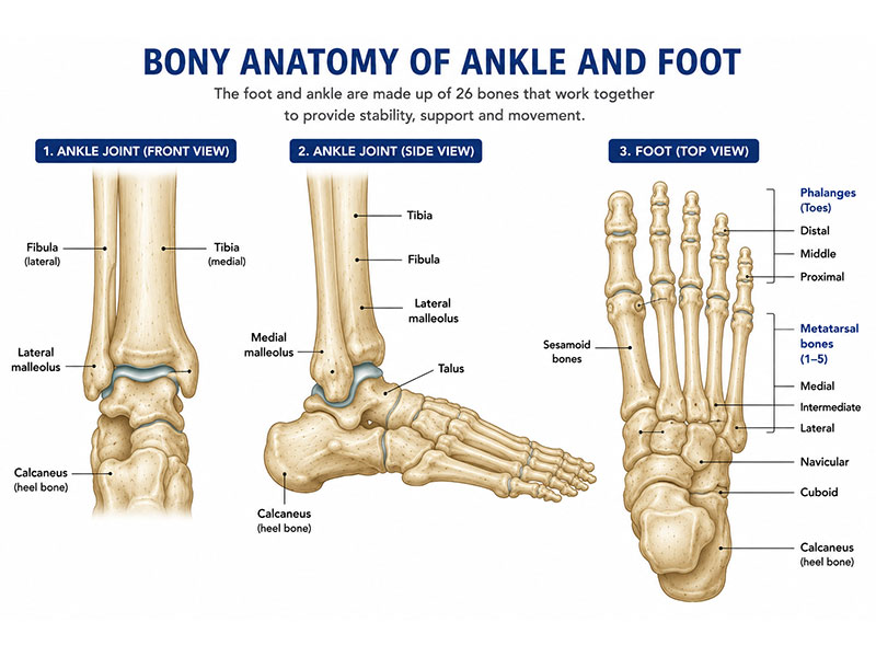
Figure 1. Bony anatomy of the ankle and foot, showing the major bones involved in foot and ankle conditions and bone tumors.
For simplicity, surgeons divide the foot into three regions.
Hindfoot
The hindfoot forms the back of the foot and includes two important bones:
- Calcaneus (heel bone): the largest bone in the foot, responsible for transmitting body weight to the ground during standing and walking.
- Talus: sits above the calcaneus and connects the foot to the tibia and fibula, forming the ankle joint.
Tumors in this region are particularly challenging because they involve major weight-bearing structures and may affect both walking and ankle movement.
Midfoot
The midfoot is composed of the:
- Navicular
- Cuboid
- Three cuneiform bones
These bones act as the structural “bridge” between the hindfoot and forefoot, maintaining the arches of the foot and contributing to stability during walking.
Forefoot
The forefoot includes:
- Five metatarsal bones
- Toe bones (phalanges)
Tumors affecting these bones may interfere with balance, push-off while walking, and shoe fitting. In many cases, however, surgery can preserve excellent function because the remaining parts of the foot compensate remarkably well.
The Ankle Joint
The ankle joint is where the tibia (shin bone), fibula, and talus meet. It allows the foot to move up and down during walking. Although primary bone tumors involving the ankle are uncommon, treatment in this area requires meticulous planning because even small changes can influence joint stability and long-term mobility.
One of the unique challenges of foot and ankle tumor surgery is preserving the plantar surface—the thick, specialized skin on the sole of the foot. This tissue is designed to withstand body weight and cannot easily be replaced. Whenever possible, surgeons aim to preserve this important weight-bearing surface.
Unlike larger bones such as the femur or tibia, the bones of the foot are relatively small and closely connected by joints. A tumor involving one bone may influence nearby joints, tendons, or soft tissues, making individualized surgical planning essential.
Doctor Explains
Removing a tumor is only one part of successful treatment. Equally important is preserving the mechanics of the foot so that patients can stand, walk, and perform everyday activities comfortably. This is why surgery for foot and ankle bone tumors should be carefully planned by a team experienced in musculoskeletal oncology.
Clinical Pearl
In foot and ankle tumor surgery, preserving function is just as important as removing the tumor. Successful surgery balances cancer control with maintaining weight-bearing, stability, and mobility.
Did You Know?
Although the foot contains only about one-quarter of your body weight, it contains nearly one-quarter of all the bones in the human body. This remarkable complexity explains why reconstruction after foot and ankle tumor surgery is highly individualized.
Symptoms of Foot & Ankle Bone Tumors
The symptoms of a foot or ankle bone tumor vary depending on the type of tumor, its size, and its location. Many benign tumors cause only mild discomfort or are discovered incidentally during imaging performed for another reason. Others may gradually weaken the bone or interfere with walking.
Because foot pain is extremely common and is more often caused by conditions such as plantar fasciitis, arthritis, tendon injuries, or stress fractures, bone tumors are not always suspected initially. However, symptoms that persist or progressively worsen deserve further evaluation.
The most common symptom is persistent pain. Unlike pain from a minor injury, tumor-related pain often develops gradually and may continue despite rest, medication, or changes in footwear. Some patients notice discomfort that becomes worse at night or interrupts sleep.
Swelling or a visible lump may develop if the tumor enlarges or extends toward the surface of the bone. Depending on its location, the swelling may make it difficult to wear normal shoes or may become noticeable only after several months.
Some tumors weaken the bone enough to cause a pathological fracture—a break that occurs through bone already weakened by disease rather than by a significant injury. Occasionally, the fracture is the first indication that a tumor is present.
Tumors located near joints may cause:
- Stiffness
- Reduced ankle movement
- Difficulty climbing stairs
- Pain while standing for prolonged periods
- Altered walking pattern (limp)
Rarely, tumors compress nearby nerves, leading to numbness, tingling, or weakness in the toes or foot.
Common Symptoms
- Persistent foot or ankle pain
- Swelling around the foot or ankle
- A slowly enlarging lump
- Pain at night
- Difficulty walking or limping
- Reduced joint movement
- Difficulty wearing shoes
- Pathological fracture
Red Flag
Seek medical evaluation if you experience:
- Persistent pain lasting more than 6 weeks without improvement
- Increasing swelling or a growing lump
- Night pain that repeatedly wakes you from sleep
- A fracture occurring after minimal injury
- Foot pain accompanied by an abnormal X-ray
Patient Tip
Many people attribute persistent foot pain to overuse, ill-fitting shoes, or aging. If symptoms continue despite appropriate treatment—or if imaging reveals an unexplained bone lesion—consult a specialist before assuming it is a routine foot problem.
Reassurance
Persistent foot pain does not automatically mean cancer. Most cases are caused by common conditions such as plantar fasciitis, arthritis, tendon disorders, or stress injuries. However, persistent or unexplained symptoms should be properly evaluated so that, if a bone tumor is present, it can be diagnosed and treated at an early stage.
Types of Foot & Ankle Bone Tumors
Bone tumors of the foot and ankle are uncommon, and most are benign (non-cancerous). However, some benign tumors behave aggressively, while malignant tumors require prompt treatment. Because treatment varies greatly depending on the diagnosis, every suspicious bone lesion should be evaluated before surgery is planned.
Benign Bone Tumors
Benign tumors do not spread to other parts of the body. Many remain stable and only require observation, while others may cause pain, weaken bone, or increase the risk of fracture, making surgery necessary.
Common benign tumors include:
- Osteochondroma – a cartilage-covered bony outgrowth.
- Enchondroma – a benign cartilage tumor within the bone.
- Chondroblastoma – a rare tumor occurring near joints in younger patients.
- Aneurysmal Bone Cyst (ABC) – a blood-filled bone lesion that can expand and weaken bone.
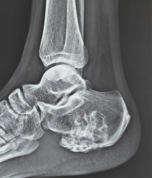
Figure 2. Osteochondroma of the ankle—a benign bone tumor arising near the ankle joint.
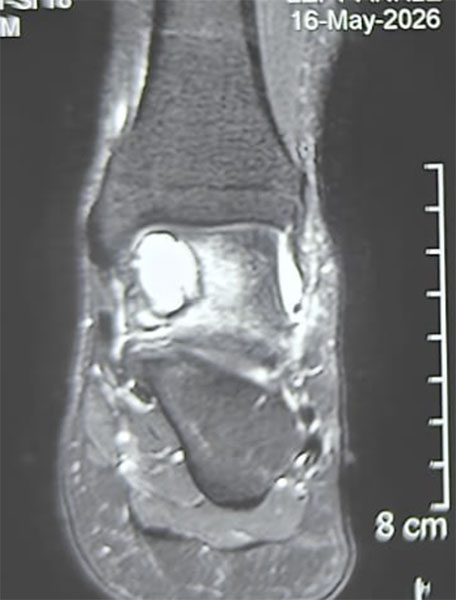
Figure 3. Chondroblastoma of the ankle—a benign bone tumor requiring careful evaluation and treatment planning.
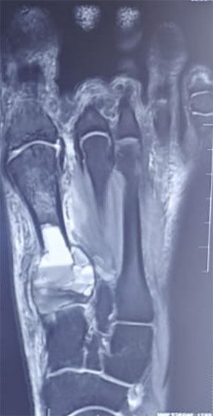
Figure 4. Aneurysmal bone cyst of the ankle and foot—a benign but locally aggressive bone lesion.
Aggressive Benign Tumors
Some benign tumors grow aggressively within the bone and have a higher risk of recurrence. The most important example is the Giant Cell Tumor of Bone, which may destroy surrounding bone and occasionally spread to the lungs. These tumors require specialized surgical planning to preserve function while minimizing recurrence.
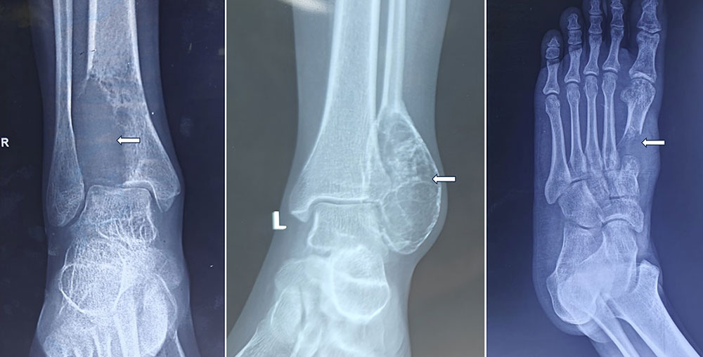
Figure 5. Giant cell tumor of the ankle—a locally aggressive bone tumor requiring specialized treatment.
Malignant Bone Tumors
Malignant tumors are cancers capable of invading nearby tissues and spreading to other organs.
The most important primary malignant bone tumors include:
- Osteosarcoma
- Ewing Sarcoma
- Chondrosarcoma
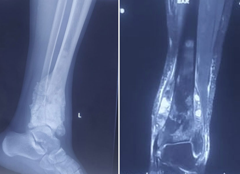
Figure 6. Osteosarcoma of the ankle—a malignant bone tumor requiring multidisciplinary cancer treatment.
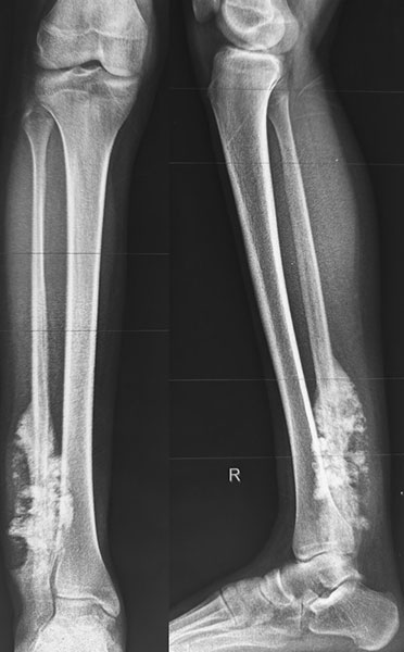
Figure 7. Osteosarcoma involving the distal tibia, illustrating the complex challenges of bone tumor surgery around the ankle.
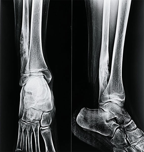
Figure 8. Ewing sarcoma of the ankle and foot—a malignant bone tumor requiring specialized oncological management.
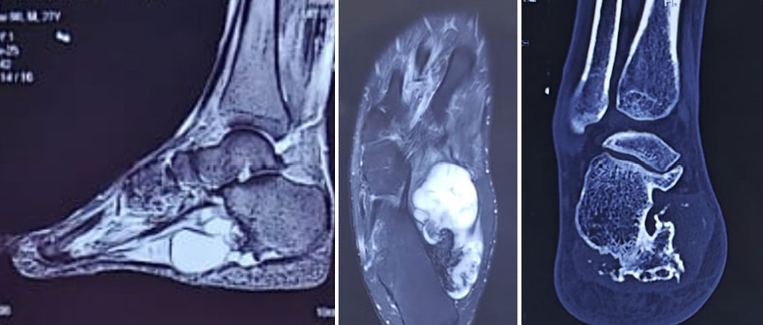
Figure 9. Chondrosarcoma around the ankle.
Detailed MRI and CT imaging helped define the tumour’s extent and its relationship with the surrounding bone and soft tissues, allowing careful planning of limb-salvage surgery.
Although uncommon in the foot and ankle, these tumors require treatment by a multidisciplinary sarcoma team.
Table. Common Foot & Ankle Bone Tumors
Clinical Pearl
Similar-looking bone lesions on an X-ray may require completely different treatments. A precise diagnosis is essential before surgery.
Reassurance
Most foot and ankle bone tumors are benign, and even many malignant tumors can now be treated with successful limb-preserving surgery.
Diagnosis
Accurate diagnosis is the foundation of successful treatment. The aim is not only to identify the tumor but also to determine its type, extent, and biological behavior, allowing the most appropriate treatment to be planned.
Evaluation usually begins with a clinical examination followed by imaging studies.
X-rays
An X-ray is the first investigation and provides valuable information about the location, size, and appearance of the lesion. It often allows an experienced orthopaedic oncologist to narrow the possible diagnosis.
Magnetic Resonance Imaging (MRI)
MRI provides detailed images of bone, muscles, tendons, nerves, and blood vessels. It defines:
- Tumor size
- Joint involvement
- Soft tissue extension
- Relationship to major nerves and vessels
- Surgical planning
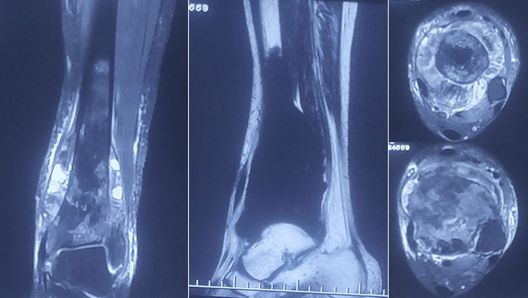
Figure 10. MRI – Seeing the tumour in three dimensions.
MRI provides a detailed map of the tumour, showing its extent within the bone and its relationship with the surrounding muscles, tendons and soft tissues—information that is critical before surgery.
Computed Tomography (CT)
A CT scan provides excellent detail of bone architecture and is particularly useful for tumors involving the calcaneus, talus, distal tibia, or distal fibula. CT of the chest may also be required to detect lung metastases in malignant tumors.
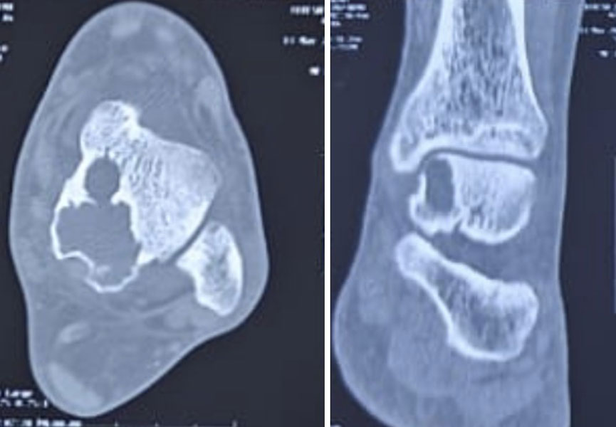
Figure 11. CT Scan – Understanding the bone before removing the tumour.
CT imaging provides a detailed view of the bone destruction and remaining cortical architecture, helping determine the extent of resection and plan reconstruction.
PET-CT and Bone Scan
For selected malignant tumors, PET-CT or bone scans help determine whether the disease has spread elsewhere in the body.
Biopsy
A biopsy is the only investigation that confirms the diagnosis. Most patients undergo a core needle biopsy, while an open biopsy is reserved for selected cases.
Because the biopsy tract is removed during definitive surgery, it should ideally be performed by the same team that will treat the tumor.
Doctor Explains
A poorly planned biopsy can complicate later surgery. Whenever possible, it should be performed by the specialist team providing definitive treatment.
Red Flag
Never remove or curette a suspicious bone lesion without establishing the diagnosis.
Treatment Planning
Treatment is individualized and depends on:
- Tumor type
- Benign or malignant nature
- Tumor grade
- Size and location
- Joint involvement
- Relationship to nerves and blood vessels
- Patient age and activity level
- Overall health
- Patient preferences
Patients with malignant tumors are usually discussed in a multidisciplinary team (MDT) meeting involving orthopaedic oncologists, radiologists, pathologists, medical oncologists, radiation oncologists, and plastic surgeons. Some tumors require chemotherapy or radiotherapy in addition to surgery.
Doctor Insight
Modern orthopaedic oncology aims to remove the tumor safely while preserving as much normal anatomy and function as possible.
Limb Salvage Surgery
The primary goal of modern bone tumor surgery is limb salvage—removing the tumor completely while preserving a functional foot or ankle whenever it is oncologically safe.
The decision between limb salvage and amputation depends on:
- Ability to achieve complete tumor removal
- Preservation of major nerves and blood vessels
- Remaining healthy bone
- Expected functional outcome
- Patient goals and lifestyle
Amputation is now reserved for carefully selected situations where limb preservation cannot provide safe tumor control or a functional limb.
Table. Limb Salvage vs Amputation
Clinical Pearl
Limb salvage is recommended only when it offers both effective tumor control and a durable, functional limb.
Reassurance
Most patients with foot and ankle bone tumors do not require amputation. Advances in surgical techniques allow limb preservation in many carefully selected cases.
How Much Bone Needs to Be Removed?
The extent of surgery is determined by surgical margins, not simply by the visible size of the tumor.
A surgical margin is a small rim of healthy tissue removed with the tumor to reduce the risk of leaving microscopic disease behind.
- Benign tumors may be treated with curettage or limited resection.
- Malignant tumors usually require wide resection, removing the tumor together with a surrounding cuff of healthy tissue.
Modern MRI and CT imaging allow surgeons to define these margins accurately, helping preserve as much healthy bone and joint as possible.
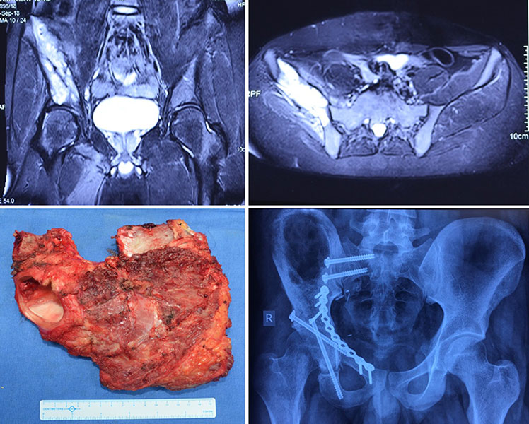
Figure 12. Giant Cell Tumour — Curettage.
Treating a giant cell tumour while preserving the ankle: After carefully assessing the lesion, the tumour was treated with extended curettage and reconstruction, aiming to control the disease while preserving as much normal bone and joint function as possible.
Doctor Explains
Surgeons remove only the amount of bone necessary to achieve safe tumor clearance while maximizing long-term function.
Foot & Ankle Tumor Resection
The exact operation depends on which bone is involved and whether the tumor extends into the ankle joint or surrounding soft tissues. Because the foot and ankle contain numerous small bones, joints, tendons, and neurovascular structures packed into a limited space, every operation is individualized. The goal is always to remove the tumor completely while preserving as much normal anatomy and function as possible.
Distal Tibia Tumor Resection
The distal tibia forms the upper part of the ankle joint and is one of the body’s primary weight-bearing bones. Tumors in this location are uncommon but present significant reconstructive challenges because surgery must balance complete tumor removal with preservation of ankle stability.
When the tumor is confined to the bone, surgeons may remove only the affected segment while preserving surrounding muscles, tendons, nerves, and blood vessels. If the ankle joint is involved, reconstruction may require procedures such as ankle arthrodesis (surgical fusion of the ankle joint), biological reconstruction using bone grafts, or, in selected cases, custom-made endoprosthetic replacement.
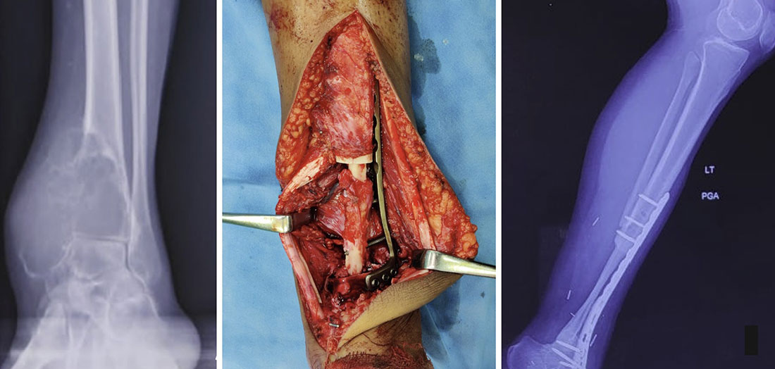
Figure 13. Distal Tibia Resection, Fibular Reconstruction & Ankle Fusion.
When removing the tumour is only the first step: Following wide resection of a distal tibia tumour, the limb was reconstructed using a fibular graft and ankle fusion. The goal was not simply to remove the tumour, but to create a stable, functional limb for the long term.
Distal Fibula Tumor Resection
The distal fibula forms the outer part of the ankle and acts as an important stabilizer of the ankle joint. Although it bears less body weight than the tibia, preserving or reconstructing the distal fibula is often important for maintaining long-term ankle stability.
Depending on the extent of the tumor, treatment may involve partial fibular resection alone or reconstruction using ligament repair, tendon transfers, or biological grafts to restore ankle stability.
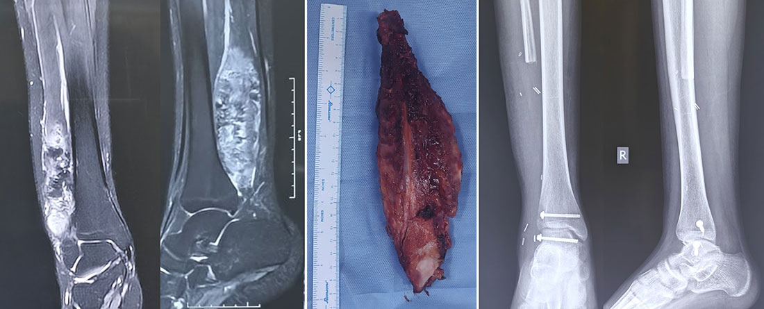
Figure 14. Distal Fibula Osteosarcoma.
Limb-salvage surgery for osteosarcoma of the distal fibula: The tumour was removed with a wide surgical margin while carefully planning reconstruction around the ankle, allowing the limb to be preserved.
Calcaneal (Heel Bone) Tumor Resection
The calcaneus (heel bone) is the largest bone of the foot and bears much of the body’s weight during standing and walking. Because of its critical role in weight-bearing, tumors involving the calcaneus require careful planning to preserve heel function whenever possible.
Depending on the diagnosis, surgery may range from curettage for selected benign lesions to partial or total calcaneal resection for more aggressive tumors. Reconstruction may involve bone grafts, custom implants, or specialized biological techniques.
Talus Tumor Resection
The talus forms both the ankle and subtalar joints, making it essential for smooth ankle movement. Tumors in this bone are rare but technically demanding because removal can significantly affect joint function.
Treatment options include curettage, partial talar resection, or complete talectomy in selected cases, followed by reconstruction or fusion depending on the remaining bone and joint involvement.
Metatarsal Bone Resection
The metatarsal bones form the framework of the forefoot and play an important role in weight-bearing and balance during walking. Tumors involving the metatarsals are uncommon and may be benign or malignant. Surgical treatment aims to remove the tumor completely while preserving as much of the foot’s stability and function as possible.
Depending on the extent of disease, surgery may involve partial metatarsal resection or ray resection, in which the affected metatarsal and its corresponding toe are removed. Reconstruction is not always necessary, particularly when a single ray is excised, as the remaining metatarsals often provide adequate support. However, larger defects or resections involving the first metatarsal may require bone grafting or other reconstructive procedures to maintain foot alignment and weight-bearing.
The functional outcome after metatarsal resection depends not only on the amount of bone removed but also on which metatarsal is involved. Resections of the first and fifth metatarsals have a greater impact on foot biomechanics and may require more careful reconstructive planning.
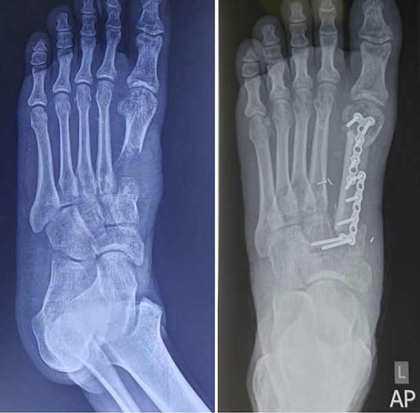
Figure 15. Giant Cell Tumour of the First Metatarsal.
Preserving the foot after tumour removal: A giant cell tumour involving the first metatarsal required resection of the affected bone. Reconstruction with a fibular graft restored the structural framework of the foot while aiming to preserve useful function.
Clinical Pearl
Tumors around the ankle joint require careful reconstruction because even small changes in bone alignment can significantly affect long-term walking, balance, and joint function.
Reconstruction After Foot & Ankle Tumor Surgery
Removing a bone tumor is only one part of successful treatment. The second challenge is reconstruction—restoring the stability, alignment, and function of the foot and ankle after the tumor has been removed.
Not every patient requires reconstruction. The need depends on which bone is removed, how much bone is removed, whether the ankle joint is involved, and how much surrounding soft tissue can be preserved. The goal is to create a foot that is not only free of disease but also as comfortable and functional as possible.
The foot and ankle present unique reconstructive challenges because they must support the entire body weight while allowing balance, flexibility, and efficient walking. Unlike larger bones such as the femur, even relatively small defects around the ankle can significantly affect long-term function.
Doctor Insight
The best reconstruction is not always the most complex one. Surgeons choose the option that offers the safest cancer treatment while providing the most durable and functional result for the individual patient.
When Is Reconstruction Required?
Reconstruction is usually considered when tumor removal results in:
- Loss of an important weight-bearing bone
- Instability of the ankle joint
- Large defects in the distal tibia or calcaneus
- Loss of articular (joint) surfaces
- Significant soft tissue defects requiring coverage
The reconstructive plan is finalized before surgery and often involves collaboration between orthopaedic oncologists, plastic surgeons, and rehabilitation specialists.
When Reconstruction May Not Be Necessary
Some tumors can be removed without rebuilding the bone.
Examples include:
- Small benign tumors treated by curettage
- Limited resections that preserve the structural stability of the foot
- Selected distal fibular resections where adequate ankle stability can be maintained
In these situations, preserving normal anatomy often provides the best functional outcome.
Clinical Pearl
Reconstruction should be performed only when it improves long-term function. Preserving healthy tissues is often more valuable than replacing bone unnecessarily.
Biological Reconstruction
Biological reconstruction uses living bone to restore skeletal continuity after tumor removal. Depending on the clinical situation, this may involve autografts (bone taken from the patient’s own body), allografts (donor bone), or vascularized bone grafts supplied with their own blood vessels.
These techniques are particularly useful in younger patients because living bone has the potential to heal, remodel, and integrate with the surrounding skeleton.
Read more: Biological Reconstruction After Bone Tumor Surgery.
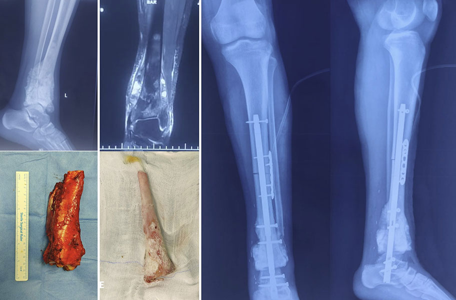
Figure 16. Distal Tibia Osteosarcoma — Extracorporeal Irradiation
Removing the tumour while preserving the patient’s own bone: After wide resection of a distal tibia osteosarcoma, the resected bone was treated with extracorporeal irradiation and reimplanted. This biological reconstruction allowed the patient’s own bone to become part of the limb-salvage reconstruction.
Ankle Arthrodesis (Ankle Fusion)
When tumors involve the distal tibia, talus, or ankle joint, preserving a mobile ankle is not always possible.
Ankle arthrodesis, also called ankle fusion, joins the tibia to the remaining foot using metal implants and, when required, bone grafts. Although ankle movement is lost, fusion often provides a stable, pain-free limb capable of weight-bearing and everyday activities.
For many patients with distal tibial tumors, ankle fusion remains one of the most reliable reconstructive options.
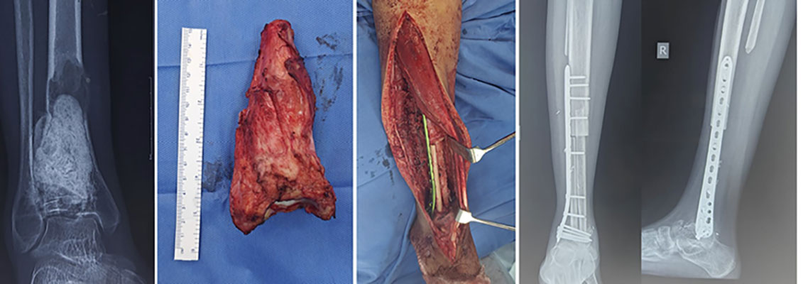
Figure 17. Recurrent Giant Cell Tumour
A challenging recurrence, treated with limb salvage: Recurrent giant cell tumour of the distal tibia required en-bloc resection of the involved bone. Reconstruction using double fibular grafts and ankle fusion provided a stable foundation for the preserved limb.
Endoprosthetic and Custom Implant Reconstruction
Advances in implant technology have expanded the role of custom-made endoprostheses and patient-specific 3D-printed implants for selected foot and ankle tumors.
These implants are designed using CT and MRI data to match the patient’s anatomy and replace the portion of bone removed during surgery.
Although not suitable for every patient, they may help preserve limb length and restore joint alignment in carefully selected cases.
Read more: 3D-Printing & Navigation in Bone Tumors.
Soft Tissue Reconstruction
Successful reconstruction is not limited to replacing bone. Adequate soft tissue coverage is equally important for wound healing, infection prevention, and long-term function.
Depending on the location of the tumor, reconstruction may require:
- Local muscle flaps
- Rotational flaps
- Microsurgical free tissue transfer
- Skin grafts
Plastic surgeons often work alongside orthopaedic oncologists during these complex procedures.
Expected Function After Reconstruction
Recovery varies according to the type of surgery performed, but the primary aim is to achieve a stable, painless, and functional limb.
Some patients regain near-normal walking, whereas others may require:
- Custom footwear
- Orthotic support
- Walking aids during rehabilitation
- Long-term physiotherapy
Most patients can return to everyday activities, although high-impact sports may not always be advisable after major reconstruction.
Doctor Explains
There is no universally “best” reconstruction. The ideal solution depends on the diagnosis, tumor location, expected lifespan of the reconstruction, patient age, activity level, and personal goals.
Patient Tip
Before surgery, ask your surgeon:
- Why is this reconstruction recommended for me?
- Will I be able to bear weight normally?
- Will I need special footwear or braces?
- What activities should I avoid in the long term?
Reassurance
Reconstruction after foot and ankle tumor surgery has advanced significantly over the past two decades. Many patients regain independent walking and return to work and daily activities with appropriate rehabilitation, although recovery is gradual and individualized.
Recovery, Rehabilitation and Long-Term Follow-up
Successful treatment does not end with surgery. Recovery is a gradual process that focuses on restoring mobility, strength, balance, and confidence while monitoring for tumor recurrence and ensuring that any reconstruction continues to function well.
Most patients begin rehabilitation within the first few days after surgery under the guidance of a multidisciplinary rehabilitation team.
Immediately After Surgery
The early recovery period focuses on:
- Pain control
- Wound healing
- Preventing blood clots
- Protecting the reconstruction
- Gentle movement exercises
Depending on the type of reconstruction, patients may need to avoid weight-bearing for several weeks while the bone or soft tissues heal.
Walking Again
Walking usually progresses in stages:
- Non-weight-bearing with crutches or a walker
- Partial weight-bearing as healing permits
- Full weight-bearing after confirmation of adequate healing
- Progressive strengthening and balance training
The exact timeline depends on the reconstruction performed and should never be rushed.
Physiotherapy
Physiotherapy plays a vital role in recovery by helping patients:
- Restore muscle strength
- Improve ankle and foot function
- Retrain balance and coordination
- Adapt to changes in walking mechanics
- Return safely to work and recreational activities
Some patients may also benefit from custom orthotics or specialized footwear.
Returning to Daily Life
Many patients eventually return to independent living, office-based work, driving, and low-impact recreational activities. Recovery after major limb salvage surgery, however, continues for many months, and improvements often occur well beyond the first postoperative year.
Table. Typical Recovery Timeline
*Recovery varies depending on the diagnosis, surgical procedure, and reconstruction.
Long-Term Follow-up
Regular follow-up is essential to:
- Detect local recurrence early
- Monitor for spread in malignant tumors
- Assess bone healing
- Evaluate implant function
- Identify complications before they become serious
The frequency of follow-up depends on the tumor type, but patients with malignant tumors generally require closer surveillance during the first few years after treatment.
Clinical Pearl
Rehabilitation is an integral part of treatment—not an optional extra. Long-term function depends as much on structured rehabilitation as it does on the surgery itself.
Did You Know?
Functional recovery after limb salvage surgery often continues for 12 to 18 months, even after the surgical wounds have completely healed.
Reassurance
Every patient’s recovery is different. It is normal for progress to occur gradually, and temporary setbacks do not necessarily indicate a problem. Maintaining regular follow-up and actively participating in rehabilitation are key to achieving the best possible outcome.
When Should You Seek a Specialist Opinion?
You should consider consulting an orthopaedic oncologist if you have:
- Persistent foot or ankle pain without a clear cause
- An unexplained bone lesion on an X-ray or MRI
- A confirmed diagnosis of a bone tumor
- A recommendation for biopsy or surgery and would like a second opinion
- A recurrent bone tumor or complications after previous treatment
Early specialist evaluation can help establish the correct diagnosis and ensure that treatment is planned appropriately from the outset.
Frequently Asked Questions (FAQs)
1. What are the symptoms of a bone tumor in the foot or ankle?
The most common symptoms include persistent pain, swelling, a gradually enlarging lump, difficulty walking, or pain that does not improve with rest or routine treatment. Some tumors may weaken the bone and cause a pathological fracture. While most foot pain is not caused by a bone tumor, persistent or unexplained symptoms should be evaluated by a specialist.
2. Are all foot and ankle bone tumors cancerous?
No. Most bone tumors of the foot and ankle are benign (non-cancerous). However, some benign tumors can behave aggressively, while malignant tumors require prompt treatment. Imaging and, when necessary, a biopsy are needed to establish the correct diagnosis.
3. How are foot and ankle bone tumors diagnosed?
Diagnosis usually begins with X-rays, followed by MRI to define the extent of the tumor. CT scans, PET-CT, or bone scans may be required in selected cases. A biopsy is the only way to confirm the exact diagnosis and should ideally be performed by the specialist team treating the tumor.
4. Can foot and ankle bone tumors be treated without amputation?
Yes. Limb salvage surgery is possible for many patients and aims to remove the tumor while preserving a functional foot or ankle. Amputation is reserved for selected cases where safe tumor removal or satisfactory limb function cannot otherwise be achieved.
5. What is limb salvage surgery for foot and ankle bone tumors?
Limb salvage surgery involves removing the tumor with an adequate margin of healthy tissue while preserving as much normal bone, joints, muscles, nerves, and blood vessels as possible. Reconstruction may be required to restore stability and walking function.
6. What reconstruction options are available after foot and ankle tumor surgery?
Reconstruction depends on the location and size of the defect. Options include bone grafts, ankle fusion (arthrodesis), biological reconstruction, custom-made implants, 3D-printed prostheses, and soft tissue flap reconstruction. The most appropriate technique is individualized for each patient.
7. How long does it take to recover after foot and ankle bone tumor surgery?
Recovery varies depending on the procedure performed. Most patients begin rehabilitation within a few days of surgery, while walking progresses gradually over several weeks. Functional recovery often continues for 6–12 months, especially after major reconstructive surgery.
8. Will I be able to walk normally after surgery?
Many patients regain independent walking after treatment. The final outcome depends on the tumor location, the amount of bone removed, the type of reconstruction performed, and adherence to physiotherapy. Some patients may require custom footwear or orthotic support.
9. Can a foot or ankle bone tumor come back after surgery?
Yes. Some benign and malignant tumors have a risk of local recurrence even after successful treatment. Regular follow-up with clinical examination and imaging helps detect recurrence early and improves the chances of successful management.
10. Which bone tumors commonly occur in the foot and ankle?
Common benign tumors include osteochondroma, enchondroma, chondroblastoma, aneurysmal bone cyst, and giant cell tumor. Malignant tumors include osteosarcoma, Ewing sarcoma, and chondrosarcoma. Rarely, cancers from other organs can spread to the bones of the foot and ankle.
11. When should I see an orthopaedic oncologist for foot or ankle pain?
You should seek specialist evaluation if you have persistent foot pain lasting more than six weeks, an unexplained bone lesion on X-ray or MRI, recurrent swelling, a painful lump, or a suspected bone tumor. Early diagnosis improves treatment options and may increase the likelihood of successful limb salvage.
12. Why should foot and ankle bone tumors be treated by an orthopaedic oncology specialist?
These tumors are rare and often require complex surgical planning. An orthopaedic oncologist works with a multidisciplinary team to ensure accurate diagnosis, carefully planned biopsy, complete tumor removal, appropriate reconstruction, and long-term follow-up. Treatment at a specialist center is associated with better oncological and functional outcomes.
Medical Disclaimer
The information on this page is provided for educational purposes only and is not a substitute for professional medical advice, diagnosis, or treatment. Every bone tumor is unique, and treatment should be individualized based on clinical evaluation, imaging, and biopsy findings. If you have persistent foot or ankle pain, swelling, or have been diagnosed with a bone tumor, consult an orthopaedic oncologist or a specialist musculoskeletal oncology team for appropriate evaluation and management. Never disregard professional medical advice or delay seeking treatment based on information provided on this website.
References
- Zhao Z, Yan T, Guo W, Yang R, Tang X, Wang W. Surgical options and reconstruction strategies for primary bone tumors of distal tibia: A systematic review of complications and functional outcome. J Bone Oncol. 2019;14:100209.
- Mavrogenis AF, Abati CN, Romagnoli C, Ruggieri P. Similar survival but better function for patients after limb salvage versus amputation for distal tibia osteosarcoma. Clin Orthop Relat Res. 2012;470(6):1735-1748.
- Abudu A, Grimer RJ, Tillman RM, Carter SR. Endoprosthetic replacement of the distal tibia and ankle joint for aggressive bone tumours. Int Orthop. 1999;23(5):291-294.
- Ebeid W, Amin S, Abdelmegid A, Refaat Y, Ghoneimy A. Reconstruction of distal tibial defects following resection of malignant tumours by pedicled vascularised fibular grafts. Acta Orthop Belg. 2007;73(3):354-359.
- Mascard E, Gaspar N, Brugières L, Glorion C, Pannier S, Gomez-Brouchet A. Malignant tumours of the foot and ankle. EFORT Open Rev. 2017;2(5):261-271. Published 2017 May 11.
- Biz C, Angelini A, Fantoni I, Cerchiaro M, Longhi V, Ruggieri P. Benign bone and soft tissue tumors of the foot. EFORT Open Rev. 2024;9(11):1060-1076. Published 2024 Nov 8.
 +91 93558 33005