Quick Answer
Some bone tumors are uncommon growths that develop within or on bones. They include a wide range of conditions—from benign (non-cancerous) tumors such as osteochondroma and osteoid osteoma, to locally aggressive tumors such as aneurysmal bone cysts, and malignant (cancerous) tumors such as chordoma and adamantinoma. Multiple myeloma, although a blood cancer, frequently affects bones and is therefore included in this guide.
Not every bone tumor requires surgery. Some can be safely monitored, while others require procedures such as curettage (surgical removal of the tumor from within the bone), minimally invasive treatments, radiotherapy, or systemic medications. The best treatment depends on the exact diagnosis, symptoms, tumor location, and overall health of the patient.
Quick Summary
Key Points
- Rare bone tumors include benign, locally aggressive, and malignant conditions.
- Many bone tumors are not cancer and have an excellent prognosis.
- Persistent bone pain, swelling, or an unexplained fracture should be evaluated promptly.
- Imaging studies such as X-rays, MRI (Magnetic Resonance Imaging), and CT (Computed Tomography) often provide important diagnostic clues.
- A biopsy (removal of a small tissue sample for laboratory examination) is performed only when necessary.
- Treatment ranges from observation to surgery, minimally invasive procedures, radiotherapy, or systemic therapy.
- Whenever possible, management should involve an orthopaedic oncologist, a surgeon who specializes in tumors of bone and soft tissues.
Reassurance Box
Receiving a diagnosis of a rare bone tumor can be frightening. However, “rare” does not mean untreatable. Many rare bone tumors are benign, and even those requiring treatment can often be managed successfully with modern multidisciplinary care.
What Are Bone Tumors?
A bone tumor is an abnormal growth of cells within or on a bone. While many people associate the word tumor with cancer, a tumor simply means an abnormal mass of tissue. Bone tumors can be benign (non-cancerous), locally aggressive (unlikely to spread but capable of damaging nearby bone and joints), or malignant (cancerous and capable of invading surrounding tissues or spreading to other parts of the body).
Bone tumors account for only a small proportion of musculoskeletal diseases. Because they are uncommon, many patients—and even some healthcare professionals—may encounter them only rarely. Accurate diagnosis is therefore essential before any treatment is planned.
Some rare bone tumors cause significant pain, while others are discovered incidentally, meaning they are found unexpectedly during an X-ray or MRI performed for another reason. A small, painless lesion may require only observation, whereas a painful or aggressive tumor may need surgery or additional treatment.
The tumors discussed in this guide include:
- Cartilage tumors, which arise from cartilage (the smooth, flexible tissue that covers the ends of bones and forms part of the growing skeleton).
- Bone-forming tumors, which produce abnormal bone tissue.
- Bone cysts, which are fluid- or blood-filled cavities within bone.
- Fibrous dysplasia, a developmental disorder in which normal bone is replaced by fibrous tissue.
- Adamantinoma, a rare low-grade malignant bone tumor.
- Chordoma, a malignant tumor arising from remnants of the embryonic notochord (the structure that helps form the spine during early development).
- Multiple myeloma, a cancer of plasma cells that commonly weakens bones and causes skeletal complications.
Although these conditions differ greatly, they share one important principle: the correct diagnosis must always come before treatment.
Doctor Explains
A bone tumor is named according to the type of tissue from which it arises. For example:
- Osteo- refers to bone.
- Chondro- refers to cartilage.
- Fibro- refers to fibrous tissue.
- -oma usually indicates a benign tumor, although there are important exceptions such as myeloma and lymphoma, which are cancers.
Understanding the name often provides clues about the tumor’s origin and behavior.
Table 1. Categories of Rare Bone Tumors
Clinical Pearl
The appearance of a bone lesion on an X-ray alone is rarely enough to determine treatment. The patient’s age, symptoms, imaging findings, and, when required, biopsy results must all be considered together.
How Are Bone Tumors Classified?
Bone tumors are classified according to the type of cells from which they arise and how they behave biologically. This classification helps doctors predict how a tumor is likely to grow, whether it may recur after treatment, and which treatment is most appropriate.
From a patient’s perspective, the most useful way to understand bone tumors is to divide them into four broad groups.
1. Cartilage Tumors
These tumors develop from cartilage, the smooth connective tissue found at the ends of bones and within growth plates. Most cartilage tumors discussed in this article are benign, although some can resemble low-grade cartilage cancers on imaging and require specialist evaluation.
Examples include:
- Osteochondroma
- Enchondroma
- Chondroblastoma
- Chondromyxoid Fibroma
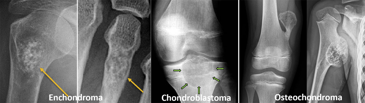
Figure 1. Common cartilage-forming bone tumors, including enchondroma, chondroblastoma and osteochondroma, seen on X-rays.
2. Bone-forming Tumors
These tumors arise from cells that normally produce bone.
Most are benign, but they often cause pain because they stimulate nearby bone and nerves.
Examples include:
- Osteoid Osteoma
- Osteoblastoma
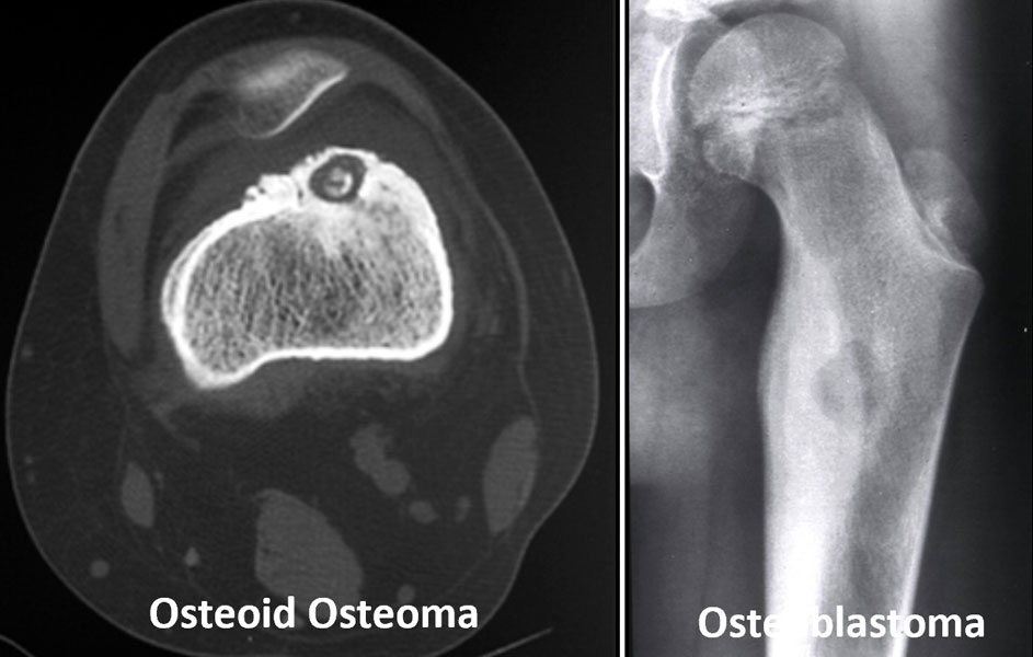
Figure 2. Benign bone-forming tumors, including osteoid osteoma and osteoblastoma, shown on X-ray and CT imaging.
3. Bone Cysts and Developmental Bone Disorders
Not every bone lesion is a true tumor.
Some conditions represent abnormal bone development or fluid-filled cavities rather than uncontrolled tumor growth.
Examples include:
- Unicameral Bone Cyst
- Aneurysmal Bone Cyst
- Fibrous Dysplasia
Although benign, these conditions may weaken bone and increase the risk of fracture.
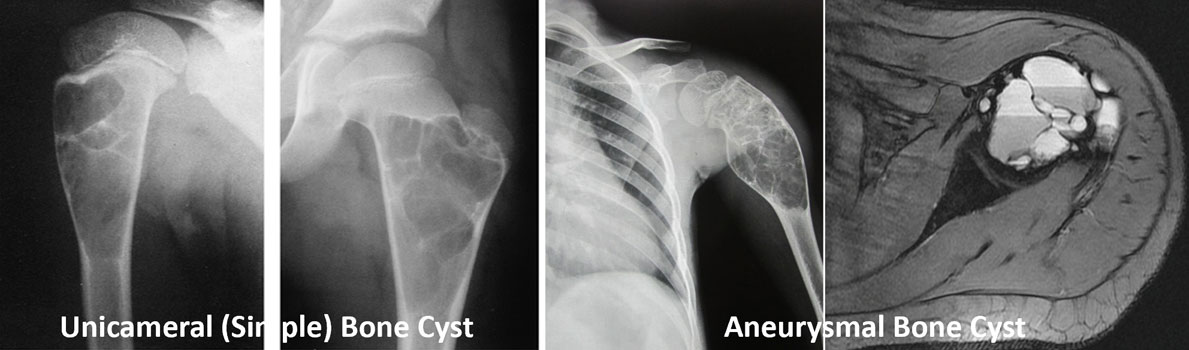
Figure 3. Common types of bone cysts, including simple (unicameral) bone cyst and aneurysmal bone cyst, seen on X-ray and MRI.
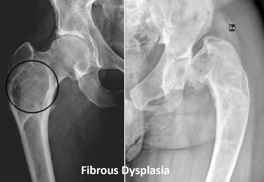
Figure 4. Fibrous dysplasia of the femur, a benign bone disorder that can weaken and deform the affected bone.
4. Malignant Bone Tumors and Bone Diseases
This group includes tumors that behave as cancers.
Some arise directly from bone, while others originate elsewhere but significantly affect the skeleton.
Examples include:
- Adamantinoma
- Chordoma
- Multiple Myeloma
Unlike benign tumors, these conditions often require multidisciplinary treatment involving orthopaedic oncologists, medical oncologists, radiation oncologists, radiologists, and pathologists.
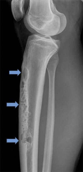
Figure 5. Adamantinoma of the tibia, a rare bone tumor affecting the shin bone, seen on X-ray imaging.
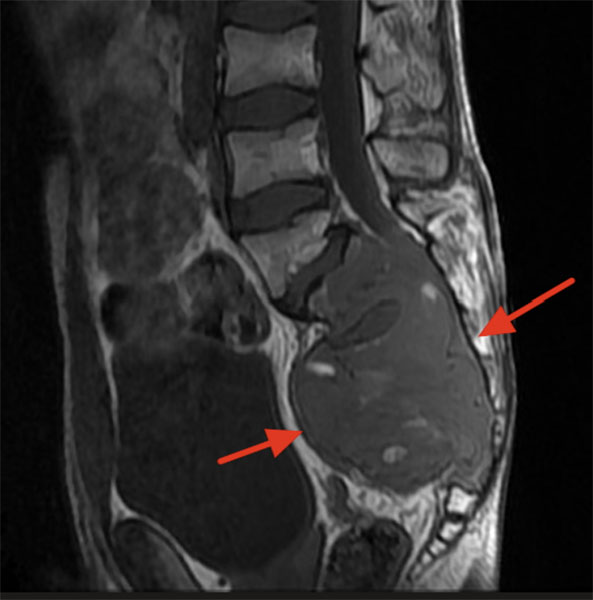
Figure 6. MRI showing a chordoma of the sacrum, a rare malignant bone tumor arising in the lower spine.
Doctor Insight
Tumor names can sound intimidating, but they do not indicate severity on their own. Two tumors with similar names may behave very differently. Treatment decisions are based on the tumor’s biological behaviour—not simply its name.
Patient Tip
Ask your doctor for the exact diagnosis rather than simply being told you have a “bone tumor.” Knowing the specific tumor type will help you understand your treatment options and expected outcome.
Did You Know?
Many rare bone tumors are discovered incidentally during imaging performed after a sports injury or minor accident. These incidental findings are often benign and may never require surgery.
Cartilage Tumors
Cartilage tumors arise from cartilage, the smooth, flexible connective tissue that covers the ends of bones, forms joints, and contributes to bone growth during childhood. They are among the most common benign bone tumors encountered in orthopaedic practice.
The biological behavior of cartilage tumors varies considerably. Some remain stable throughout life and never require treatment, while others may grow, weaken the bone, or occasionally resemble malignant cartilage tumors on imaging. For this reason, accurate diagnosis by an experienced orthopaedic oncologist is essential before deciding on treatment.
Most cartilage tumors have an excellent prognosis, and many patients can be managed safely with observation alone.
Doctor Explains
Cartilage tumors are named according to the type of cartilage-producing cells from which they arise. Although several tumors contain cartilage, they behave very differently. An osteochondroma, for example, is usually harmless, whereas a chondroblastoma is locally aggressive and generally requires surgery.
Osteochondroma
What is an Osteochondroma?
An osteochondroma is the most common benign bone tumor. It is a bony projection covered by a cap of cartilage that grows from the surface of a bone, usually near a growth plate (the area of growing bone in children and adolescents).
Rather than being a true tumor in the traditional sense, many experts consider osteochondroma to be a developmental growth abnormality. Most stop growing once skeletal maturity is reached.
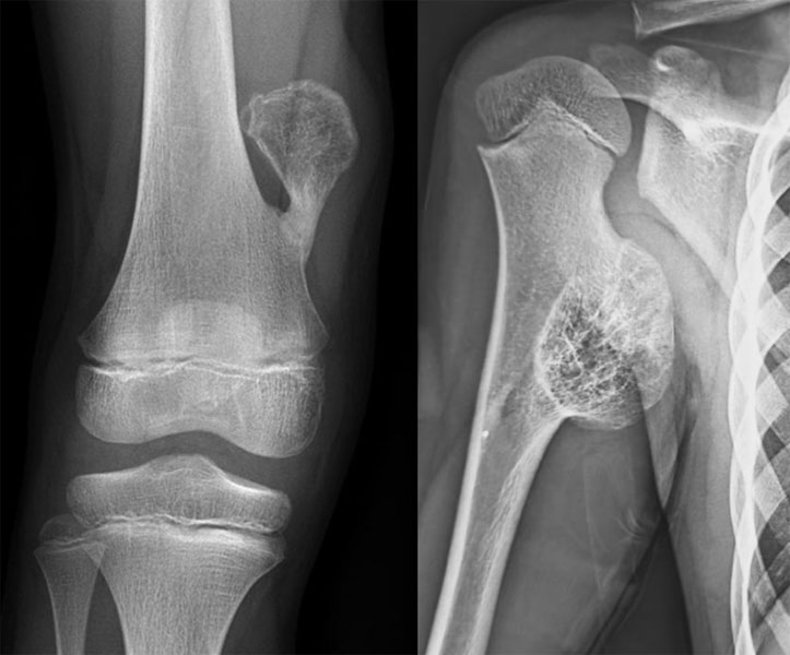
Figure 7. Osteochondroma, a common benign bone growth arising near the growth plate of a growing bone.
Who is affected?
Osteochondromas usually develop during childhood and adolescence and are often diagnosed before the age of 20 years. They commonly occur around the:
- Knee (distal femur and proximal tibia)
- Shoulder (proximal humerus)
- Pelvis
Some individuals have a hereditary condition called Multiple Hereditary Exostoses (MHE), in which multiple osteochondromas develop throughout the skeleton.
Symptoms
Many osteochondromas cause no symptoms and are discovered incidentally.
When symptoms occur, they may include:
- A painless hard lump
- Cosmetic prominence
- Pain due to irritation of nearby muscles or tendons
- Limited joint movement
- Pressure on nearby nerves or blood vessels (rare)
Diagnosis
Diagnosis is usually straightforward with X-rays, which show a bony outgrowth that is continuous with the underlying bone. MRI may be recommended if the cartilage cap appears unusually thick or if malignant transformation is suspected.
Treatment
Most osteochondromas do not require treatment.
Surgery is considered when:
- The tumor causes persistent pain.
- It interferes with joint movement.
- It compresses nerves or blood vessels.
- There is concern for malignant transformation.
- Cosmetic appearance significantly affects the patient.
Complete surgical removal is usually curative.
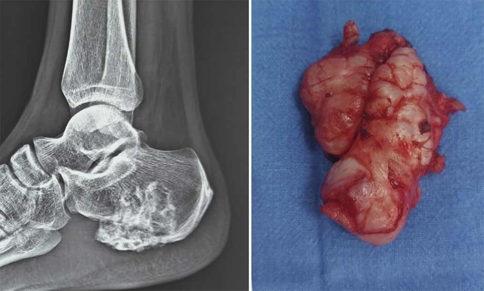
Figure 8. Surgical removal of an osteochondroma when symptoms or concern about malignant transformation warrant excision.
Prognosis
The outlook is excellent. Recurrence after complete removal is uncommon, and the risk of malignant transformation in a solitary osteochondroma is very low, although it is slightly higher in patients with Multiple Hereditary Exostoses.
Enchondroma
What is an Enchondroma?
An enchondroma is a benign cartilage tumor that develops inside the bone marrow cavity. Unlike osteochondromas, which grow outward from the bone surface, enchondromas remain within the bone.
They are commonly found in the small bones of the hand, but may also occur in the humerus, femur, or tibia.
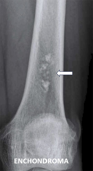
Figure 9. Enchondroma of the femur, a benign cartilage tumor that can often be safely monitored with regular imaging.
Who is affected?
Enchondromas are most frequently diagnosed in young and middle-aged adults. Many are discovered accidentally during imaging for an unrelated injury.
Symptoms
Most enchondromas are painless.
Symptoms may occur if the lesion:
- Weakens the bone
- Causes a pathological fracture (a fracture through weakened bone)
- Becomes unusually large
Diagnosis
Typical imaging findings on X-rays often establish the diagnosis. MRI may be used when the diagnosis is uncertain or when distinguishing an enchondroma from a low-grade cartilage cancer.
Treatment
Observation with periodic imaging is appropriate for most asymptomatic enchondromas.
Surgery may be recommended when:
- The lesion causes pain attributable to the tumor.
- There is a pathological fracture.
- Imaging raises concern for malignancy.
- The diagnosis remains uncertain.
The usual operation involves curettage (scraping the tumor from inside the bone) followed by filling the cavity with bone graft or bone substitute when required.
Prognosis
Most enchondromas remain stable throughout life and have an excellent prognosis. Regular follow-up may be advised for selected lesions, particularly those in long bones.
Chondroblastoma
What is a Chondroblastoma?
A chondroblastoma is a rare benign but locally aggressive cartilage tumor that usually develops near the end of a growing bone, close to a joint. “Locally aggressive” means that although the tumor does not spread to distant organs, it can damage nearby bone and cartilage if left untreated.
Who is affected?
Chondroblastomas occur predominantly in children, adolescents, and young adults, often before the growth plates have completely closed.
Common locations include:
- Upper end of the humerus (shoulder)
- Distal femur (knee)
- Proximal tibia
- Proximal femur (hip)
Symptoms
Patients typically present with:
- Persistent joint pain
- Swelling
- Reduced range of motion
- Occasional joint stiffness
Because the tumor lies close to a joint, symptoms are often mistaken for a sports injury.
Diagnosis
X-rays usually show a well-defined lesion near the end of the bone. MRI helps determine the extent of the tumor and its relationship to the surrounding cartilage and soft tissues. A biopsy is often performed before treatment to confirm the diagnosis.
Treatment
Surgery is recommended for most chondroblastomas.
The standard treatment is:
- Curettage
- High-speed burr to remove microscopic tumor cells
- Bone graft or bone substitute to fill the cavity
Prognosis
Most patients recover well after surgery. However, chondroblastomas have a higher recurrence rate than many other benign tumors, making regular follow-up important during the first few years after treatment.
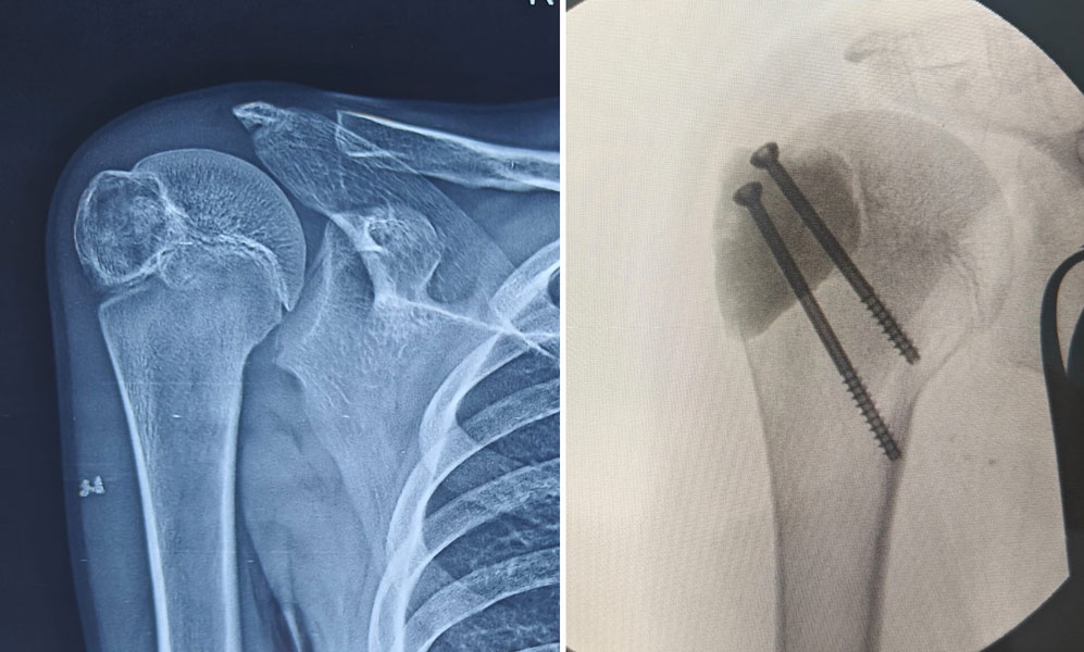
Figure 10. Chondroblastoma of the shoulder region treated with curettage and filling of the bone cavity with bone cement.
Chondromyxoid Fibroma
What is Chondromyxoid Fibroma?
Chondromyxoid fibroma is an extremely rare benign cartilage tumor composed of cartilage, fibrous tissue, and myxoid (gel-like) tissue. Although benign, it can slowly enlarge and weaken the surrounding bone.
Who is affected?
It usually occurs in adolescents and young adults.
The most common sites include:
- Upper tibia
- Distal femur
- Small bones of the foot
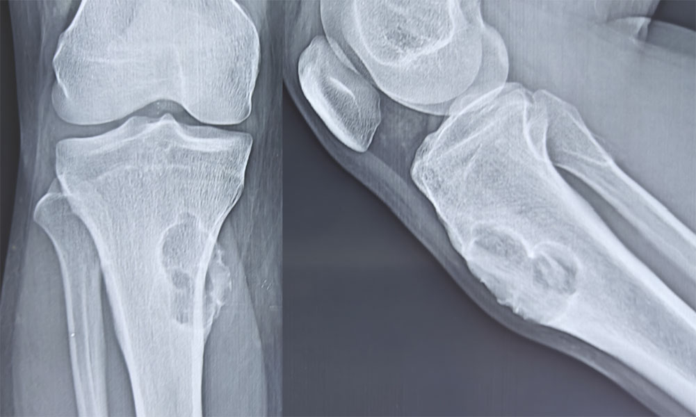
Figure 11. Chondromyxoid fibroma , a rare benign cartilage-forming bone tumor seen on X-ray imaging.
Symptoms
Symptoms develop gradually and may include:
- Mild to moderate pain
- Localized swelling
- Tenderness
- Occasionally a pathological fracture
Diagnosis
Imaging often suggests the diagnosis, but biopsy is usually required because the tumor can resemble other bone lesions.
Treatment
The preferred treatment is surgical curettage combined with local adjuvants and bone grafting or bone substitute reconstruction. In selected cases, complete excision may be recommended.
Prognosis
The long-term outlook is excellent. Although recurrence is possible, especially after incomplete removal, malignant transformation is exceptionally rare.
Table 2. Comparison of Common Cartilage Tumors
Clinical Pearl
Not every cartilage tumor requires surgery. Many osteochondromas and enchondromas can be managed safely with observation, whereas chondroblastoma and chondromyxoid fibroma usually require surgical treatment because of their locally aggressive behavior.
Doctor Insight
Distinguishing a benign cartilage tumor from a low-grade cartilage cancer can sometimes be challenging. The diagnosis is based on a combination of the patient’s symptoms, imaging findings, and, when necessary, biopsy results rather than any single test.
Patient Tip
If your doctor recommends observation rather than surgery, it does not mean your condition is being ignored. Many cartilage tumors remain unchanged for years, and regular follow-up imaging is often the safest and most appropriate approach.
Did You Know?
Osteochondroma accounts for approximately one-third of all benign bone tumors, making it the most common benign bone tumor encountered in clinical practice.
Reassurance Box
Most cartilage tumors discussed in this section are benign and have an excellent long-term prognosis. With appropriate diagnosis and follow-up, the majority of patients maintain normal daily activities, and many never require surgery.
Bone-forming Tumors
Bone-forming tumors arise from cells that normally produce bone tissue. Most are benign, but they often cause pain because they stimulate surrounding bone and nearby nerves. Unlike many cartilage tumors, which are frequently discovered incidentally, bone-forming tumors usually come to medical attention because of persistent symptoms.
The two most important benign bone-forming tumors are osteoid osteoma and osteoblastoma. Although their names are similar, they differ in size, behaviour, and treatment.
Osteoid Osteoma
What is an Osteoid Osteoma?
An osteoid osteoma is a small benign bone-forming tumor, usually measuring less than 2 cm in diameter. Despite its small size, it can cause severe pain because it produces high levels of prostaglandins—naturally occurring chemicals that increase pain and inflammation.
Who is affected?
Osteoid osteoma most commonly affects:
- Children
- Adolescents
- Young adults
It usually develops in the:
- Femur (thigh bone)
- Tibia (shin bone)
- Spine (less commonly)
Symptoms
The classic symptom is:
- Persistent pain that is worse at night
A distinctive feature is that the pain is often dramatically relieved by non-steroidal anti-inflammatory drugs (NSAIDs) such as ibuprofen or naproxen.
Some patients may also develop:
- Limping
- Muscle wasting from disuse
- Painful scoliosis (when the tumor is located in the spine)
Diagnosis
X-rays may show a small central lesion called a nidus (the core of the tumor), surrounded by dense reactive bone.
A CT scan is usually the best imaging test because it clearly identifies the nidus. MRI may be helpful in selected cases but can sometimes make the lesion appear larger than it actually is because of surrounding inflammation.
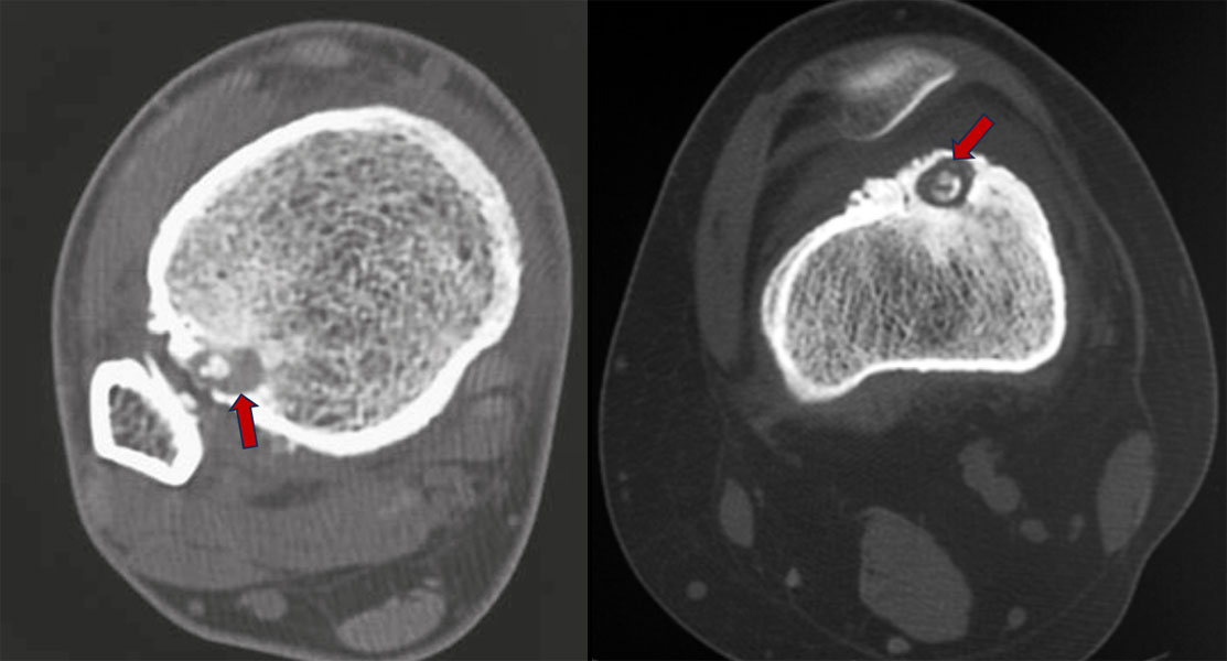
Figure 12. CT scan showing the nidus of an osteoid osteoma, the small focus responsible for the characteristic pain of this benign bone tumor.
Treatment
Small, mildly symptomatic lesions may occasionally be managed with pain medication while waiting for spontaneous resolution, but many patients prefer definitive treatment.
The current standard treatment is CT-guided radiofrequency ablation (RFA). During this minimally invasive procedure, a needle is placed into the tumor under CT guidance, and heat is used to destroy the nidus without the need for open surgery.
Open surgical removal is now reserved for selected situations where ablation is not feasible.
-for-osteoid-osteoma.jpg "Radiofrequency ablation (RFA) for osteoid osteoma")
Figure 13. Radiofrequency ablation (RFA) for osteoid osteoma, a minimally invasive treatment that destroys the tumor nidus using heat.
Prognosis
The prognosis is excellent. Most patients experience rapid pain relief after treatment and return to normal activities within a few weeks.
Osteoblastoma
What is an Osteoblastoma?
An osteoblastoma is a rare benign bone-forming tumor that resembles osteoid osteoma but is generally larger than 2 cm and may behave more aggressively. Although benign, it can progressively destroy surrounding bone if left untreated.
Who is affected?
Osteoblastomas usually occur in:
Common locations include:
Symptoms
Symptoms typically include:
- Persistent localized pain
- Swelling
- Reduced movement
- Neurological symptoms if the spine is involved due to pressure on nearby nerves
Unlike osteoid osteoma, pain is less predictably relieved by NSAIDs.
Diagnosis
X-rays, CT, and MRI help determine the size and extent of the tumor. Because imaging findings may overlap with other bone tumors, a biopsy is often performed before treatment.
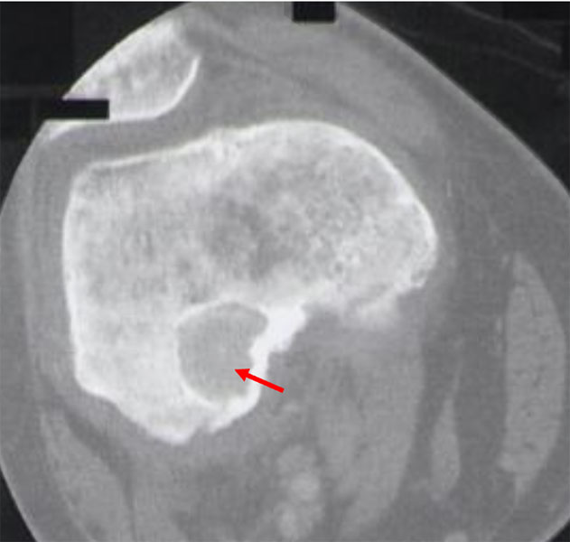
Figure 14. Osteoblastoma of the distal femur, a rare benign but locally aggressive bone tumor seen on X-ray.
Treatment
Most osteoblastomas require surgery.
Treatment usually consists of:
- Curettage with local adjuvants, or
- Complete surgical excision for larger or recurrent tumors
Complex tumors involving the spine or pelvis should be managed in specialized musculoskeletal oncology centers.
Prognosis
Most patients recover well after surgery. Local recurrence is uncommon but occurs more frequently than with osteoid osteoma, emphasizing the importance of follow-up.
Table 3. Osteoid Osteoma vs Osteoblastoma
Did You Know?
CT-guided radiofrequency ablation has replaced open surgery for most osteoid osteomas because it provides excellent pain relief with smaller incisions, faster recovery, and high success rates.
Bone Cysts
Bone cysts are fluid- or blood-filled cavities within bone. Although they are often grouped with bone tumors, they are not true tumors because they do not arise from uncontrolled cell growth. Instead, they represent abnormal bone cavities that may weaken the surrounding bone.
Many bone cysts occur in children and adolescents and are discovered after pain or a fracture.
Unicameral Bone Cyst
What is a Unicameral Bone Cyst?
A unicameral bone cyst (UBC), also called a simple bone cyst, is a fluid-filled cavity that usually develops near the growth plate of a long bone.
Who is affected?
Most patients are:
The cyst most commonly affects:
- Proximal humerus
- Proximal femur
Symptoms
Many cysts produce no symptoms until they weaken the bone enough to cause a pathological fracture.
Symptoms may include:
- Mild pain
- Swelling
- Fracture after minor injury
Diagnosis
X-rays usually provide the diagnosis. MRI is occasionally performed when the diagnosis is uncertain.
-bone-cyst-of-the-proximal-femur.jpg "Simple (unicameral) bone cyst of the proximal femur")
Figure 15. Simple (unicameral) bone cyst of the proximal femur, a benign fluid-filled bone lesion that may require treatment if it weakens the bone.
Treatment
Treatment depends on:
- Patient age
- Size of the cyst
- Fracture risk
- Presence of symptoms
Options include:
- Observation
- Steroid or bone marrow injections
- Curettage with bone grafting
- Internal fixation if a fracture is present
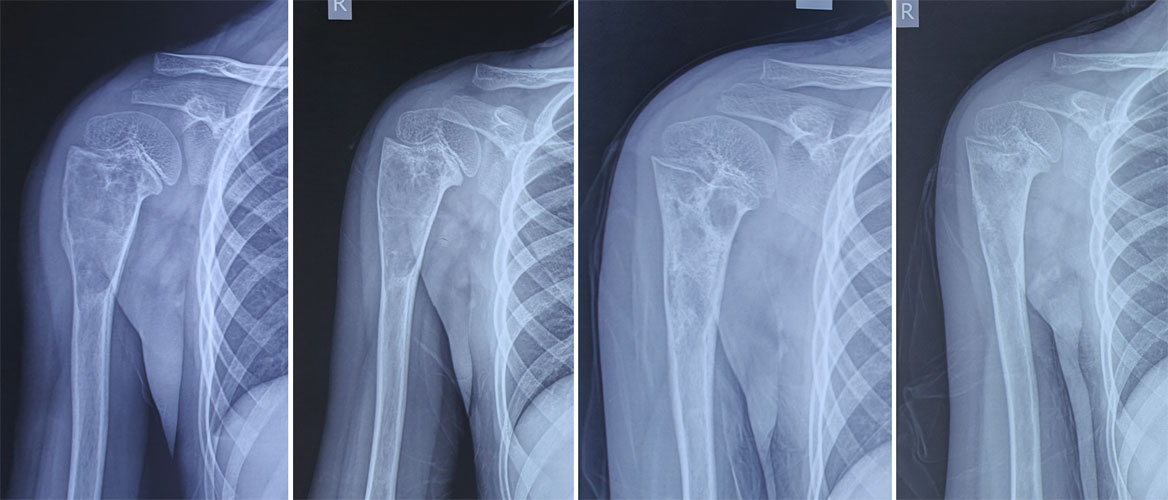
Figure 16. Healing of a simple bone cyst after steroid injection, demonstrating progressive filling and strengthening of the affected bone over time.
Prognosis
Many cysts gradually heal as skeletal maturity is reached, although recurrence may occur during childhood.
Aneurysmal Bone Cyst
What is an Aneurysmal Bone Cyst?
An aneurysmal bone cyst (ABC) is a benign but locally aggressive bone lesion consisting of multiple blood-filled cavities separated by thin fibrous walls.
Despite its name, it is not an aneurysm and not a true cyst.
Who is affected?
ABCs usually occur in:
- Children
- Adolescents
- Young adults
Common sites include:
Symptoms
Patients may develop:
- Progressive pain
- Swelling
- Reduced joint movement
- Pathological fracture
- Neurological symptoms if the spine is involved
Diagnosis
MRI typically shows multiple fluid-fluid levels, a characteristic feature of ABCs. Biopsy is frequently performed to exclude other tumors that may resemble an aneurysmal bone cyst.
.jpg "MRI of an aneurysmal bone cyst (ABC)")
Figure 17. MRI of an aneurysmal bone cyst (ABC) showing multiple fluid-fluid levels, a characteristic imaging feature of this benign bone tumor.
Treatment
Treatment options include:
- Extended curettage
- Bone grafting
- Selective arterial embolization in selected cases
- Image-guided sclerotherapy for carefully chosen lesions
The choice depends on the size, location, and accessibility of the lesion.
MRI typically shows multiple fluid-fluid levels, a characteristic feature of ABCs. Biopsy is frequently performed to exclude other tumors that may resemble an aneurysmal bone cyst.
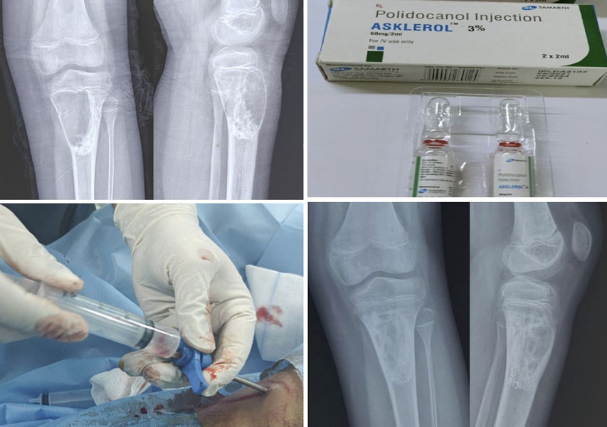
Figure 18. Healing of an aneurysmal bone cyst after sclerotherapy , a minimally invasive treatment that promotes gradual healing of the cyst.
Prognosis
Most patients have excellent long-term outcomes, although recurrence is more common than with simple bone cysts, particularly in younger children.
Table 4. Comparison of Bone Cysts
Reassurance Box
Hearing the word “cyst” can be worrying, but most bone cysts are benign and respond well to treatment. Many children recover completely and return to normal activities after healing.
Fibrous Dysplasia
What is Fibrous Dysplasia?
Fibrous dysplasia is not a true bone tumor, but a developmental bone disorder in which normal bone is replaced by fibrous (scar-like) tissue. This abnormal tissue weakens the bone and may lead to deformity or fractures.
The condition may affect:
- A single bone (monostotic fibrous dysplasia), or
- Multiple bones (polyostotic fibrous dysplasia)
Symptoms
Many patients remain symptom-free.
Others may develop:
- Bone pain
- Recurrent fractures
- Bowing of long bones
- Limb deformity
Diagnosis
X-rays often demonstrate a characteristic ground-glass appearance, which reflects the abnormal mixture of fibrous tissue and immature bone. CT or MRI may be required in selected cases.
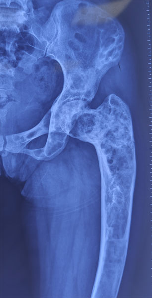
Figure 19. Fibrous dysplasia causing deformity of the femur, illustrating how this benign bone disorder can lead to bone weakness and deformity.
Treatment
Treatment depends on symptoms.
Options include:
- Observation
- Pain management
- Bisphosphonate medications for selected patients
- Surgery for fractures, deformity, or severe pain
Prognosis
Most patients lead active lives with appropriate monitoring. Malignant transformation is extremely rare.
Clinical Pearl
Fibrous dysplasia is a developmental disorder rather than a cancer. Treatment is directed at relieving symptoms, preventing fractures, and maintaining normal function.
Adamantinoma
What is Adamantinoma?
Adamantinoma is a rare, low-grade malignant bone tumor that most commonly develops in the tibia (shin bone). “Low-grade” means that the tumor generally grows slowly but still has the potential to invade nearby tissues and spread if not treated appropriately.
Who is affected?
Adamantinoma usually affects:
- Young adults
- Middle-aged adults
Symptoms
Patients commonly present with:
- Persistent pain
- Swelling over the tibia
- Gradually enlarging mass
- Occasionally a pathological fracture
Diagnosis
X-rays and MRI help define the extent of the tumor, but a biopsy is required to confirm the diagnosis.
Treatment
The standard treatment is wide surgical excision, in which the tumor is removed together with a margin of healthy tissue. Limb-salvage surgery is possible in most patients and is preferred whenever it can achieve complete tumor removal.
Radiotherapy and chemotherapy have only a limited role in most cases.
Prognosis
With complete surgical removal, long-term survival is generally good. Lifelong follow-up is recommended because recurrence or metastasis can occur many years after initial treatment.
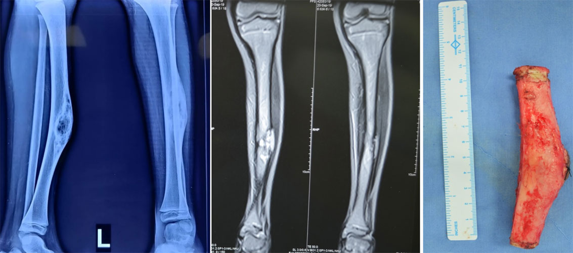
Figure 20. Adamantinoma of the tibia treated with wide surgical excision, removing the tumor with a margin of healthy bone to achieve local tumor control.
Doctor Insight
Adamantinoma is rare and should be treated in a specialised orthopaedic oncology centre. Careful surgical planning is essential because the first operation offers the best chance of long-term cure.
Patient Tip
If you have been diagnosed with a rare malignant bone tumor such as adamantinoma, do not hesitate to seek a second opinion at a specialist bone tumor centre before surgery. This can help ensure the most appropriate treatment plan.
Chordoma
What is a Chordoma?
A chordoma is a rare malignant (cancerous) bone tumor that develops from remnants of the notochord, a temporary structure present during early fetal development that helps form the spine. Although the notochord normally disappears before birth, small remnants may persist and rarely give rise to a chordoma later in life.
Chordomas grow slowly, but they are locally aggressive, meaning they can invade nearby bone, nerves, and other important structures. Because they often arise close to the brain, spinal cord, or major nerves, they can be challenging to treat.
Who is affected?
Chordomas most commonly occur in adults between 40 and 70 years of age.
The usual locations are:
- Sacrum (base of the spine) – most common
- Skull base (clivus) – behind the nose and beneath the brain
- Mobile spine (neck, chest, or lower back)
Symptoms
Symptoms depend on the tumor’s location and usually develop gradually.
They may include:
- Persistent back or neck pain
- A slowly enlarging swelling
- Numbness or weakness in the legs
- Difficulty walking
- Changes in bladder or bowel control
- Headache or double vision (for skull base tumors)
Because these symptoms are common to many other conditions, diagnosis is often delayed.
Diagnosis
Diagnosis usually begins with MRI, which provides detailed images of the tumor and its relationship to nearby nerves and blood vessels. CT scans help evaluate bone destruction, while a carefully planned biopsy confirms the diagnosis.
Treatment
Successful treatment requires a multidisciplinary team.
Depending on the tumor location, treatment may include:
- Surgery to remove the tumor completely whenever possible
- High-dose radiotherapy, particularly proton beam or carbon ion therapy in selected centers
- Long-term follow-up with regular MRI scans
Because chordomas tend to recur locally, achieving complete removal during the first operation offers the best chance of long-term disease control.
Prognosis
The outlook varies depending on the tumor’s size, location, and completeness of removal. Although chordomas have a tendency to recur, advances in surgical techniques and highly focused radiotherapy have significantly improved long-term outcomes.
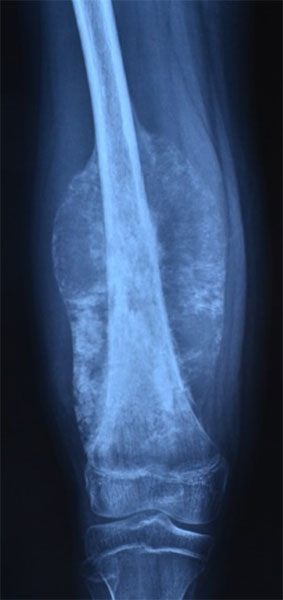
Figure 21. MRI showing a sacral chordoma before surgery (left), and postoperative X-ray following surgical removal of the tumor (right).
Doctor Explains
Chordomas usually grow slowly, but “slow-growing” does not mean harmless. Their location near critical nerves and the spinal cord makes careful surgical planning more important than the speed of growth.
Clinical Pearl
Patients with suspected chordoma should ideally be evaluated in a specialized bone and soft tissue tumor center before any biopsy or surgery.
Reassurance Box
Although chordoma is a rare cancer, treatment has improved considerably over the past two decades. Modern imaging, specialized surgery, and advanced radiotherapy have enabled many patients to achieve long-term disease control while preserving function.
Multiple Myeloma
What is Multiple Myeloma?
Multiple myeloma is not a primary bone tumor. Instead, it is a blood cancer that develops from plasma cells, a type of white blood cell responsible for producing antibodies that help fight infections.
Abnormal plasma cells accumulate in the bone marrow (the soft tissue inside bones where blood cells are produced), weakening the surrounding bone and increasing the risk of fractures.
Because bones are commonly affected, patients with multiple myeloma often first seek medical attention for bone pain or a fracture.
Who is affected?
Multiple myeloma usually affects:
- Adults over 60 years of age
It commonly involves:
- Spine
- Pelvis
- Ribs
- Skull
- Long bones
Symptoms
Symptoms may include:
- Persistent bone pain, especially in the back or ribs
- Fatigue due to anemia
- Recurrent infections
- Unexplained weight loss
- Pathological fractures
- High calcium levels causing excessive thirst, constipation, or confusion
- Kidney dysfunction
Diagnosis
Diagnosis combines:
- Blood tests
- Urine tests
- Bone marrow biopsy
- Whole-body imaging (low-dose CT, MRI, or PET-CT)
Unlike most bone tumors, diagnosis relies heavily on laboratory investigations in addition to imaging.
Treatment
Treatment is coordinated by a hematologist (a doctor specializing in blood disorders).
Depending on the patient’s condition, treatment may include:
- Chemotherapy
- Targeted therapy
- Immunotherapy
- Stem cell transplantation in selected patients
- Bone-strengthening medications such as bisphosphonates
- Surgery or radiotherapy for fractures or painful bone lesions
Prognosis
Although multiple myeloma is generally considered a chronic disease rather than a curable one, advances in modern therapy have greatly improved survival and quality of life. Many patients now live for many years with good disease control.
Doctor Insight
Although both chordoma and multiple myeloma affect bone, they are fundamentally different diseases. Chordoma is treated primarily with surgery and radiotherapy, whereas multiple myeloma is managed with systemic medical therapy.
How Are These Bone Tumors Diagnosed?
Diagnosing a bone tumor involves more than interpreting an X-ray. Doctors combine information from your medical history, physical examination, imaging studies, and, when necessary, a biopsy to establish the correct diagnosis and develop an individualized treatment plan.
Medical History and Physical Examination
Your doctor will ask about:
- When the pain began
- Whether pain is worse at night
- History of swelling or a lump
- Previous fractures
- Family history of bone disorders
- General symptoms such as fever or weight loss
During the examination, your doctor will assess:
- Tenderness
- Swelling
- Joint movement
- Limb function
- Nerve and blood vessel function
Imaging Studies
Different imaging tests provide different types of information.
- X-ray: Usually the first investigation and often sufficient to suggest the diagnosis.
- MRI (Magnetic Resonance Imaging): Best for evaluating the extent of the tumor and its relationship to muscles, nerves, blood vessels, and joints.
- CT (Computed Tomography): Excellent for assessing bone detail and identifying small lesions such as osteoid osteoma.
- PET-CT or Bone Scan: Used selectively for malignant tumors to assess disease elsewhere in the body.
Biopsy
A biopsy involves removing a small tissue sample for microscopic examination by a pathologist.
Not every bone tumor requires a biopsy. Many benign lesions have characteristic imaging features that allow a confident diagnosis without surgery. When a biopsy is necessary, it should be carefully planned because the biopsy pathway may need to be removed during definitive surgery if the tumor is malignant.
Biopsy in Bone & Soft Tissue Tumors
Clinical Pearl
MRI should generally be performed before a biopsy whenever possible. This provides the most accurate assessment of the tumor and helps the surgeon plan the safest biopsy approach.
General Principles of Treatment
Treatment is based on the type of tumor, its biological behaviour, location, symptoms, and overall health of the patient. There is no single treatment that is appropriate for every bone tumor.
Depending on the diagnosis, treatment may include:
- Observation: Appropriate for many asymptomatic benign lesions.
- Minimally invasive procedures: Such as radiofrequency ablation for osteoid osteoma.
- Surgery: To remove tumors causing symptoms, weakening bone, or demonstrating aggressive behaviour.
- Radiotherapy: Important for selected malignant tumors such as chordoma.
- Systemic therapy: Including chemotherapy, targeted therapy, immunotherapy, or other medications for diseases such as multiple myeloma.
Whenever possible, treatment decisions should be made by a multidisciplinary team that includes orthopaedic oncologists, radiologists, pathologists, medical oncologists, radiation oncologists, and rehabilitation specialists.
Doctor Insight
Modern treatment focuses not only on removing the tumor but also on preserving function, minimizing complications, and maintaining the best possible quality of life.
Recovery and Long-Term Follow-up
Recovery depends on the type of tumor and the treatment performed. Patients managed with observation usually continue normal daily activities while attending scheduled follow-up visits. Recovery after surgery varies from a few weeks following minimally invasive procedures to several months after complex reconstruction.
Long-term follow-up may include:
- Clinical examination
- Periodic X-rays or MRI
- Assessment of bone healing
- Monitoring for recurrence
- Evaluation of implants or bone grafts when present
Attending follow-up appointments is important even when you feel well, as some tumors can recur before symptoms develop.
Patient Tip
Keep copies of your imaging studies and operative reports. They can be valuable if you move, seek a second opinion, or require future treatment.
Reassurance Box
Most patients with benign bone tumors return to school, work, sports, and other daily activities after appropriate treatment or observation. Even patients with more complex conditions often achieve excellent functional outcomes with specialized care and rehabilitation.
When Should You See an Orthopaedic Oncologist?
Although many bone lesions are benign, specialist assessment is recommended when:
- The diagnosis is uncertain.
- Imaging suggests an aggressive lesion.
- A biopsy is being considered.
- Surgery is planned.
- The tumor is located in the pelvis, spine, or around a major joint.
- There is a pathological fracture.
- Symptoms continue despite treatment.
Early referral helps ensure an accurate diagnosis and appropriate treatment while reducing the risk of unnecessary or poorly planned procedures.
Red Flag Box
Seek prompt medical attention if you develop:
- Persistent bone pain lasting several weeks
- Pain that regularly wakes you at night
- Rapidly enlarging swelling
- An unexplained fracture after minor trauma
- New weakness, numbness, or bowel/bladder symptoms
- Persistent pain after treatment or new swelling near a previous tumor site
Frequently Asked Questions (FAQs)
Are all rare bone tumors cancer?
No. Most rare bone tumors are benign (non-cancerous). Conditions such as osteochondroma, enchondroma, osteoid osteoma, osteoblastoma, unicameral bone cyst, aneurysmal bone cyst, and fibrous dysplasia do not spread to other parts of the body. However, some tumors, including adamantinoma and chordoma, are malignant and require specialized treatment.
What is the most common benign cartilage tumor?
Osteochondroma is the most common benign cartilage tumor and also the most common benign bone tumor overall. It usually develops during childhood or adolescence near the growth plates of long bones and often requires no treatment unless it causes symptoms.
Can an osteochondroma turn into cancer?
Malignant transformation of a solitary osteochondroma is uncommon. The risk is slightly higher in people with Multiple Hereditary Exostoses (MHE). New pain, rapid enlargement after skeletal maturity, or a thick cartilage cap may require further evaluation.
Is an enchondroma dangerous?
Most enchondromas are harmless and remain stable throughout life. Many are discovered incidentally on X-rays. Treatment is usually unnecessary unless the lesion causes pain, weakens the bone, or raises concern for a cartilage cancer.
Why does an osteoid osteoma cause severe night pain?
An osteoid osteoma produces high levels of prostaglandins, chemicals that stimulate pain receptors. This explains why the pain is often worse at night and improves dramatically after taking anti-inflammatory medications such as ibuprofen or naproxen.
Is radiofrequency ablation better than surgery for osteoid osteoma?
For most patients, CT-guided radiofrequency ablation (RFA) has become the preferred treatment because it is minimally invasive, provides rapid pain relief, has a high success rate, and usually allows a quicker recovery than open surgery.
What is the difference between osteoid osteoma and osteoblastoma?
Although both are benign bone-forming tumors, osteoblastomas are generally larger, may behave more aggressively, and are less likely to respond to pain medication. Osteoid osteomas are usually treated with radiofrequency ablation, whereas osteoblastomas more commonly require surgery.
Can a bone cyst heal without surgery?
Yes. Many unicameral (simple) bone cysts gradually heal as a child reaches skeletal maturity. Small, asymptomatic cysts can often be monitored with periodic X-rays. Surgery is considered if there is pain, repeated fractures, or a high risk of fracture.
Is an aneurysmal bone cyst actually a cyst?
Not exactly. Despite its name, an aneurysmal bone cyst (ABC) is not a true cyst. It is a benign but locally aggressive bone lesion made up of blood-filled spaces that can enlarge and weaken the surrounding bone.
Can fibrous dysplasia spread to other bones?
No. Fibrous dysplasia does not spread from one bone to another. However, some people are born with the form that affects multiple bones (polyostotic fibrous dysplasia), while others have involvement of only a single bone.
Does fibrous dysplasia increase the risk of fractures?
Yes. Fibrous dysplasia replaces normal bone with weaker fibrous tissue, making the affected bone more susceptible to fractures and deformity, particularly in weight-bearing bones such as the femur.
Is chondroblastoma a cancer?
No. Chondroblastoma is a benign tumor. However, it is considered locally aggressive, meaning it can damage nearby bone and joint cartilage if left untreated. Surgical removal is therefore recommended in most patients.
Why does chondromyxoid fibroma often require surgery?
Although chondromyxoid fibroma is benign, it can continue to enlarge, weaken the bone, and recur if incompletely removed. Surgical curettage or excision usually provides excellent long-term results.
How serious is adamantinoma?
Adamantinoma is a rare, slow-growing malignant bone tumor. Although it generally has a better prognosis than many other bone cancers, it requires complete surgical removal because incomplete treatment increases the risk of recurrence and spread.
Can chordoma be cured?
Many chordomas can be successfully controlled, especially when diagnosed early and treated with complete surgical removal combined with specialized radiotherapy. However, long-term follow-up is important because chordomas can recur, sometimes many years after treatment.
Why is multiple myeloma included in a guide on bone tumors?
Multiple myeloma is a blood cancer, not a primary bone tumor. However, it commonly weakens bones, causes painful bone lesions, and increases the risk of fractures. Patients often first present with bone symptoms, making it an important condition to understand alongside other bone tumors.
Will every bone tumor need a biopsy?
No. Many benign bone tumors have characteristic appearances on imaging and can be diagnosed confidently without a biopsy. When the diagnosis is uncertain or cancer is suspected, a biopsy helps determine the exact type of tumor and guide treatment.
How often should I have follow-up scans for a bone tumor?
The frequency of follow-up depends on the diagnosis. Some benign tumors require only one or two follow-up visits, whereas locally aggressive or malignant tumors may need regular imaging for several years to monitor for recurrence.
When should I see an orthopaedic oncologist?
You should consider specialist evaluation if:
- The diagnosis is uncertain.
- A biopsy has been recommended.
- Surgery is being planned.
- The lesion involves the pelvis or spine.
- Imaging suggests an aggressive tumor.
- You have a pathological fracture.
- You have been diagnosed with a rare malignant bone tumor.
What is the outlook for people with rare bone tumors?
The prognosis depends on the specific diagnosis. Most benign bone tumors have an excellent outlook and many never require treatment. Even patients with rare malignant tumors such as chordoma or adamantinoma often achieve good long-term outcomes when managed by an experienced multidisciplinary team.
References
- World Health Organization. WHO Classification of Soft Tissue and Bone Tumours. 5th ed. Lyon, France: International Agency for Research on Cancer (IARC); 2020.
- NCCN Clinical Practice Guidelines in Oncology (NCCN Guidelines®). Bone Cancer. National Comprehensive Cancer Network. Latest available version.
- Kapoor, L., Kumar, V.S. and Khan, S.A., 2026. Limb Salvage Surgery in Bone Tumors. CRC Press, Taylor & Francis Group.
- Campanacci M. Bone and Soft Tissue Tumors. 2nd ed. Vienna: Springer-Verlag; 1999.
- Gibbs CP Jr, Peabody TD, Simon MA. Principles of Musculoskeletal Oncology. In: Campbell’s Operative Orthopaedics. 14th ed. Elsevier; 2021.
- Enneking WF. Musculoskeletal Tumor Surgery. New York: Churchill Livingstone; 1983.
- American Academy of Orthopaedic Surgeons (AAOS). OrthoInfo: Bone Tumors. Available at: https://orthoinfo.aaos.org
- Musculoskeletal Tumor Society (MSTS). Clinical Resources and Educational Materials. Available at: https://msts.org
- International Society of Limb Salvage (ISOLS). Consensus Statements and Educational Resources. Available at: https://www.isols.org
Medical Disclaimer
The information provided in this article is intended for educational purposes only and should not replace professional medical advice, diagnosis, or treatment. Every benign bone tumor is unique, and treatment decisions depend on the type of tumor, its location, symptoms, imaging findings, and individual patient factors. Always consult a qualified orthopaedic oncologist or your healthcare provider for personalized medical advice. Never ignore professional medical guidance or delay seeking medical attention because of information you have read online.
 +91 93558 33005