A bone tumor biopsy is a procedure in which a small sample of tissue is removed from a bone or soft tissue tumor and examined under a microscope to determine the exact diagnosis.
Although imaging tests such as X-rays, MRI (Magnetic Resonance Imaging), and CT (Computed Tomography) can strongly suggest the nature of a tumor, a biopsy is usually required to confirm the diagnosis before treatment begins.
A carefully planned biopsy provides crucial information about:
- Whether the tumor is benign (non-cancerous) or malignant (cancerous)
- The exact type of tumor
- The tumor grade (how aggressive it appears)
- Additional molecular or genetic features that may influence treatment
Modern biopsy techniques are highly accurate and, when performed correctly, are safe and do not compromise future treatment.
One important principle in orthopaedic oncology is that the biopsy should ideally be planned by the same specialist team that will perform the definitive surgery, because the biopsy pathway influences later surgical planning and limb salvage.
Quick Summary
- A biopsy is the gold standard for diagnosing most bone and soft tissue tumors.
- Imaging should almost always be completed before the biopsy.
- The biopsy helps determine the exact diagnosis and guides treatment planning.
- Most biopsies are performed using a core needle biopsy, which is minimally invasive.
- A properly planned biopsy does not spread cancer.
- The biopsy should ideally be performed at the centre where definitive treatment will be undertaken.
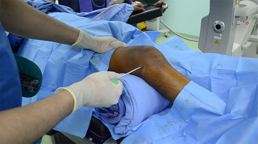
Figure 1. Core needle biopsy of a bone tumor performed under image guidance for accurate tissue diagnosis.
Being told that you need a biopsy can be worrying.
Many patients immediately wonder:
- Does this mean I have cancer?
- Will the biopsy be painful?
- Can the biopsy spread the tumor?
- Why can’t the doctor simply remove the lump?
These are understandable concerns.
A biopsy is one of the most important steps in diagnosing a bone or soft tissue tumor because it tells doctors exactly what the tumor is. Different tumors can look similar on imaging but require completely different treatments. A biopsy ensures that treatment is based on an accurate diagnosis rather than assumptions.
In orthopaedic oncology, biopsy is much more than a diagnostic procedure. It is the first step of definitive treatment planning.
Every biopsy must be carefully planned because the path taken by the biopsy needle or incision—known as the biopsy tract—is considered potentially contaminated with tumor cells and is usually removed during definitive surgery. For this reason, the biopsy should be performed in a way that does not interfere with future limb salvage or reconstruction.
This is why specialists often recommend that the biopsy be carried out at the same centre where the final treatment will be performed.
Fortunately, advances in imaging, image-guided biopsy techniques, pathology, and molecular diagnostics have made modern biopsy procedures highly accurate and safe. Most patients undergo the procedure without major complications and return home the same day or after a short observation period.
This guide explains why biopsy is necessary, how it is planned, the different biopsy techniques, what happens to the tissue sample after it is collected, how the results guide treatment, and what you can expect during recovery.
Whether you are a patient, caregiver, or referring doctor, understanding these principles will help you make informed decisions and appreciate why careful biopsy planning is one of the most important steps in successful musculoskeletal oncology care.
Clinical Pearl
The first biopsy is often the best opportunity to establish an accurate diagnosis. A carefully planned biopsy not only confirms the nature of the tumor but also helps preserve future treatment options, including limb salvage surgery.
Doctor Explains
Why Can’t Doctors Remove the Tumor Without a Biopsy?
It may seem logical to remove the lump first and identify it later. However, different tumors require very different treatments.
For example:
- Some tumors require chemotherapy before surgery.
- Others are treated with surgery alone.
- Some benign tumors do not require extensive surgery at all.
- Certain soft tissue tumors require different surgical margins than bone tumors.
A biopsy allows your medical team to choose the right treatment for the right diagnosis, avoiding unnecessary or inappropriate surgery.
Did You Know?
More than 90% of musculoskeletal tumors can be accurately diagnosed using a properly planned core needle biopsy, making it the preferred biopsy technique for many bone and soft tissue tumors.
Patient Tip
Before your biopsy, ask your doctor:
- Has all the necessary imaging been completed?
- What type of biopsy is recommended?
- Who will perform the biopsy?
- Will the biopsy tract be planned with future surgery in mind?
- When can I expect the results?
Understanding these steps can make the process less stressful and help you participate in treatment planning.
If You Have Been Advised to Undergo a Biopsy
Needing a biopsy does not automatically mean you have cancer.
Many bone and soft tissue lumps are benign. However, because imaging alone cannot always distinguish benign from malignant tumors, a biopsy is often the safest way to establish a definitive diagnosis.
A properly planned biopsy is:
- A routine and well-established procedure.
- Usually minimally invasive.
- Carefully performed to minimize complications.
- An essential step in choosing the most appropriate treatment.
Waiting for biopsy results can be emotionally difficult, but obtaining an accurate diagnosis is the foundation of effective treatment.
Key Facts Box
Why is a properly planned biopsy so important?
Think Before You Biopsy
A biopsy may appear to be a simple procedure, but in musculoskeletal oncology it is one of the most carefully planned steps in the entire treatment journey.
Unlike biopsies performed for many other conditions, a bone or soft tissue tumor biopsy must be planned with future surgery already in mind.
This is because the biopsy is not an isolated diagnostic test—it is the first step of definitive treatment.
A properly planned biopsy helps establish the diagnosis while preserving future treatment options, including limb salvage surgery. Conversely, a poorly planned biopsy can complicate surgery, increase the extent of tissue that must be removed, and in some cases reduce the possibility of preserving the limb.
For these reasons, biopsy planning is usually undertaken by the same multidisciplinary team that will provide definitive treatment.
Why MRI Should Always Be Performed Before Biopsy
One of the most important principles in orthopaedic oncology is:
MRI should almost always be performed before the biopsy.
MRI provides detailed information about:
- The exact size of the tumor.
- Extension within the bone.
- Involvement of nearby muscles.
- Relationship to major nerves and blood vessels.
- Joint involvement.
- Areas most suitable for tissue sampling.
This information helps the surgeon and radiologist select the safest biopsy pathway while ensuring that representative tumor tissue is obtained.
Doctor Explains
Why Can a Biopsy Affect MRI?
Even a carefully performed biopsy causes a small amount of bleeding and inflammation around the tumor.
Although this is a normal part of healing, these changes may make it more difficult to distinguish the true tumor from biopsy-related changes on subsequent MRI scans.
Completing MRI first provides the clearest roadmap for surgical planning.
Clinical Pearl
MRI before biopsy is an internationally accepted principle in musculoskeletal oncology because it provides the most accurate assessment of tumor extent and helps guide safe biopsy planning.
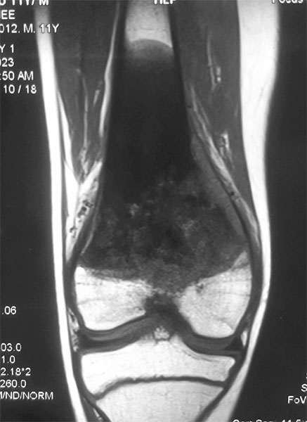
Figure 2. MRI before biopsy helps define the bone tumor and plan a safe and appropriate biopsy approach.
Why the Biopsy Should Ideally Be Performed at the Treating Centre
Many patients wonder why they are advised to travel to a specialist sarcoma centre for what appears to be a relatively simple procedure.
The reason is that every biopsy influences future surgery.
The route taken by the biopsy needle or surgical incision is known as the biopsy tract. Because tumor cells may be deposited along this pathway, the biopsy tract is usually removed together with the tumor during definitive surgery.
If the biopsy tract is placed incorrectly, additional healthy muscles, skin, or other tissues may also need to be removed to achieve safe surgical margins.
This can make limb salvage surgery more complex and may affect the reconstruction required.
For this reason, biopsy planning is closely coordinated with the anticipated surgical approach.
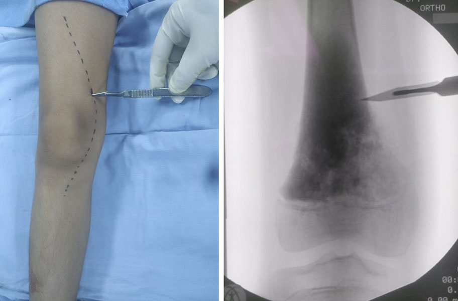
Figure 3. A carefully planned biopsy tract is aligned with the future surgical incision to facilitate safe tumor removal.
Doctor Insight
In orthopaedic oncology, we often say:
“The biopsy should be performed as though the definitive surgery has already been planned.”
This approach helps ensure that the biopsy tract can be removed safely during tumor resection without compromising future reconstruction or limb preservation.
The Multidisciplinary Team Plans the Biopsy
A bone tumor biopsy is rarely planned by a single doctor in isolation.
Instead, several specialists contribute to the decision.
These commonly include:
- Orthopaedic oncologist
- Musculoskeletal radiologist
- Musculoskeletal pathologist
- Medical oncologist
- Radiation oncologist (when appropriate)
Together they determine:
- Whether biopsy is required.
- Which biopsy technique is most appropriate.
- Which part of the tumor should be sampled.
- The safest biopsy pathway.
- Whether additional molecular testing is likely to be required.
This collaborative approach improves diagnostic accuracy and helps avoid unnecessary procedures.
Did You Know?
💡 The diagnosis of many bone and soft tissue tumors is based not only on the biopsy itself but also on correlation between imaging, pathology, and clinical findings. This multidisciplinary review is one of the reasons specialist sarcoma centres achieve high diagnostic accuracy.
Common Biopsy Mistakes That Can Affect Treatment
Most biopsies are performed safely. However, certain avoidable errors can complicate subsequent treatment.
Examples include:
Performing the Biopsy Before MRI
This may obscure the true tumor boundaries and complicate surgical planning.
Poorly Planned Biopsy Tract
An incorrectly positioned biopsy tract may require removal of additional normal tissues during definitive surgery.
Excision of an Undiagnosed Soft Tissue Lump
Removing an unexplained lump without appropriate imaging or biopsy may result in incomplete tumor removal and necessitate further surgery.
Internal Fixation Before Diagnosis
Fixing a pathological fracture before establishing the diagnosis can contaminate surrounding tissues and make definitive treatment more challenging.
Delayed Referral to a Specialist Centre
Early referral allows biopsy planning, imaging, pathology, and surgery to be coordinated from the outset.
Seek Specialist Advice Before Biopsy If:
- A bone tumor is suspected on X-ray or MRI.
- A soft tissue lump is increasing in size.
- Surgery has been recommended without a tissue diagnosis.
- You have a pathological fracture through a suspected tumor.
- You are unsure whether biopsy has been planned by the treating surgical team.
Early specialist involvement helps protect future treatment options.
Questions to Ask Before Your Biopsy
Being informed before the procedure can reduce anxiety and help you participate in treatment decisions.
Consider asking:
- Why do I need a biopsy?
- What type of biopsy is recommended?
- Has my MRI already been completed?
- Who will perform the biopsy?
- Will the biopsy be image-guided?
- How long will the results take?
- Will additional molecular or genetic tests be required?
- Will the biopsy be planned with future surgery in mind?
Patient Tip
If you have already undergone imaging elsewhere, bring all original MRI, CT, and X-ray (not only the written reports) to your consultation. Reviewing the actual often provides important information for biopsy planning.
“How is the biopsy actually performed, and which type is best for me?”
Types of Biopsy in Bone & Soft Tissue Tumors
There is no single biopsy technique that is suitable for every patient.
The best biopsy method depends on several factors, including:
- The location of the tumor
- Whether the tumor is in bone or soft tissue
- Its size and depth
- Relationship to nerves and blood vessels
- Information seen on MRI and CT scans
- The amount of tissue required for diagnosis
- Future surgical planning
In most patients, a core needle biopsy provides sufficient tissue to establish the diagnosis while minimizing trauma to surrounding tissues. However, some situations require an open (incisional) biopsy or image-guided techniques to obtain an adequate sample safely.
The choice is individualized and made after careful review of the imaging studies.
Core Needle Biopsy
A core needle biopsy is the preferred biopsy technique for most bone and soft tissue tumors.
During the procedure, a special hollow needle is inserted into the tumor to remove several small cylinders (cores) of tissue. These tissue samples preserve the microscopic architecture of the tumor, allowing the pathologist to make an accurate diagnosis.
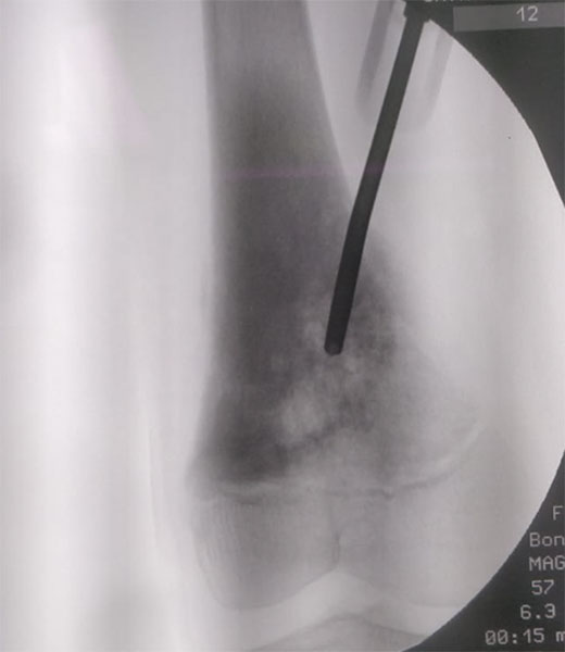
Figure 4. Core needle biopsy of a bone tumor using a biopsy needle to obtain tissue samples for diagnosis.
The procedure is usually performed under:
- Local anaesthesia
- Sedation (when required)
- General anaesthesia in selected patients, particularly children
Image guidance is frequently used to ensure that the needle reaches the most representative part of the tumor while avoiding important nerves and blood vessels.
Advantages
- Minimally invasive
- Small skin incision
- Less pain after the procedure
- Lower risk of complications
- Usually performed as a day-care procedure
- High diagnostic accuracy in experienced centres
Limitations
- Small tissue sample
- Occasionally insufficient for diagnosis
- Repeat biopsy may rarely be required
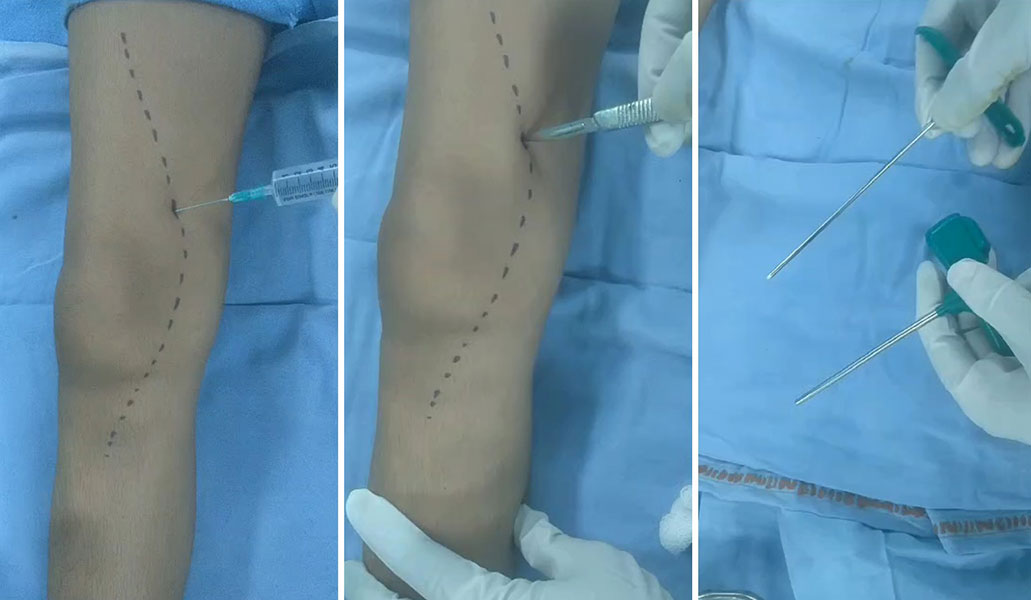
Figure 5A. The biopsy site and tract are carefully planned and marked before needle insertion.
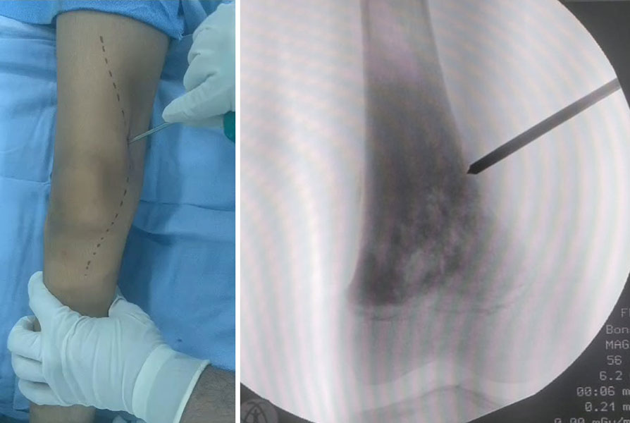
Figure 5B. The biopsy needle is advanced along the planned tract toward the bone tumor.
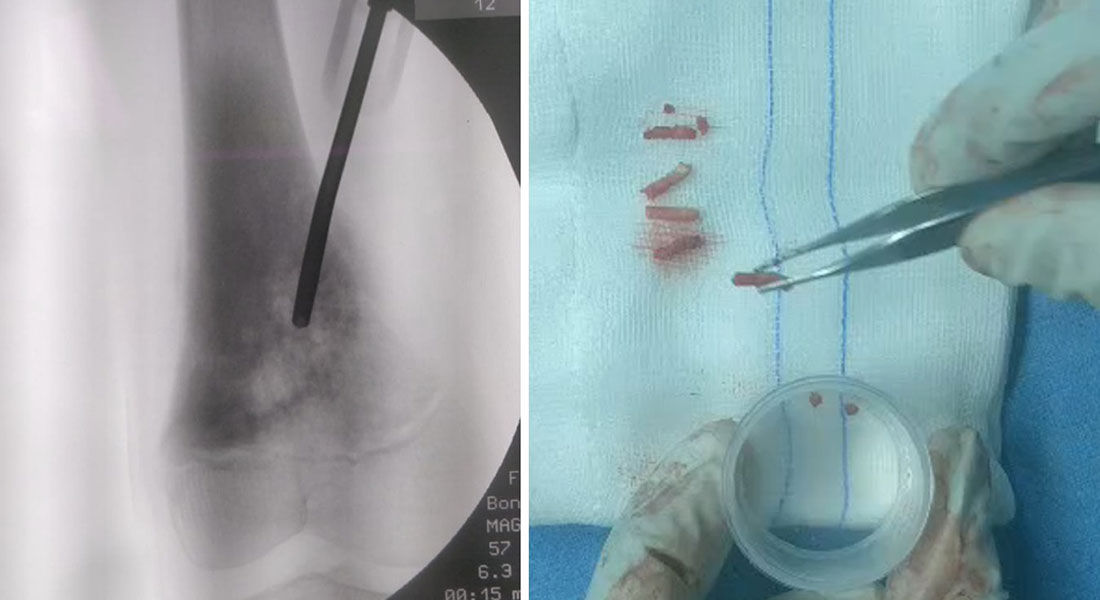
Figure 5C. Tissue cores are obtained through the biopsy needle for pathological examination.
Doctor Explains
Why Are Several Tissue Samples Taken?
One small sample may not represent the entire tumor.
Taking multiple cores from different areas increases the likelihood of obtaining enough tissue for:
- Histopathology
- Immunohistochemistry
- Molecular testing (when required)
This improves diagnostic accuracy without significantly increasing risk.
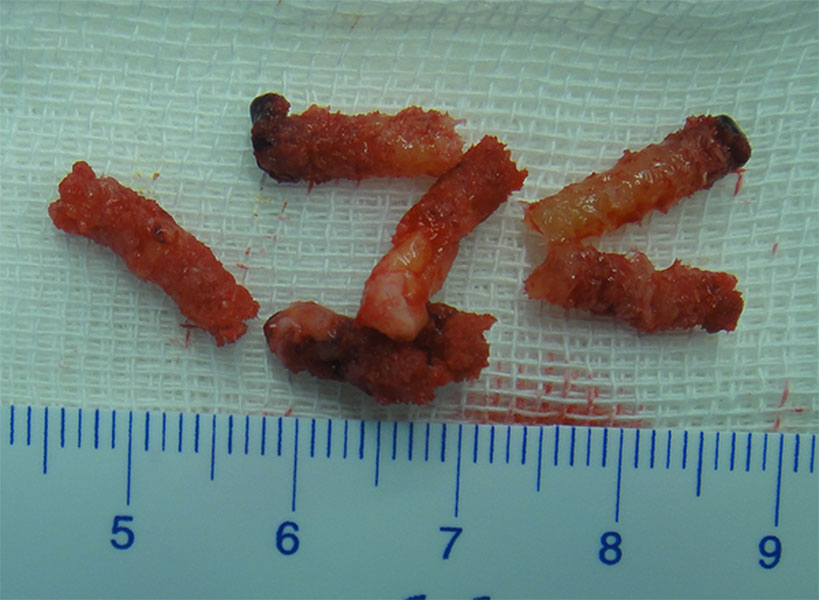
Figure 6. Multiple tissue cores are obtained during a core needle biopsy to improve diagnostic accuracy.
Open (Incisional) Biopsy
An open biopsy, also called an incisional biopsy, involves making a small surgical incision to remove a larger tissue sample.
It is generally reserved for situations where:
- A previous core needle biopsy is inconclusive.
- The tumor is difficult to access safely with a needle.
- Larger tissue samples are required for diagnosis.
Unlike an excisional biopsy, the goal is not to remove the entire tumor, but to obtain enough tissue for accurate diagnosis.
Because open biopsy requires a surgical incision, careful planning of the incision is essential so that it can be removed during definitive tumor surgery.
Advantages
- Larger tissue sample
- Useful when previous biopsy is nondiagnostic
- Helpful for selected uncommon tumors
Limitations
- More invasive
- Larger incision
- Slightly longer recovery
- Greater potential for wound-related complications
Clinical Pearl
When an open biopsy is required, the incision should be planned as though it will become part of the definitive tumor resection. Proper incision placement is essential for successful limb salvage.
Image-Guided Biopsy
Some tumors are located deep within the body or close to important structures, making image guidance essential.
Image guidance allows the biopsy needle to be directed precisely into the desired part of the tumor while minimizing injury to surrounding tissues.
CT-Guided Biopsy
CT (Computed Tomography)-guided biopsy is particularly useful for:
- Deep bone tumors
- Pelvic tumors
- Spine lesions
- Small lesions that cannot be easily felt
CT provides excellent visualization of bone and allows accurate needle placement.
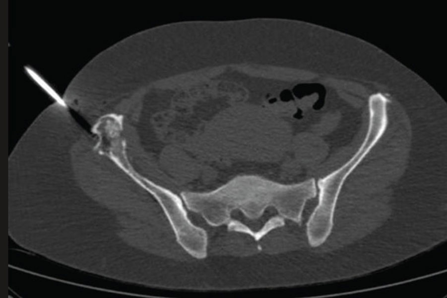
Figure 7. CT-guided biopsy of a pelvic bone tumor allows precise needle placement and safe tissue sampling.
Ultrasound-Guided Biopsy
Ultrasound-guided biopsy is commonly used for:
- Superficial soft tissue tumors
- Lesions close to the skin
- Selected soft tissue masses around the limbs
It offers the advantage of real-time visualization without exposing the patient to ionizing radiation.
Did You Know?
💡 Image guidance not only improves biopsy accuracy but also helps avoid important nerves, blood vessels, and uninvolved tissues, reducing the risk of complications.
Core Needle Biopsy vs Open Biopsy
Both techniques are valuable, but they serve different purposes.
Comparison of Biopsy Techniques
Both procedures are highly effective when appropriately selected and performed by an experienced team.
Which Biopsy Is Best?
There is no universally superior biopsy technique.
The “best” biopsy is the one that:
- Provides enough tissue for an accurate diagnosis.
- Minimizes complications.
- Preserves future treatment options.
- Fits safely within the planned surgical approach.
In most patients, this is a core needle biopsy, but certain situations require an open biopsy to obtain sufficient tissue.
The decision is based on the individual characteristics of the tumor rather than a fixed rule.
Doctor Insight
Modern image-guided core needle biopsy has become the preferred first-line technique for most musculoskeletal tumors because it combines high diagnostic accuracy with minimal disruption of surrounding tissues. Open biopsy remains an important option when additional tissue is needed or previous sampling has been inconclusive.
Patient Tip
Ask your doctor:
- Why is this biopsy technique recommended for me?
- Will image guidance be used?
- How many tissue samples will be taken?
- Will I need local or general anaesthesia?
- What happens if the biopsy does not provide a definite diagnosis?
Understanding the reasons behind the chosen technique can help reduce anxiety before the procedure.
Does Needing an Open Biopsy Mean Something Is Wrong?
No.
An open biopsy is sometimes recommended simply because a larger tissue sample is needed to reach an accurate diagnosis.
Choosing an open biopsy does not necessarily mean that the tumor is more serious or that the previous treatment has failed. The goal is always to obtain the information needed to plan the safest and most effective treatment.
What Happens After the Biopsy?
Once the biopsy has been completed, the work has only just begun.
The tissue sample is carefully processed and examined to determine the exact diagnosis. This process involves much more than simply looking at the tissue under a microscope. In many cases, additional laboratory tests are required to identify the precise type of tumor and guide treatment.
For patients, this waiting period can be one of the most stressful parts of the diagnostic journey. Understanding what happens behind the scenes can make it easier to appreciate why accurate diagnosis takes time.
From Biopsy to Diagnosis: The Journey of Your Tissue Sample
After the biopsy, the tissue is sent to a pathology laboratory, where doctors trained in diagnosing diseases by examining tissues—called pathologists—begin a detailed evaluation.
The process usually includes several steps:
- Preparing the tissue.
- Microscopic examination.
- Additional specialized tests when needed.
- Correlation with imaging findings.
- Final diagnosis.
- Multidisciplinary treatment planning.
Each step contributes important information, ensuring that treatment is based on the most accurate diagnosis possible.
Histopathology: Looking at the Tissue Under the Microscope
The first stage of analysis is histopathology, which means examining thin sections of tissue under a microscope.
Before this can be done, the tissue is:
- Preserved in a special solution to prevent deterioration.
- Embedded in wax to support the specimen.
- Cut into extremely thin slices.
- Stained with dyes that highlight different cellular structures.
The pathologist studies:
- The appearance of the tumor cells.
- Their arrangement.
- The surrounding tissue.
- Features that suggest whether the tumor is benign or malignant.
In many cases, histopathology alone is sufficient to establish the diagnosis.
Doctor Explains
What Does a Pathologist Do?
A pathologist is a doctor who specializes in diagnosing disease by examining tissues and cells under the microscope.
For bone and soft tissue tumors, the pathologist works closely with the orthopaedic oncologist and radiologist. The diagnosis is based not only on what the tissue looks like but also on where the tumor is located, how it appears on imaging, and the patient’s clinical history.
Immunohistochemistry: Identifying the Tumor More Precisely
Sometimes tumors look very similar under the microscope.
In these situations, immunohistochemistry (IHC) may be required.
Immunohistochemistry uses special antibodies that attach to specific proteins within tumor cells. This helps identify the exact type of tumor and distinguish between conditions that may appear similar on routine microscopy.
For example, IHC may help differentiate:
- Ewing sarcoma from lymphoma.
- Different soft tissue sarcomas.
- Metastatic tumors from primary bone tumors.
Not every patient requires immunohistochemistry, but when it is needed, it significantly improves diagnostic accuracy.
Clinical Pearl
Modern diagnosis of bone and soft tissue tumors relies on the combination of imaging, histopathology, immunohistochemistry, and clinical assessment—not on any single test alone.
Molecular Testing: Looking at the Tumor’s Genetic Features
For certain tumors, additional molecular testing may be recommended.
These tests examine specific genetic changes within the tumor cells that can:
- Confirm the diagnosis.
- Distinguish between similar tumors.
- Guide treatment decisions.
- Provide prognostic information in selected situations.
Examples include identifying characteristic genetic alterations seen in some sarcomas.
Not every patient requires molecular testing, and your doctor will advise whether it is appropriate in your case.
Did You Know?
💡 Many bone and soft tissue tumors have characteristic genetic changes that help doctors confirm the diagnosis with greater confidence, especially when the microscopic appearance is uncertain.
Why Do Biopsy Results Take Several Days?
Patients often expect biopsy results within 24 hours.
However, obtaining an accurate diagnosis requires careful processing of the tissue and, in some cases, additional specialized tests.
The time taken depends on:
- The complexity of the tumor.
- Whether immunohistochemistry is required.
- Whether molecular testing is necessary.
- Review by specialist musculoskeletal pathologists.
- Referral to expert laboratories for rare tumors.
Although waiting can be frustrating, taking the time to reach the correct diagnosis is far more important than receiving a rapid but incomplete report.
Waiting for Biopsy Results Can Be Difficult
Many patients find the period between the biopsy and the diagnosis emotionally challenging.
Remember that the waiting time is usually spent performing additional tests to ensure the diagnosis is as accurate as possible.
These results form the foundation of your entire treatment plan, so careful evaluation is well worth the time.
How Imaging and Pathology Work Together
One of the unique aspects of musculoskeletal oncology is that the biopsy result is never interpreted in isolation.
The final diagnosis is reached by combining:
- Clinical examination.
- X-rays.
- MRI findings.
- CT scans (when applicable).
- Histopathology.
- Immunohistochemistry.
- Molecular testing (if required).
This process is known as radiological-pathological correlation.
Occasionally, if the biopsy findings do not match the imaging appearance, the multidisciplinary team may recommend additional review or even a repeat biopsy to ensure that the diagnosis is correct.
Doctor Insight
An accurate diagnosis depends on concordance between the clinical picture, imaging, and pathology. If these do not agree, the answer is not to accept uncertainty but to investigate further until the diagnosis is clear.
The Multidisciplinary Tumor Board
Once all investigations are complete, the case is discussed by a multidisciplinary tumor board.
This typically includes:
- Orthopaedic oncologist.
- Musculoskeletal radiologist.
- Musculoskeletal pathologist.
- Medical oncologist.
- Radiation oncologist.
- Plastic or vascular surgeon when required.
Together, they review:
- The biopsy results.
- Imaging studies.
- Stage of the disease.
- Overall health of the patient.
- Treatment options.
The treatment plan is then tailored to the individual patient rather than based solely on the biopsy report.
When Is a Repeat Biopsy Needed?
Most biopsies provide enough information to establish a diagnosis.
However, a repeat biopsy may occasionally be necessary if:
- The sample is too small.
- The tissue is not representative of the tumor.
- The findings do not match the imaging studies.
- Additional tissue is required for specialized testing.
Needing another biopsy does not necessarily mean the tumor is more serious. It simply means that more information is required to make the correct diagnosis.
Patient Tip
If your doctor recommends a repeat biopsy, ask why it is necessary. In many cases, it is performed to obtain additional tissue for specialized testing or to clarify findings that do not fully explain the imaging results.
Recovery and Safety After a Bone Tumor Biopsy
Most patients recover quickly after a bone or soft tissue tumor biopsy. Compared with definitive tumor surgery, a biopsy is usually a minor procedure, and many patients return home the same day or after a short hospital stay.
Recovery depends on:
- The type of biopsy performed
- The location of the tumor
- Whether image guidance was used
- Your general health
- Whether the biopsy involved bone or soft tissue
Although mild discomfort is common, serious complications are uncommon when the procedure is carefully planned and performed by an experienced team.
What Should I Expect After the Procedure?
Immediately after the biopsy, you will be monitored for a short period before returning home or to your hospital room.
You may notice:
- Mild pain or tenderness around the biopsy site.
- Bruising or slight swelling.
- A small dressing covering the incision or needle entry site.
- Temporary stiffness if the biopsy was performed near a joint.
Most patients find that these symptoms improve over the next few days with simple pain medication and rest.
If general anaesthesia or sedation was used, you may feel drowsy for several hours and should arrange for someone to accompany you home.
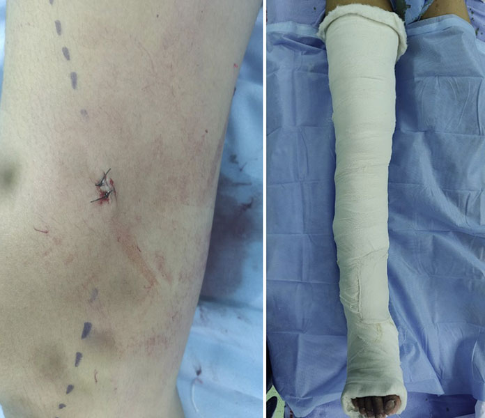
Figure 8. After biopsy, meticulous wound closure and limb immobilization help protect the biopsy site during healing.
Patient Tip
Follow your biopsy care instructions carefully. Keep the dressing clean and dry, avoid unnecessary strain on the affected limb, and attend your scheduled follow-up appointment even if you are feeling well.
Caring for the Biopsy Site
Your healthcare team will provide specific instructions based on the type of biopsy you have undergone.
General recommendations include:
- Keep the dressing clean and dry for the recommended period.
- Avoid soaking the wound until your doctor advises it is safe.
- Take pain medication only as prescribed.
- Avoid strenuous activity involving the affected limb until permitted.
- Watch for signs of infection or excessive bleeding.
For biopsies involving weight-bearing bones, your doctor may recommend temporarily reducing weight-bearing or using crutches to protect the bone while the biopsy site heals.
When Can I Return to Normal Activities?
Most patients resume light daily activities within a few days.
However, the exact timing depends on:
- The location of the biopsy.
- Whether the biopsy involved bone or soft tissue.
- The size of the tissue sample obtained.
- Your planned treatment.
Avoid heavy lifting, vigorous exercise, or contact sports until your treating team confirms that it is safe to do so.
Remember that recovery from the biopsy is only one part of your overall treatment journey. Once the diagnosis is confirmed, your doctors will discuss the next steps.
Clinical Pearl
The goal after biopsy is not simply wound healing but protecting the biopsy site until the diagnosis is confirmed and definitive treatment begins.
Possible Risks and Complications
Like any medical procedure, a biopsy carries some risks. Fortunately, significant complications are uncommon.
Potential complications include:
Mild Pain
Temporary discomfort is expected and usually improves within a few days.
Bleeding or Bruising
Small amounts of bruising are common. Significant bleeding is uncommon but may occur in highly vascular tumors or patients taking blood-thinning medications.
Infection
Infection after biopsy is uncommon. If it occurs, it is usually treated successfully with antibiotics and, rarely, additional procedures.
Fracture
Bone tumors can weaken the bone before the biopsy is performed.
Although uncommon, a fracture may occur if the bone is already severely weakened. Your doctor may recommend activity restrictions to reduce this risk.
Nondiagnostic Biopsy
Occasionally, the tissue sample is insufficient or does not represent the tumor adequately.
This does not necessarily indicate a serious problem—it simply means additional tissue may be needed to establish a definitive diagnosis.
Does a Biopsy Spread Cancer?
This is one of the most common concerns patients have.
The reassuring answer is:
A properly planned biopsy performed using accepted oncological principles does not increase the risk of cancer spreading throughout the body.
In musculoskeletal oncology, the biopsy tract is carefully planned because a small number of tumor cells may remain along the pathway used to obtain the tissue sample.
This is not the same as cancer spreading (metastasis).
To address this possibility, the biopsy tract is routinely removed during definitive tumor surgery. This practice has been part of standard orthopaedic oncology surgery for many years and is one of the reasons careful biopsy planning is so important.
Current international guidelines continue to support biopsy as the safest and most reliable method of establishing a diagnosis before treatment.
Doctor Explains
What Is the Difference Between Biopsy Tract Contamination and Cancer Spread?
These two concepts are often confused.
- Biopsy tract contamination refers to the possibility that a small number of tumor cells may be present along the pathway of the biopsy needle or incision.
- Metastasis means that cancer has spread to distant organs such as the lungs or other bones.
A carefully planned biopsy does not cause distant spread of cancer. The biopsy tract is deliberately included within the planned surgical resection to minimise any local risk.
Should I Be Worried About Having a Biopsy?
No.
Without a biopsy, it is often impossible to determine the exact type of tumor or choose the most appropriate treatment.
The benefits of obtaining an accurate diagnosis greatly outweigh the small risks associated with the procedure. Modern biopsy techniques have an excellent safety record when performed by experienced teams.
Seek Immediate Medical Attention If You Experience:
- Heavy bleeding from the biopsy site.
- High fever with worsening wound pain.
- Rapidly increasing swelling.
- Sudden severe pain after a bone biopsy.
- Loss of movement or sensation in the affected limb.
These symptoms require urgent medical evaluation.
Doctor Insight
Most patients recover uneventfully after biopsy. The most important part of the recovery period is not the wound itself but ensuring that the biopsy results are reviewed promptly and incorporated into a well-planned multidisciplinary treatment strategy.
Frequently Asked Questions (FAQs)
Does every bone tumor need a biopsy?
No. Some benign bone lesions have very characteristic imaging features and may not require a biopsy. However, if there is any uncertainty about the diagnosis or if cancer is suspected, a biopsy is usually necessary before treatment.
Why can’t my doctor remove the tumor without a biopsy?
Different tumors require different treatments. A biopsy confirms the diagnosis and helps determine whether surgery, chemotherapy, radiotherapy, or observation is the most appropriate approach.
Does a biopsy mean I have cancer?
No. Many bone and soft tissue tumors are benign. A biopsy is performed to establish an accurate diagnosis, not because cancer has already been confirmed.
Is a bone tumor biopsy painful?
Most patients experience only mild discomfort. Local anaesthesia, sedation, or general anaesthesia is used depending on the procedure, and pain after the biopsy is usually well controlled with simple medications.
Does a biopsy spread cancer?
No. Current evidence shows that a properly planned biopsy does not cause cancer to spread throughout the body. The biopsy tract is carefully planned and is usually removed during definitive surgery.
Which is better: core needle biopsy or open biopsy?
For most bone and soft tissue tumors, core needle biopsy is the preferred first-line technique because it is accurate, minimally invasive, and has a quicker recovery. Open biopsy is reserved for selected situations where additional tissue is needed.
Why is MRI performed before the biopsy?
MRI accurately shows the size and extent of the tumor and helps doctors plan the safest biopsy pathway. Performing MRI before biopsy also avoids biopsy-related changes that can interfere with image interpretation.
How long does the biopsy procedure take?
The procedure itself is usually completed within 30–60 minutes, although the total hospital visit may be longer because of preparation and post-procedure observation.
Will I need to stay overnight in the hospital?
Most biopsies are performed as day-care procedures, allowing patients to return home the same day. Some patients, particularly children or those undergoing more complex procedures, may require a short hospital stay.
When will I receive the biopsy results?
Initial pathology results are often available within several days. If additional tests such as immunohistochemistry or molecular analysis are required, the final report may take longer.
Why are my biopsy results taking longer than expected?
Complex tumors sometimes require additional laboratory tests or review by specialist musculoskeletal pathologists. Although this increases the waiting time, it also improves diagnostic accuracy.
What happens if the biopsy is inconclusive?
Occasionally, the tissue sample does not provide enough information for a definite diagnosis. Your doctor may recommend a repeat biopsy or additional investigations to obtain the necessary information.
Can children safely undergo a biopsy?
Yes. Bone tumor biopsies are routinely performed in children by experienced multidisciplinary teams. General anaesthesia is commonly used to ensure the procedure is safe and comfortable.
Will I need stitches?
Most core needle biopsies require only a tiny skin incision that may not need stitches. Open biopsies usually require sutures, which are removed or dissolve depending on the type used.
Can I eat before the biopsy?
This depends on the type of anaesthesia planned. If sedation or general anaesthesia is required, you may need to fast beforehand. Your healthcare team will provide specific instructions.
Should I stop my blood-thinning medications?
Do not stop any medication without medical advice. Your doctor will review your medications and advise whether temporary adjustment is necessary before the biopsy.
When can I drive after the biopsy?
If only local anaesthesia was used, many patients can resume driving once they are comfortable and can safely control the vehicle. If sedation or general anaesthesia was given, avoid driving until your healthcare team advises it is safe.
Can I exercise after a biopsy?
Light activities can usually be resumed within a few days. Heavy lifting, vigorous exercise, or high-impact activities should be avoided until your doctor confirms that the biopsy site has healed adequately.
Should I seek a second opinion before or after the biopsy?
A second opinion is reasonable if the diagnosis is uncertain, if major surgery has been recommended, or if you would like confirmation that the biopsy and treatment plan follow current specialist practice.
Why should the biopsy and surgery ideally be performed at the same centre?
The biopsy tract must usually be removed during definitive surgery. Performing both procedures at the same specialist centre allows the biopsy to be planned with future surgery in mind, helping preserve treatment options and reduce complications.
Myths and Facts
When Should You Consult an Orthopaedic Oncologist?
You should seek evaluation by an orthopaedic oncologist if:
- A bone tumor is suspected on X-ray, MRI, or CT scan.
- You have a persistent bone pain or an unexplained soft tissue lump.
- A biopsy has been recommended and you would like to understand the available options.
- A biopsy has been performed but the diagnosis remains uncertain.
- You have been advised to undergo major surgery or amputation and would like a second opinion.
- A pathological fracture has occurred through a suspected bone tumor.
- Your biopsy or pathology findings do not match the imaging results.
Early referral helps ensure that the biopsy, diagnosis, and definitive treatment are coordinated appropriately.
References
This article is based on current evidence and internationally accepted guidance, including:
- National Comprehensive Cancer Network (NCCN). Bone Cancer Guidelines (latest available version).
- National Comprehensive Cancer Network (NCCN). Soft Tissue Sarcoma Guidelines.
- ESMO–EURACAN–GENTURIS Clinical Practice Guidelines for Bone Sarcomas.
- ESMO Clinical Practice Guidelines for Soft Tissue Sarcomas.
Medical Disclaimer
The information provided on this page is intended for educational purposes only and should not replace consultation with a qualified healthcare professional. Every patient with a suspected bone or soft tissue tumor has unique clinical circumstances, and decisions regarding biopsy and treatment should be individualized after assessment by a multidisciplinary sarcoma team.
If you have persistent bone pain, a suspicious bone lesion, an unexplained soft tissue mass, or have been advised to undergo a biopsy, consult an experienced orthopaedic oncologist. Early specialist evaluation helps ensure accurate diagnosis, appropriate biopsy planning, and the best opportunity for successful treatment.
About the Author
Dr. Love Kapoor, MS (Orthopaedics), Fellowship in Musculoskeletal Oncology (AIIMS, New Delhi), Former Assistant Professor, Orthopaedic Oncology, AIIMS, New Delhi.
Orthopaedic Oncologist specializing in bone tumors, soft tissue sarcomas, limb salvage surgery, bone reconstruction, mega prosthesis surgery, and metastatic bone disease.
Meet Your Orthopaedic Oncologist
Choosing treatment for a bone or soft tissue tumor often involves complex decisions. An orthopaedic oncologist specializes in diagnosing and treating tumors affecting the bones, joints, and soft tissues while working closely with medical oncologists, radiation oncologists, radiologists, pathologists, plastic surgeons, vascular surgeons, physiotherapists, and rehabilitation specialists.
Dr. Love Kapoor is an orthopaedic oncologist with dedicated fellowship training in musculoskeletal oncology. His clinical practice focuses on:
- Primary bone tumors
- Soft tissue sarcomas
- Limb salvage surgery
- Bone reconstruction
- Mega prosthesis surgery
- Management of metastatic bone disease
- Complex revision surgery after tumor reconstruction
His approach emphasizes:
- Accurate diagnosis before treatment
- Multidisciplinary decision-making
- Evidence-based care
- Limb preservation whenever oncologically safe
- Personalized treatment planning
- Long-term functional recovery
If you have been diagnosed with a bone tumor, have been advised to undergo amputation, or would like a second opinion regarding limb salvage surgery, consultation with an experienced orthopaedic oncologist can help you better understand your treatment options.
 +91 93558 33005