Quick Summary
- GCTB is not a bone cancer, but it requires treatment.
- It usually affects young adults (20–40 years).
- The knee is the most commonly affected site.
- The aim of treatment is to remove the tumor while preserving the joint whenever safely possible.
- Most patients undergo extended curettage rather than major bone removal.
- Regular follow-up is important because the tumor can recur, particularly during the first few years after treatment.
.jpg "X-ray showing a giant cell tumor of bone (GCT) involving the lower end of the femur near the knee joint")
Figure 1. X-ray showing a giant cell tumor of bone (GCT) involving the lower end of the femur near the knee joint.
“My doctor says it’s a Giant Cell Tumor, but it’s benign. Should I be worried?”
Being told that you have a bone tumor can be frightening. Many people immediately think of cancer and worry about losing a limb or undergoing major treatment.
Fortunately, a Giant Cell Tumor of Bone is different.
Although GCTB is classified as a benign tumor, it should not be ignored. As it grows, it can gradually destroy the surrounding bone, weaken the joint, and increase the risk of a fracture. This is why timely diagnosis and appropriate treatment are important.
The encouraging news is that most giant cell tumors can be treated while preserving the patient’s own joint. Modern surgical techniques, careful preoperative planning, and advances in bone reconstruction allow many patients to regain excellent function after treatment.
One of the distinctive features of GCTB is that it develops close to joints, particularly around the knee. Because of this location, treatment focuses not only on removing the tumor but also on maintaining joint function and preserving mobility.
This guide explains every stage of the journey—from recognising the symptoms and understanding the diagnosis to treatment options, recovery, recurrence, and long-term follow-up. Whether you are a patient, family member, or referring healthcare professional, our aim is to provide clear, evidence-based information to help you make informed decisions with confidence.
Clinical Pearl
Although Giant Cell Tumor of Bone is benign, it is locally aggressive. Early treatment helps preserve bone, protect the joint, and reduce the complexity of surgery.
Doctor Explains
Is Giant Cell Tumor the Same as Bone Cancer?
No.
A Giant Cell Tumor of Bone is not considered a bone cancer because it does not usually spread throughout the body like malignant tumors such as osteosarcoma or Ewing sarcoma.
However, it can enlarge, destroy bone, recur after treatment, and, in rare cases, spread to the lungs. For these reasons, it should always be evaluated and treated by a team experienced in bone tumors.
Did You Know?
💡 Approximately half of all Giant Cell Tumors of Bone occur around the knee, making persistent pain or swelling near the knee in young adults an important reason to seek medical evaluation.
Patient Tip
If you have been told that you have a bone cyst, bone lesion, or giant cell tumor, ask for copies of your X-rays, MRI scans, and reports. Keeping your records organised makes specialist consultations and second opinions much easier.
If You Have Just Been Diagnosed
Receiving a diagnosis of a bone tumor can feel overwhelming, but there are several reassuring points to remember:
- Giant Cell Tumor of Bone is usually not cancer.
- Many patients can be treated with joint-preserving surgery.
- The goal is not only to remove the tumor but also to help you return to normal daily activities.
- Surgery should be carefully planned rather than performed as an emergency.
- Regular follow-up allows early detection and treatment if the tumor recurs.
Try to focus on one step at a time. The first priority is confirming the diagnosis and planning the most appropriate treatment for your individual situation.
- Bone Tumors: Symptoms, Diagnosis, Treatment & Types
- Orthopaedic Oncologist
- Osteosarcoma
- Ewing Sarcoma
- Bone Tumor Biopsy (to be linked later when discussing diagnosis)
Why Does Giant Cell Tumor of Bone Develop?
Unlike many diseases, the exact cause of Giant Cell Tumor of Bone (GCTB) is still unknown. Most patients have no identifiable risk factors, and there is usually nothing they did—or could have done—to prevent it.
Researchers believe GCTB develops because of genetic changes within the tumor cells, rather than inherited genetic disorders. One of the most important discoveries has been mutations in the H3F3A gene, which are found in the majority of giant cell tumors and help distinguish GCTB from other bone lesions.
Importantly, these mutations occur within the tumor itself and are not usually inherited. This means that GCTB is not considered a hereditary condition, and routine screening of family members is not required.
Did You Know?
💡 Nearly 90–95% of Giant Cell Tumors of Bone carry an H3F3A mutation, making it one of the defining molecular features of this tumor.
Who Is Most Commonly Affected?
GCTB has a characteristic age distribution.
It most often affects:
- Young adults between 20 and 40 years
- Men and women almost equally
- Individuals who have reached skeletal maturity (after the growth plates have closed)
The tumor is uncommon in children because it rarely develops before the skeleton has finished growing.
Unlike osteosarcoma, which typically occurs during adolescence, GCTB usually appears after skeletal maturity, making age an important diagnostic clue.
Where does Giant Cell Tumor develop?
One of the most distinctive features of GCTB is its location.
Giant Cell Tumor usually develop near a joint.
Unlike many other bone tumors that arise in the shaft or metaphysis of long bones, GCTB usually develops in the epiphysis—the end of the bone immediately beneath the joint surface.
Common sites include:
- Distal femur (lower end of the thigh bone)
- Proximal tibia (upper end of the shin bone)
- Distal radius (near the wrist)
- Proximal humerus (near the shoulder)
- Sacrum
- Proximal femur (less common)
Because the tumor is located close to the joint cartilage, untreated lesions may gradually weaken the supporting bone and affect joint function. This is why treatment aims not only to remove the tumor but also to preserve the native joint whenever possible.
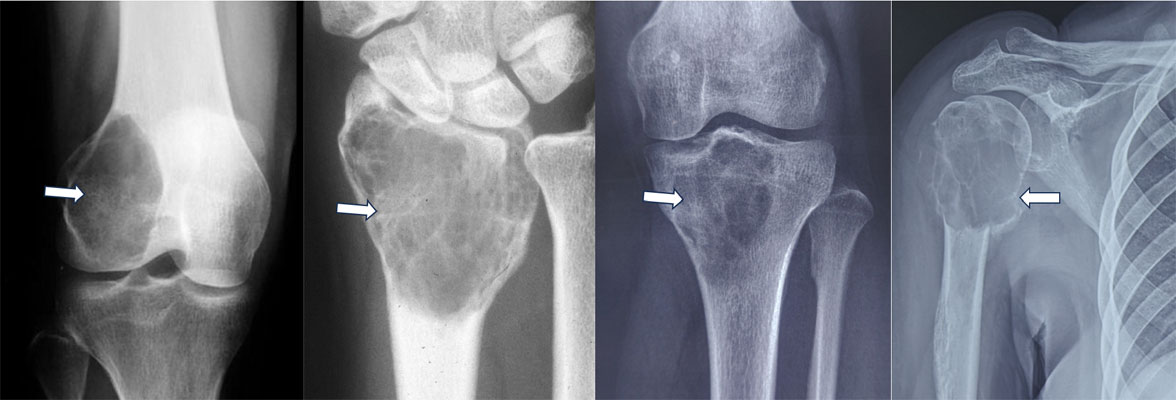
Figure 2. Giant cell tumor of bone can occur in different locations, commonly around the knee, wrist and shoulder
Clinical Pearl
The close relationship between Giant Cell Tumor and the joint surface makes early diagnosis particularly important. Treating the tumor before significant bone destruction occurs increases the likelihood of preserving the patient’s own joint.
Is Giant Cell Tumor Related to an Injury?
Many patients first notice pain after a fall, sporting activity, or minor injury and wonder whether the injury caused the tumor.
Current evidence shows that trauma does not cause Giant Cell Tumor of Bone.
Instead, an injury may simply draw attention to a tumor that was already present by prompting an X-ray or MRI.
What are the symptoms of Giant Cell Tumor of Bone?
The symptoms of Giant Cell Tumor of Bone (GCTB) usually develop gradually over weeks or months. Because the tumor often grows close to a joint, the symptoms may initially resemble more common conditions such as a ligament injury, tendon problem, arthritis, or overuse injury.
The most common symptom is persistent pain around a joint, particularly around the knee or wrist.
Persistent Joint Pain
Pain is the earliest and most frequent symptom.
It typically:
- Develops gradually
- Is localized to one area
- Worsens with activity
- Improves only partially with rest
- Becomes progressively more frequent over time
Unlike a minor injury, the pain usually does not resolve completely with pain medication or physiotherapy.
Swelling Around the Joint
As the tumor enlarges, patients may notice:
- Swelling near the affected joint
- A visible or palpable lump
- Tenderness over the bone
- Fullness around the joint
Swelling usually develops after the onset of pain.
Joint Stiffness and Reduced Movement
Because GCTB develops immediately beneath the joint surface, it may affect joint function.
Patients commonly notice:
- Difficulty bending or straightening the joint
- Pain during movement
- Reduced flexibility
- Difficulty squatting, climbing stairs, or lifting objects (depending on the site)
Pathological Fracture
As the tumor destroys bone, the affected bone becomes weaker.
Occasionally, the first presentation is a pathological fracture, meaning the bone breaks after minimal trauma or even during normal daily activities.
Although uncommon, this requires urgent specialist evaluation.
Pathological Fractures in Bone Tumors
Symptoms According to Tumor Location
Around the Knee
The most common presentation includes:
- Knee pain
- Swelling
- Limping
- Difficulty climbing stairs
- Pain while squatting
Around the Wrist
Patients may experience:
- Wrist pain
- Reduced grip strength
- Swelling
- Difficulty lifting objects
Sacrum
Sacral GCTs may present differently, causing:
- Persistent lower back pain
- Pain radiating to the buttock or leg
- Sitting discomfort
- Occasionally bowel or bladder symptoms if the tumor becomes very large
When Should You See an Orthopaedic Oncologist?
You should seek specialist evaluation if you have:
- Persistent pain lasting more than 4–6 weeks
- Swelling around a bone or joint
- An unexplained bone lesion on an X-ray
- A pathological fracture
- A suspected Giant Cell Tumor on MRI or biopsy
- Recurrence after previous treatment
Early referral helps ensure accurate diagnosis and appropriate treatment planning.
Internal Link: → What Is an Orthopaedic Oncologist?
Clinical Pearl
Giant Cell Tumor often presents with persistent joint pain rather than dramatic symptoms. Early imaging before significant bone destruction develops offers the best opportunity for joint-preserving surgery.
Doctor Insight
One of the most common reasons for delayed diagnosis is that patients receive repeated treatment for a “sports injury” or “arthritis” without obtaining appropriate imaging. While these conditions are far more common than bone tumors, symptoms that persist despite conservative treatment deserve reassessment.
Did You Know?
💡 Many Giant Cell Tumors are diagnosed only after an X-ray obtained for persistent joint pain. Early imaging can identify the tumor before it causes extensive bone damage.
Patient Tip
If your pain has persisted despite physiotherapy, pain medication, or activity modification, ask your doctor whether an X-ray is appropriate. A simple X-ray is often the first step in identifying a bone lesion.
Diagnosis of Giant Cell Tumor of Bone
Diagnosing a Giant Cell Tumor of Bone (GCTB) requires a combination of clinical evaluation, imaging, and biopsy. No single investigation can confirm the diagnosis on its own.
Clinical Evaluation
Important points include:
- Duration and progression of pain
- Swelling around the joint
- Limitation of movement
- Previous fracture or injury
- Any prior treatment for the lesion
During examination, your doctor assesses:
- Tenderness
- Joint movement
- Limb function
- Soft tissue swelling
- Signs of a pathological fracture
Although clinical evaluation raises suspicion, imaging and biopsy are required to establish the diagnosis.
Imaging Studies
X-ray – The First Investigation
A plain X-ray is usually the first test performed.
Typical features include:
- A well-defined osteolytic (bone-destroying) lesion
- Location at the end of the bone (epiphysis)
- Thinning of the surrounding bone (cortex)
- Expansion of the bone
- Usually no internal calcification
These findings strongly suggest GCTB but are not diagnostic.
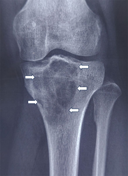
Figure 3. X-rays showing giant cell tumors of bone at different sites, with the characteristic expansile bone destruction highlighted.
Internal Link: → X-ray Evaluation of Bone Tumors
MRI – Planning Treatment
MRI is the most important investigation after the X-ray.
It helps determine:
- The exact size of the tumor
- Extension into surrounding soft tissues
- Condition of the joint surface
- Remaining subchondral bone
- Presence of pathological fracture
MRI is essential for surgical planning, particularly when joint-preserving surgery is being considered.
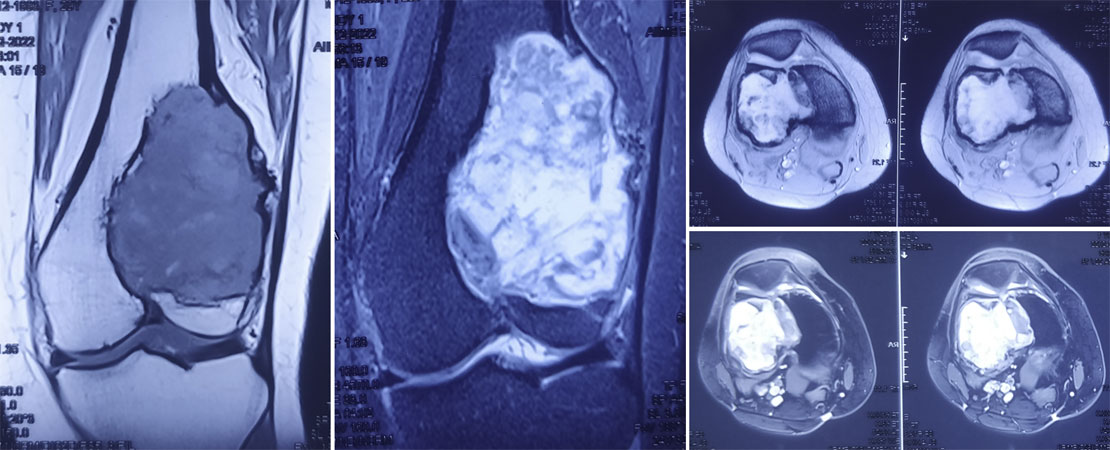
Figure 4. MRI provides a detailed view of the giant cell tumor, showing its extent within the bone and surrounding soft tissues
Internal Link: → MRI for Bone Tumors
CT Scan
CT is not required for every patient but may be helpful in:
- Assessing cortical bone destruction
- Evaluating complex anatomical sites (e.g., pelvis or sacrum)
- Planning reconstruction
- Assessing the lungs when pulmonary metastasis is suspected
- Determining whether the joint can be preserved.
- Plan the safest and most effective surgical treatment.
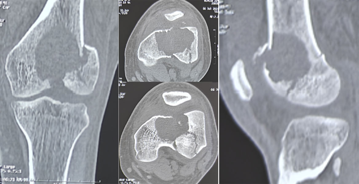
Figure 5. CT scan showing the extent of bone destruction caused by a giant cell tumor of bone, helping plan the appropriate surgical treatment
Why Is Biopsy Necessary?
Although imaging may strongly suggest a Giant Cell Tumor, a biopsy is required before definitive treatment.
A biopsy confirms:
- The diagnosis
- That the lesion is benign
- That another bone tumor is not being mistaken for GCTB
Most biopsies are performed using a core needle, often under imaging guidance.
The biopsy should be carefully planned because an improperly placed biopsy can complicate subsequent surgery.
Internal Link: → Bone Tumor Biopsy
Clinical Pearl
Every suspected Giant Cell Tumor should undergo biopsy before definitive surgery. Treatment should never be based on imaging findings alone.
Doctor Explains
Why Can’t the Tumor Be Removed Immediately?
Some bone tumors closely resemble Giant Cell Tumor on imaging but require very different treatment.
A biopsy ensures that the correct diagnosis is made before surgery, helping avoid inappropriate treatment and allowing the operation to be planned safely.
Campanacci Classification
After diagnosis, Giant Cell Tumors are commonly described using the Campanacci grading system, which estimates the extent of bone destruction seen on X-rays.
Although the grade does not determine the diagnosis, it helps surgeons plan treatment.
Grade I
- Well-defined lesion
- Cortex remains intact
- Minimal bone expansion
Usually easier to treat while preserving bone.
Grade II
- Thinned and expanded cortex
- No obvious cortical breach
- More extensive bone involvement
Often managed with extended curettage.
Grade III
- Cortical destruction
- Extension into surrounding soft tissues
- More aggressive local disease
May require more complex reconstruction or, in selected cases, wide resection.
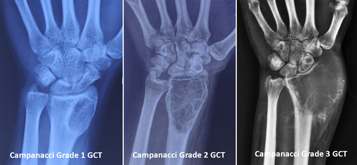
Figure 6. Campanacci grading of giant cell tumor of bone, ranging from Grade 1 with limited bone involvement to Grade 3 with cortical destruction and soft-tissue extension.
Conditions That Can Mimic Giant Cell Tumor
Several bone lesions may resemble GCTB on imaging.
Common differential diagnoses include:
- Aneurysmal Bone Cyst (ABC)
- Chondroblastoma
- Brown Tumor of Hyperparathyroidism
- Clear Cell Chondrosarcoma
- Telangiectatic Osteosarcoma
Because treatment differs significantly, biopsy is essential before surgery.
Doctor Insight
One of the most important decisions during evaluation is whether the native joint can be preserved. MRI, X-rays, and biopsy findings are reviewed together to determine the safest surgical approach while maximizing long-term joint function.
Patient Tip
Bring both your imaging studies and pathology report to your consultation—not just the written reports. Reviewing the actual images is an essential part of treatment planning.
Did You Know?
💡 Most Giant Cell Tumors are diagnosed before surgery using a combination of imaging and biopsy, allowing the operation to be carefully planned rather than performed urgently.
Does a Giant Cell Tumor Mean I Will Need Major Surgery?
Not necessarily.
Many Giant Cell Tumors are diagnosed before they cause extensive bone destruction. Early diagnosis often allows joint-preserving surgery, avoiding more extensive procedures.
The treatment plan depends on the size, location, and extent of the tumor—not simply on the diagnosis itself.
How We Diagnose Giant Cell Tumor of Bone: Why MRI and Biopsy Matter
Treatment of Giant Cell Tumor of Bone
Treatment of Giant Cell Tumor of Bone (GCTB) is individualized based on the size, location, extent of bone destruction, involvement of the joint, presence of a pathological fracture, and whether the tumor is being treated for the first time or has recurred.
The primary goals are to:
- Completely remove the tumor
- Minimize the risk of recurrence
- Preserve the native joint whenever possible
- Restore normal function
- Allow an early return to daily activities
Treatment decisions are best made by a multidisciplinary bone tumor team, including an orthopaedic oncologist, musculoskeletal radiologist, pathologist, and rehabilitation specialists.
Treatment Overview
Most patients undergo joint-preserving surgery, known as extended curettage.
Depending on the individual situation, treatment may also include:
- Bone cement (PMMA)
- Bone grafting
- Internal fixation
- Denosumab therapy (selected patients)
- Wide resection and reconstruction (selected complex cases)
The choice depends on the characteristics of the tumor rather than a single standard approach.
Extended Curettage
Extended curettage is the preferred treatment for most Giant Cell Tumors of Bone because it removes the tumor while preserving the patient’s own joint.
Unlike simple curettage, extended curettage combines several techniques to reduce the risk of recurrence.
These usually include:
- Creating a cortical window
- Thorough removal of the tumor
- High-speed burring of the cavity walls
- Use of local adjuvants in selected cases
- Reconstruction of the resulting bone defect
The exact technique is individualized according to the location and extent of the tumor.
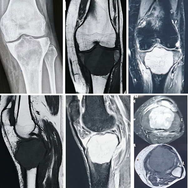
Figure 7A. Surgical exposure of a giant cell tumor with creation of a cortical window to access and remove the tumor.
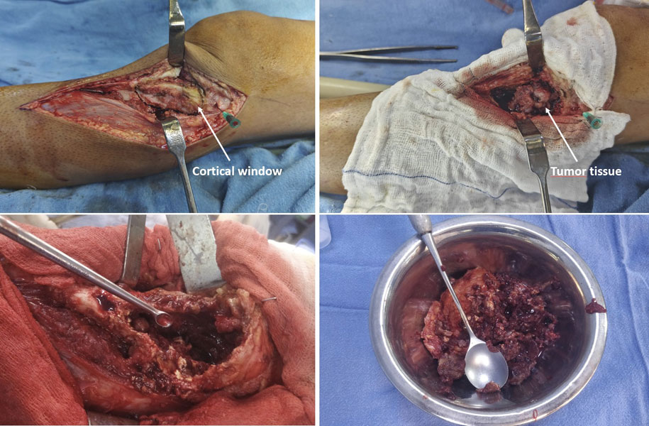
Figure 7B. Extended curettage of a giant cell tumor, carefully removing tumor tissue from the bone cavity while preserving as much healthy bone as possible
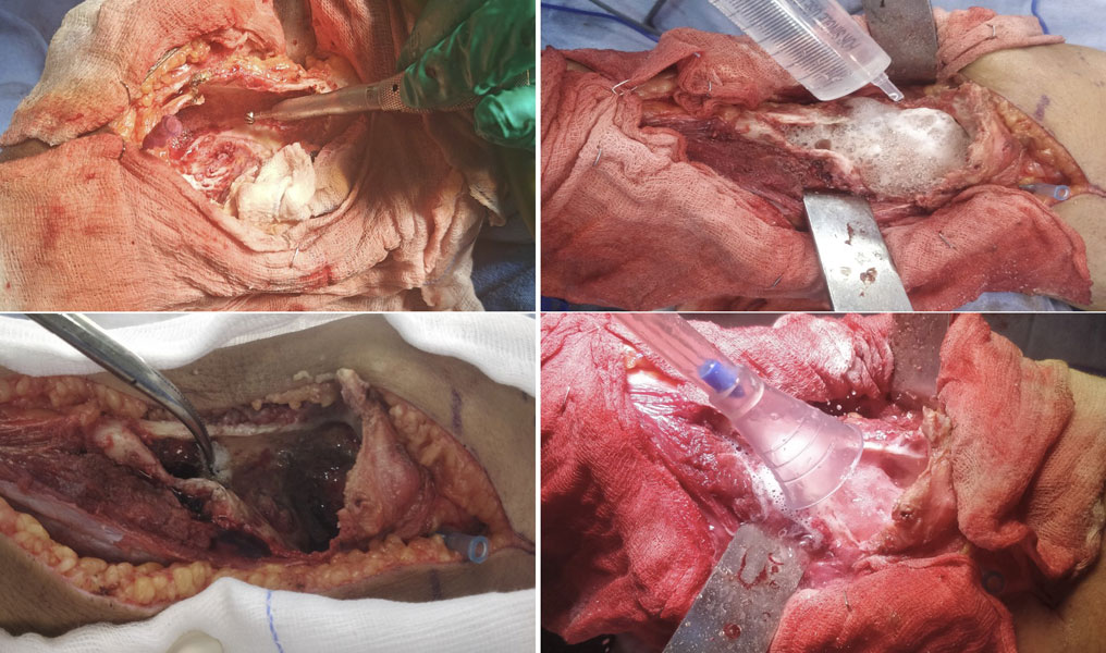
Figure 7C. Extended curettage using a high-speed burr, chemical adjuvants and thorough lavage to reduce the risk of residual tumor cells.
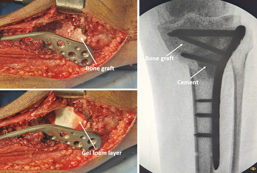
Figure 7D. Reconstruction of the bone defect after curettage using bone cement and bone graft, secured with appropriate fixation to restore strength.
Internal Link: → Extended Curettage for Bone Tumors
Clinical Pearl
Successful extended curettage depends on meticulous surgical technique. Thorough removal of microscopic tumor tissue is one of the most important factors influencing local recurrence.
Bone Cement or Bone Graft?
After the tumor has been removed, the remaining cavity must be reconstructed.
The two most common options are bone cement (PMMA) and bone graft.
Bone cement provides:
- Immediate structural support
- Earlier weight-bearing in appropriate patients
- Easier detection of recurrence on follow-up imaging
Bone graft may be preferred when:
- The defect is smaller
- Biological bone healing is desirable
- The patient is younger
- Joint preservation requires restoration of bone stock
In many cases, surgeons use a combination of both techniques.
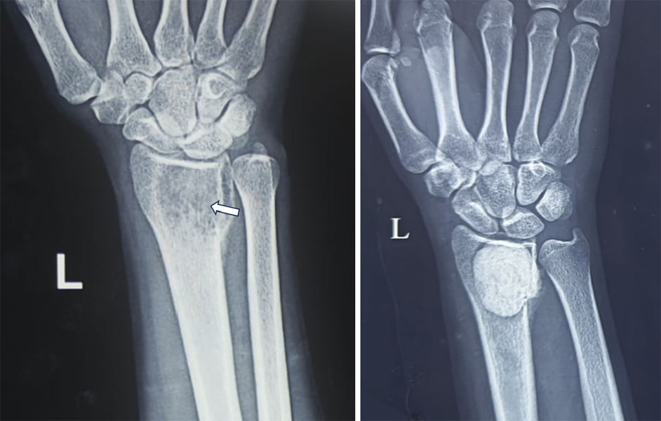
Figure 8. Extended curettage for a giant cell tumor of bone, followed by reconstruction of the resulting bone defect
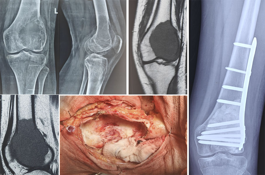
Figure 9. Giant cell tumor around the knee treated with extended curettage and bone cement reconstruction, preserving the native joint.
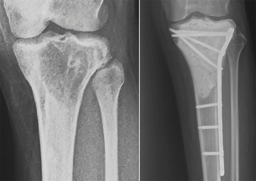
Figure 10. X-ray showing successful reconstruction after extended curettage and cementation of a giant cell tumor around the knee.
What Are Local Adjuvants?
After curettage, surgeons may use local adjuvants to destroy microscopic tumor cells that cannot be seen with the naked eye.
Depending on the clinical situation, these may include:
- High-speed burr
- Hydrogen peroxide
- Phenol
- Liquid nitrogen
- Argon beam coagulation
The choice of adjuvant varies between institutions and surgeons, and no single method is suitable for every patient.
When Is Denosumab Used?
Denosumab is a targeted medication that reduces the activity of giant cells within the tumor.
It is not routinely required for every patient but may be considered in selected situations, including:
- Large tumors where surgery would be difficult
- Tumors involving the pelvis or sacrum
- Recurrent disease
- Selected patients in whom surgery needs to be delayed or facilitated
Although Denosumab can reduce tumor activity, it does not replace surgery in most patients.
Doctor Explains
Can Medication Replace Surgery?
Usually no.
For most Giant Cell Tumors, surgery remains the definitive treatment. Medications such as Denosumab are used selectively and are often intended to support—not replace—surgical management.
When Is Wide Resection Required?
Although joint-preserving surgery is preferred, wide resection may occasionally be necessary when:
- The tumor has caused extensive bone destruction
- The joint cannot be preserved safely
- There is recurrent disease not suitable for curettage
- The tumor involves sites where curettage is unlikely to provide durable control
If wide resection is required, reconstruction may involve:
- Mega prosthesis
- Biological reconstruction
- Arthrodesis (joint fusion) in selected situations
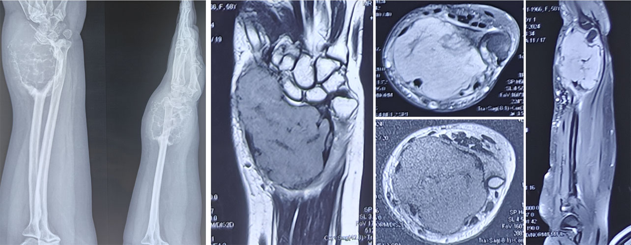
Figure 11A. A Grade 3 giant cell tumor of bone with extensive local bone destruction and soft-tissue involvement
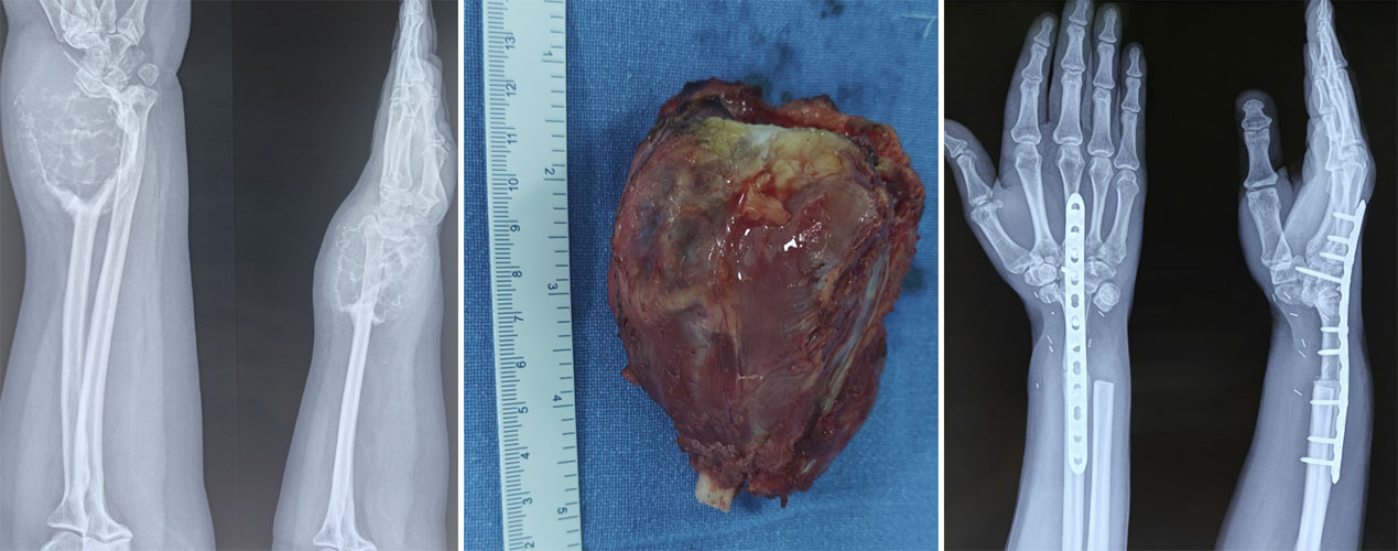
Figure 11B. Wide surgical resection of an extensive giant cell tumor of bone, removing the tumor along with the involved bone.
Common Mistakes to Avoid
Early specialist management can significantly influence the outcome.
Common mistakes include:
- Performing surgery without a biopsy
- Treating the lesion as a simple bone cyst
- Skipping preoperative MRI
- Delaying specialist referral
- Missing scheduled follow-up appointments after treatment
Doctor Insight
The objective is not simply to remove the tumor. Every treatment decision balances two equally important goals: minimizing the risk of recurrence while preserving long-term joint function. The best operation is the one that achieves both whenever safely possible.
Patient Tip
Ask your surgeon:
- Can my joint be preserved?
- Will bone cement or bone graft be used?
- What is my individual risk of recurrence?
- How often will I need follow-up imaging?
Understanding your treatment plan helps you participate in shared decision-making.
Will I Need My Joint Replaced?
In most patients, no.
Because Giant Cell Tumor is usually diagnosed before the joint surface is completely destroyed, many patients can be treated with joint-preserving surgery. More extensive reconstruction is reserved for selected situations where the tumor has caused severe bone destruction or cannot be safely removed by curettage alone.
Recovery, Rehabilitation & Long-Term Follow-up
Recovery after treatment for Giant Cell Tumor of Bone (GCTB) depends on the location of the tumor, the type of surgery performed, and the method of reconstruction. Most patients gradually return to their normal daily activities with appropriate rehabilitation and regular follow-up.
Recovery is a gradual process, and your healthcare team will guide you through each stage.
Rehabilitation After Surgery
Rehabilitation usually begins soon after surgery under the supervision of your treating team and physiotherapist.
The goals are to:
- Reduce pain and swelling
- Restore joint movement
- Rebuild muscle strength
- Improve balance and walking
- Return safely to work, school, and recreational activities
The rehabilitation programme is individualized based on the surgery performed and the affected bone.
Internal Link: → Rehabilitation After Bone Tumor Surgery
Patient Tip
Recovery is not a race. Following your physiotherapy programme consistently is more important than returning to activities too quickly.
Can Giant Cell Tumor Come Back?
Yes. Giant Cell Tumor of Bone has a recognised risk of local recurrence, meaning the tumor may return at the same site after treatment.
The risk varies depending on factors such as:
- Size and extent of the tumor
- Surgical technique
- Type of treatment performed
- Previous recurrence
Importantly, most recurrences can be successfully treated, especially when detected early through regular follow-up.
Clinical Pearl
The first few years after surgery are the most important for recurrence surveillance. Keeping follow-up appointments is one of the simplest ways patients can contribute to successful long-term outcomes.
Possible Complications
Most patients recover without major problems. However, as with any bone tumor surgery, complications can occur.
These may include:
- Local recurrence
- Infection
- Delayed wound healing
- Joint stiffness
- Fracture around the treated bone
- Degenerative arthritis in the long term (selected patients)
Your surgical team will discuss the risks relevant to your individual situation before treatment.
Prognosis
The overall outlook for Giant Cell Tumor of Bone is excellent.
Most patients:
- Preserve their limb
- Maintain good joint function
- Return to work or education
- Resume normal daily activities
Long-term outcome depends on:
- Early diagnosis
- Appropriate surgery
- Regular follow-up
- Prompt treatment if recurrence occurs
Unlike malignant bone tumors, survival is generally excellent, and treatment is primarily directed at preserving function and preventing local recurrence.
Can I Live a Normal Life After Treatment?
For most patients, yes.
Following recovery and rehabilitation, many people return to work, education, travel, and recreational activities. While periodic follow-up remains important, most patients are able to enjoy an active and independent lifestyle.
Follow-up After Treatment
Regular follow-up helps detect recurrence before symptoms develop and allows your surgeon to monitor bone healing and joint function.
Although schedules vary, follow-up commonly includes:
Typical Follow-up Schedule
At each visit, your doctor may recommend:
- Clinical examination
- X-rays of the operated bone
- Additional imaging if recurrence is suspected
- Chest imaging in selected patients
Follow-up plans are individualized according to the original tumor, treatment performed, and clinical progress.
Doctor Insight
Successful treatment does not end with surgery. Long-term follow-up allows early detection of recurrence while monitoring joint function and helping patients maintain the best possible quality of life.
Did You Know?
💡 Most recurrences occur within the first 2–3 years after treatment, which is why follow-up appointments are scheduled more frequently during this period.
Frequently Asked Questions (FAQs)
1. Is Giant Cell Tumor of Bone cancer?
No. Giant Cell Tumor of Bone (GCTB) is a benign (non-cancerous) but locally aggressive bone tumor. Although it is not considered a cancer, it can damage bone, weaken joints, and occasionally recur after treatment.
2. Why does a benign tumor need surgery?
Even though GCTB is benign, it can continue to grow and destroy surrounding bone. Early treatment helps preserve the joint, prevents fractures, and reduces the complexity of surgery.
3. Can Giant Cell Tumor spread to other parts of the body?
In most patients, it remains confined to the bone. Rarely, GCTB can spread to the lungs, even though it is considered benign. Your treating team will decide whether chest imaging is required.
4. What is the most common treatment?
The standard treatment for most patients is extended curettage, often combined with bone cement or bone grafting to reconstruct the bone.
5. Will I lose my joint?
Not usually. Modern treatment aims to preserve the native joint whenever it is safe to do so. More extensive surgery is required only in selected cases.
6. What is bone cement, and why is it used?
Bone cement (PMMA) fills the cavity after the tumor is removed, provides immediate stability, and helps detect recurrence on follow-up imaging.
7. Will I need a bone graft instead of bone cement?
Some patients benefit from bone grafting, while others benefit from bone cement. The choice depends on the size and location of the defect and your surgeon’s assessment.
8. Can the tumor come back after surgery?
Yes. GCTB has a recognised risk of local recurrence. Regular follow-up allows recurrence to be detected and treated at an early stage.
9. How long will recovery take?
Recovery varies depending on the location of the tumor and the type of surgery performed. Most patients gradually return to normal daily activities with rehabilitation.
10. Can I return to sports?
Many patients return to recreational sports after recovery. High-impact activities may need to be modified depending on the surgery performed.
11. What is Denosumab?
Denosumab is a targeted medication that may be used in selected patients to reduce tumor activity. It is not a substitute for surgery in most cases.
12. Is a biopsy always necessary?
Yes. A biopsy confirms the diagnosis and ensures that the correct treatment is planned before surgery.
13. Can Giant Cell Tumor occur in children?
It is uncommon because GCTB usually develops after skeletal maturity, when the growth plates have closed.
14. Is Giant Cell Tumor hereditary?
No. Most cases are sporadic and do not run in families.
15. Can pregnancy cause Giant Cell Tumor?
No. Pregnancy does not cause GCTB, although hormonal changes may occasionally influence symptoms in patients who already have the condition.
16. Will I need long-term follow-up?
Yes. Follow-up is important to monitor healing and detect recurrence, particularly during the first few years after treatment.
17. Can Giant Cell Tumor become malignant?
This is uncommon. Most GCTBs remain benign throughout their course, although rare malignant transformation has been reported.
18. Should I seek a second opinion?
Seeking a second opinion is reasonable, particularly before major surgery or if you have questions about your diagnosis or treatment options.
19. Can I live a normal life after treatment?
Yes. Most patients return to work, education, travel, and many recreational activities after completing treatment and rehabilitation.
20. What is the most important thing I can do after surgery?
Attend your follow-up appointments, complete your rehabilitation programme, and report any new or persistent symptoms promptly.
Myths and Facts
Key Facts
References
The information on this page is based on current evidence and internationally accepted recommendations, including:
- World Health Organization (WHO). WHO Classification of Soft Tissue and Bone Tumours, 5th Edition.
- National Comprehensive Cancer Network (NCCN). Bone Cancer Guidelines (latest available version).
- European Society for Medical Oncology (ESMO). Bone Sarcoma Clinical Practice Guidelines.
- Musculoskeletal Tumor Society (MSTS). Educational resources and consensus recommendations.
- International Society of Limb Salvage (ISOLS). Clinical guidance and educational resources.
- American Academy of Orthopaedic Surgeons (AAOS). Relevant educational material.
- Chawla S, Blay JY, Rutkowski P, et al. Denosumab in Patients with Giant-Cell Tumour of Bone: A Multicentre, Open-Label, Phase 2 Study. The Lancet Oncology.
- Errani C, Ruggieri P, Asenzio MAN, et al. Giant Cell Tumor of the Extremity: A Review of 349 Cases. Journal of Bone and Joint Surgery.
- van der Heijden L, Dijkstra PDS, van de Sande MAJ, et al. The Clinical Approach Toward Giant Cell Tumor of Bone. The Oncologist.
Medical Disclaimer
The information provided on this page is intended for educational purposes only and should not replace consultation with a qualified healthcare professional. Every patient with Giant Cell Tumor of Bone is unique, and treatment decisions should be individualized based on the location of the tumor, imaging findings, pathology, overall health, and personal circumstances.
If you have persistent bone pain, swelling, or have been diagnosed with a Giant Cell Tumor, consult an orthopaedic oncologist or a multidisciplinary bone tumor team for a comprehensive evaluation and personalized treatment plan.
 +91 93558 33005