Quick Summary
Pathological fractures differ from ordinary fractures because they are a consequence of an underlying disease, not simply an injury. Early diagnosis is essential, as the fracture may be the first sign of a previously undiagnosed bone tumor or cancer. Successful treatment requires a multidisciplinary approach that addresses both the fracture and its underlying cause.
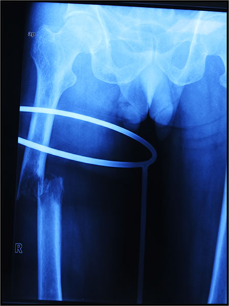
Figure 1. Pathological fracture of the thigh bone caused by cancer spread from the lung.
Understanding Pathological Fractures
Hearing the words “pathological fracture” can be frightening, particularly if the diagnosis comes unexpectedly after what seemed like a minor fall or even during routine daily activities. Many patients immediately wonder whether the fracture means they have cancer, whether they will need surgery, or whether they will be able to walk again.
Although these concerns are understandable, it is important to remember that not all pathological fractures are caused by cancer, and many can be treated successfully with modern medical and surgical techniques.
Understanding why pathological fractures occur is the first step toward understanding how they are diagnosed and treated.
What Is a Pathological Fracture?
A fracture is a break in a bone. Most fractures occur when a healthy bone is exposed to a force greater than it can withstand, such as during a road traffic accident, sports injury, or fall from a height.
A pathological fracture, in contrast, occurs because the bone itself has become abnormally weak due to an underlying disease. As a result, the bone may break during normal daily activities such as walking, climbing stairs, getting out of bed, or lifting a light object. Sometimes the fracture occurs with no obvious injury at all.
The underlying disease weakens the internal structure of the bone, making it unable to tolerate the stresses that a healthy bone would normally withstand.
Doctor Explains
The fracture is often a symptom rather than the disease itself. Simply treating the broken bone without identifying why it fractured may delay the diagnosis of an important underlying condition. For this reason, every suspected pathological fracture requires careful evaluation before treatment is planned.
How Is a Pathological Fracture Different from a Normal Fracture?
Although both involve a broken bone, their causes, diagnosis, and treatment are often very different.
Normal Fracture vs Pathological Fracture
The distinction is important because treating a pathological fracture like an ordinary fracture may result in delayed diagnosis, inappropriate surgery, or avoidable complications.
Clinical Pearl
Every fracture that occurs after minimal trauma should prompt doctors to ask an important question: “Why did this bone break?” Finding the answer is often just as important as treating the fracture itself.
Why Do Pathological Fractures Occur?
Bone is a living tissue that is constantly being renewed through a process called bone remodeling. During this process, old bone is broken down by specialized cells called osteoclasts, while new bone is formed by osteoblasts. In healthy individuals, these two processes remain in balance, allowing bones to stay strong throughout life.
Certain diseases disrupt this balance by either destroying bone faster than it can be replaced or by preventing normal bone formation. Over time, the bone becomes thinner, weaker, and more likely to fracture.
Different diseases weaken bone in different ways:
- Cancer may destroy bone directly or replace healthy bone with tumor tissue.
- Osteoporosis reduces bone density, making bones fragile.
- Bone infections can damage the normal architecture of bone.
- Some benign bone tumors and cysts create areas of weakness within otherwise healthy bone.
- Metabolic bone disorders interfere with normal bone strength and mineralization.
Regardless of the cause, the end result is the same: the bone loses its ability to withstand normal mechanical forces.
Did You Know?
Your skeleton is continuously being rebuilt throughout life. In healthy adults, this process remains balanced. Many diseases that cause pathological fractures interfere with this natural remodeling process long before a fracture actually occurs.
Are All Pathological Fractures Caused by Cancer?
No. This is one of the most common misconceptions.
While cancer is an important cause of pathological fractures—particularly in adults over 40 years of age—it is not the only cause. Many non-cancerous conditions can also weaken bone sufficiently to cause a fracture.
Broadly, the causes can be divided into malignant (cancer-related) and non-malignant (non-cancerous) conditions.
Cancer-Related Causes
Cancer weakens bone in several ways. It may begin within the bone itself (primary bone cancer) or spread to the bone from another organ (bone metastasis).
Common cancer-related causes include:
- Bone metastases from breast, prostate, lung, kidney, and thyroid cancers
- Multiple myeloma, a cancer of plasma cells within the bone marrow
- Primary bone sarcomas, such as osteosarcoma and Ewing sarcoma (less common)
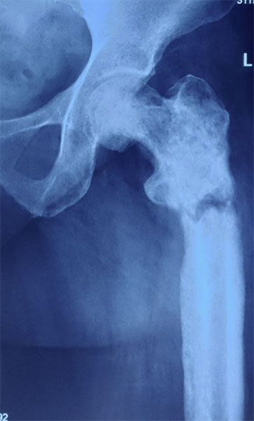
Figure 2. Pathological fracture of the thigh bone caused by cancer spread from the prostate.
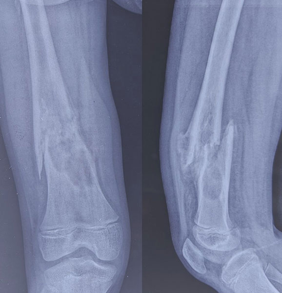
Figure 3. Pathological fractures can occur when a primary bone tumor weakens the bone.
Non-Cancer Causes
Several non-cancerous conditions can also lead to pathological fractures, including:
- Osteoporosis, the most common cause of fragile bones in older adults
- Benign bone tumors, such as giant cell tumor or enchondroma
- Bone cysts, which create hollow areas within bone
- Fibrous dysplasia, where normal bone is replaced by abnormal fibrous tissue
- Paget’s disease of bone, a disorder of abnormal bone remodeling
- Chronic bone infection (osteomyelitis)
Reassurance Box
A pathological fracture does not automatically mean you have cancer. Many patients are found to have non-cancerous conditions that weaken the bone. The purpose of further investigations is to identify the exact cause so that the most appropriate treatment can be planned.
Which Bones Are Most Commonly Affected?
Pathological fractures can occur in almost any bone, but they are most common in areas that bear body weight or are frequently subjected to mechanical stress.
The most commonly affected bones include:
- Femur (thigh bone): Particularly around the hip and upper thigh.
- Humerus (upper arm bone): Often affected by metastatic disease and multiple myeloma.
- Spine (vertebrae): Fractures may lead to severe pain or, in some cases, compression of the spinal cord.
- Pelvis: May become weakened by metastatic cancer or multiple myeloma.
- Tibia (shin bone): Less commonly affected but important because of its weight-bearing role.
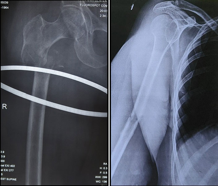
Figure 4. The thigh bone and upper arm bone are common sites for pathological fractures.
Clinical Pearl
Pain that gradually worsens over several weeks before a fracture occurs is an important warning sign of an underlying bone disease. Unlike ordinary fractures, many pathological fractures are preceded by persistent pain caused by progressive weakening of the bone.
Patient Tip
If you develop persistent bone pain, especially pain that occurs at night, worsens over time, or is associated with a fracture after a minor injury, seek medical evaluation promptly. Early diagnosis may prevent further complications and, in some cases, allow treatment before a fracture occurs.
What Are the Symptoms of a Pathological Fracture?
The symptoms of a pathological fracture depend on the underlying disease, the bone involved, and whether the bone has already fractured or is only beginning to weaken.
Unlike ordinary fractures, many pathological fractures are preceded by symptoms for weeks or even months. Recognizing these warning signs early may allow treatment before the bone breaks, reducing pain and avoiding more complex surgery.
Persistent Bone Pain
The earliest and most common symptom is persistent bone pain that gradually worsens over time.
Patients often describe the pain as:
- A deep, dull ache
- Pain that increases over weeks or months
- Pain that occurs even without activity
- Pain that becomes more noticeable at night
- Pain that is not relieved by routine pain medications
This occurs because the underlying disease gradually weakens the bone and stretches the surrounding tissues.
Unlike pain from a muscle strain or minor injury, pathological bone pain usually does not improve with rest and tends to become progressively worse.
Pain During Weight-Bearing or Movement
As the bone becomes weaker, patients may notice pain only while standing, walking, climbing stairs, or lifting objects.
This is called mechanical pain, meaning the pain occurs because the weakened bone can no longer support normal body weight or movement.
Mechanical pain often suggests that the bone has become structurally unstable and may be at risk of fracturing.
Clinical Pearl
Pain that occurs only during movement initially, but later progresses to pain at rest, often indicates that the underlying bone disease is advancing and requires prompt evaluation.
Swelling or a Visible Lump
Some patients develop swelling over the affected bone. Occasionally, a firm lump may be noticed if the underlying disease has expanded through the bone into the surrounding soft tissues.
Swelling is more commonly seen in:
- Primary bone tumors
- Benign bone tumors
- Bone infections
It is less common in many metastatic bone lesions, especially those located deep within the skeleton.
Fracture After Minimal or No Injury
One of the hallmark features of a pathological fracture is that it occurs after little or no trauma.
Examples include:
- Breaking a bone while walking
- Fracturing a bone while getting out of bed
- A fracture after lifting a light object
- A fracture following a simple twist or stumble
In contrast, healthy bones usually require a much greater force to break.
If a fracture occurs after an unusually minor injury, doctors will consider whether the bone may have been weakened by an underlying condition.
Reduced Limb Function
Patients may notice increasing difficulty using the affected limb before the fracture occurs.
Symptoms may include:
- Difficulty walking
- Limping
- Reduced ability to lift the arm
- Difficulty climbing stairs
- Reduced grip strength if the upper limb is affected
These symptoms usually reflect progressive weakening of the bone rather than injury to the muscles.
Symptoms of Spinal Pathological Fractures
When pathological fractures involve the spine, symptoms can be different because the fractured vertebra may compress nearby nerves or the spinal cord.
Symptoms may include:
- Persistent back pain
- Pain that worsens when standing
- Pain that wakes the patient from sleep
- Weakness in the arms or legs
- Numbness or tingling
- Difficulty walking
- Loss of bladder or bowel control
These symptoms require urgent medical assessment because they may indicate spinal cord compression, a condition in which pressure on the spinal cord can lead to permanent nerve damage if treatment is delayed.
Red Flag Symptoms: When Should You Seek Immediate Medical Attention?
Although many patients experience gradually worsening symptoms, certain signs require urgent medical evaluation.
Seek immediate medical attention if you experience:
- A fracture after minimal or no injury
- Sudden inability to stand or bear weight
- Severe or rapidly worsening bone pain
- New weakness or numbness in the arms or legs
- Loss of bladder or bowel control
- Sudden swelling or deformity of a limb
- Severe back pain associated with difficulty walking
Prompt assessment may prevent serious complications such as permanent nerve injury or further bone damage.
Doctor Insight
Patients often assume that persistent bone pain is caused by arthritis, muscle strain, or aging. While these are common causes of pain, bone pain that progressively worsens, occurs at night, or is followed by a fracture after minimal trauma should never be ignored. Early diagnosis can prevent complications and, in many cases, allow treatment before a fracture occurs.
Reassurance Box
Not every patient with persistent bone pain has a pathological fracture or cancer. Many common conditions, including arthritis, tendon problems, and osteoporosis, can also cause bone or joint pain. Your doctor will use your symptoms, examination, and imaging studies to determine the exact cause.
How Is a Pathological Fracture Diagnosed?
Diagnosing a pathological fracture involves more than confirming that a bone is broken. The most important step is identifying why the fracture occurred, as treatment depends on the underlying disease rather than the fracture alone.
For this reason, evaluation is usually carried out by a multidisciplinary team, which may include an orthopaedic oncologist, radiologist, pathologist, medical oncologist, and radiation oncologist.
Diagnosis generally follows a systematic pathway.
Diagnostic Pathway for a Suspected Pathological Fracture
Symptoms → Medical History → Physical Examination → Imaging → Blood Tests → Biopsy (when indicated) → Multidisciplinary Team Discussion → Treatment Planning
Medical History
Your doctor will begin by asking questions about your symptoms and overall health.
Important information includes:
- When the pain started
- Whether the pain is getting worse
- Any previous history of cancer
- Recent weight loss or fatigue
- Previous fractures
- Family history of cancer or bone disease
- Current medications, including treatment for osteoporosis
These details help narrow the possible causes and guide further investigations.
Physical Examination
During the examination, your doctor will assess:
- Tenderness over the affected bone
- Swelling or deformity
- Limb alignment
- Range of movement
- Muscle strength
- Nerve function
- Blood circulation to the affected limb
If a spinal lesion is suspected, a detailed neurological examination is essential to assess spinal cord function.
Imaging Studies
Imaging helps determine the location, extent, and possible cause of the bone abnormality.
Plain X-rays
X-rays are usually the first investigation. They confirm the fracture and may reveal features suggesting an underlying bone lesion, such as bone destruction, abnormal bone formation, or thinning of the cortex (the hard outer layer of bone).
Magnetic Resonance Imaging (MRI)
Magnetic Resonance Imaging (MRI) uses powerful magnets rather than radiation to produce highly detailed images of bones, muscles, nerves, and bone marrow.
MRI is particularly useful for:
- Assessing the full extent of a bone tumor
- Evaluating surrounding muscles and soft tissues
- Detecting additional disease within the same bone
- Planning surgery
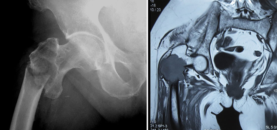
Figure 5. X-ray and MRI showing a fracture through an underlying bone lesion.
Computed Tomography (CT)
Computed Tomography (CT) uses X-rays to produce detailed cross-sectional images of the body.
CT is especially valuable for evaluating:
- Cortical bone destruction
- Complex fractures
- Pelvic lesions
- Surgical planning
PET-CT and Bone Scan
If cancer is suspected, additional imaging such as Positron Emission Tomography-Computed Tomography (PET-CT) or a bone scan may be recommended to determine whether disease is present elsewhere in the body.
Did You Know?
A fracture may sometimes be the first sign of an undiagnosed cancer. Careful imaging helps doctors determine whether the fracture is an isolated problem or part of a more widespread disease process.
Blood Tests
Blood tests cannot usually diagnose the exact cause of a pathological fracture on their own, but they provide valuable information about your general health and may offer important clues about the underlying disease.
Depending on your symptoms and medical history, your doctor may recommend tests to look for:
- Anaemia (a lower-than-normal number of red blood cells), which may occur in chronic disease or certain cancers.
- Signs of infection.
- Kidney and liver function, particularly if cancer is suspected or medications are being planned.
- Calcium and phosphate levels, as abnormalities may indicate metabolic bone disease or bone destruction.
- Markers of inflammation, which may suggest infection or inflammatory conditions.
- Tumor-specific blood tests in selected situations, such as prostate-specific antigen (PSA) in suspected prostate cancer.
- Specialized tests for multiple myeloma, including serum protein electrophoresis and free light chain assays.
Blood tests are interpreted alongside imaging and clinical findings rather than in isolation.
Doctor Explains
Blood tests are supportive investigations, not definitive diagnostic tests. A normal blood test does not rule out a bone tumor or metastatic disease, just as an abnormal result does not necessarily confirm cancer. Your doctor considers all available information before making a diagnosis.
Why Is a Biopsy Sometimes Necessary?
A biopsy is a procedure in which a small sample of tissue is removed from the abnormal area and examined under a microscope by a pathologist, a doctor who specializes in diagnosing diseases from tissue samples.
For many pathological fractures, particularly when the underlying cause is unknown, a biopsy provides the definitive diagnosis.
It helps answer critical questions such as:
- Is the lesion benign (non-cancerous) or malignant (cancerous)?
- If cancer is present, what type is it?
- Has cancer spread from another organ or started within the bone?
- Which treatment is most appropriate?
Without a tissue diagnosis, treatment may be inaccurate or incomplete.
Is a Biopsy Required for Everyone?
No.
The decision depends on the clinical situation.
A biopsy may not be necessary if:
- The patient has a known history of advanced metastatic cancer and the imaging findings are typical.
- The diagnosis is already well established through previous tissue samples.
However, a biopsy is usually recommended when:
- The patient has no known history of cancer.
- Imaging findings are uncertain.
- A primary bone tumor is suspected.
- The diagnosis will influence treatment decisions.
Your treating team carefully weighs the benefits and risks before recommending a biopsy.
Bone Biopsy
Doctor Insight
One of the most important principles in orthopaedic oncology is:
“Biopsy before fixation.”
Placing plates, screws, or rods into a bone before establishing the diagnosis may contaminate surrounding tissues, complicate future surgery, and in some cases reduce the chances of successful limb-salvage treatment.
Why Should Pathological Fractures Be Managed by an Orthopaedic Oncologist?
A pathological fracture is fundamentally different from a routine fracture because the underlying disease determines the treatment strategy.
An orthopaedic oncologist is an orthopaedic surgeon with specialized training in the diagnosis and treatment of bone and soft tissue tumors, including cancers that spread to the skeleton.
Managing these fractures requires expertise not only in fracture fixation but also in cancer surgery, biopsy planning, limb reconstruction, and multidisciplinary cancer care.
Why Specialist Care Matters
In many routine fractures, the primary goal is to stabilize the broken bone and allow it to heal.
In pathological fractures, treatment must also address questions such as:
- What disease caused the fracture?
- Will the bone heal normally?
- Does the diseased bone need to be removed?
- Is limb-salvage surgery possible?
- Is chemotherapy or radiotherapy required?
- What reconstruction will provide the best long-term function?
These decisions often require close collaboration between multiple specialists.
Routine Fracture Care vs Pathological Fracture Care
Doctor Insight
Treating every pathological fracture the same way can lead to poor outcomes. For example, a fracture caused by osteoporosis is managed very differently from one caused by metastatic cancer or a primary bone sarcoma. Establishing the correct diagnosis before definitive treatment is therefore one of the most important steps in patient care.
Treatment of Pathological Fractures
Treatment is individualized because no single approach is appropriate for every patient.
Your healthcare team considers several factors before recommending treatment, including:
- The underlying diagnosis
- Whether the condition is benign or malignant
- The location of the fracture
- The amount of bone destruction
- Your age and general health
- Your level of activity and functional goals
- Whether the disease has spread elsewhere in the body
The primary goals of treatment are to:
- Relieve pain
- Restore stability to the bone
- Allow early movement and independence
- Treat the underlying disease
- Improve quality of life
Non-Surgical Treatment
Not every pathological fracture requires surgery.
Depending on the underlying condition, treatment may include:
Pain Management
Pain is managed using a stepwise approach that may include:
- Paracetamol
- Anti-inflammatory medications (when appropriate)
- Opioid medications for severe pain
- Regional nerve blocks in selected patients
Pain control is individualized and regularly reviewed throughout treatment.
Bone-Strengthening Medications
Certain medications help reduce further bone destruction and lower the risk of future skeletal complications.
Examples include:
- Bisphosphonates, which slow bone breakdown by reducing osteoclast activity.
- Denosumab, a monoclonal antibody that blocks signals responsible for excessive bone resorption.
These medications are commonly used in metastatic bone disease, multiple myeloma, giant cell tumor of bone, and osteoporosis, depending on the clinical situation.
Radiotherapy
Radiotherapy uses carefully targeted high-energy radiation to destroy cancer cells.
It may be recommended to:
- Relieve pain
- Control local tumor growth
- Reduce the risk of progression
- Treat residual disease after surgery
Not every patient requires radiotherapy, and the decision depends on the diagnosis and treatment plan.
Surgical Treatment
Surgery is considered when the fracture causes significant pain, instability, loss of function, or when the bone is unlikely to heal without operative treatment.
The type of surgery depends on the underlying disease rather than the fracture alone.
Possible surgical approaches include:
- Internal fixation using plates, screws, or intramedullary nails
- Bone cement augmentation to improve stability in selected cases
- Curettage (surgical removal of tumor tissue) for some benign lesions
- Wide tumor resection for selected malignant tumors
- Endoprosthetic (tumor prosthesis) reconstruction when large segments of bone must be removed
Each option has specific indications, advantages, and limitations.
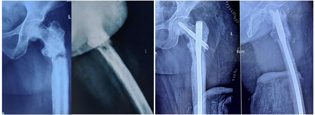
Figure 6. Stabilizing a pathological fracture helps restore strength and allows early movement.
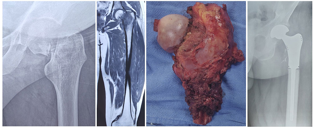
Figure 7. Tumor removal followed by reconstruction with a tumor prosthesis for early mobilization.
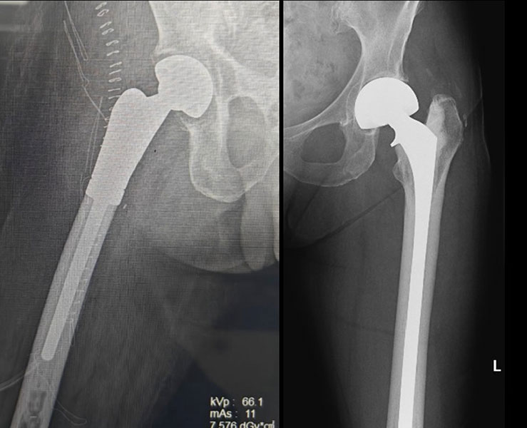
Figure 8. Tumor prosthesis reconstruction can help patients bear weight early and regain function.
Quick Summary
The best treatment for a pathological fracture is the one that addresses both the broken bone and the disease responsible for weakening it. Accurate diagnosis, multidisciplinary planning, and individualized care are essential for achieving the best possible outcome.
Can a Pathological Fracture Be Prevented?
One of the most important advances in orthopaedic oncology is that many pathological fractures can be prevented if the weakened bone is identified before it breaks. In fact, preventing a fracture is often easier, safer, and associated with better outcomes than treating a fracture after it has occurred.
Doctors refer to a bone that is at high risk of breaking but has not yet fractured as having an impending pathological fracture.
Recognizing an impending fracture provides a valuable opportunity to stabilize the bone before a painful and potentially disabling fracture occurs.
What Is an Impending Pathological Fracture?
An impending pathological fracture is a bone that has become significantly weakened by an underlying disease and is considered likely to fracture during normal daily activities or after only minor trauma.
Unlike an established pathological fracture, the bone is still intact, but its strength has been reduced to the point where it may no longer safely support normal mechanical loads.
The underlying disease may be:
- Bone metastasis (cancer that has spread to the bone)
- A primary bone tumor
- Multiple myeloma
- A benign but locally aggressive bone tumor
- Other conditions that substantially weaken bone
An impending fracture should be regarded as a warning sign rather than an emergency, providing an opportunity for planned treatment before a fracture develops.
Doctor Explains
Think of a weakened bone like a cracked bridge. It may still support traffic today, but without timely repair, it is much more likely to collapse. Similarly, a bone weakened by disease may appear intact on an X-ray but can fracture during everyday activities if left untreated.
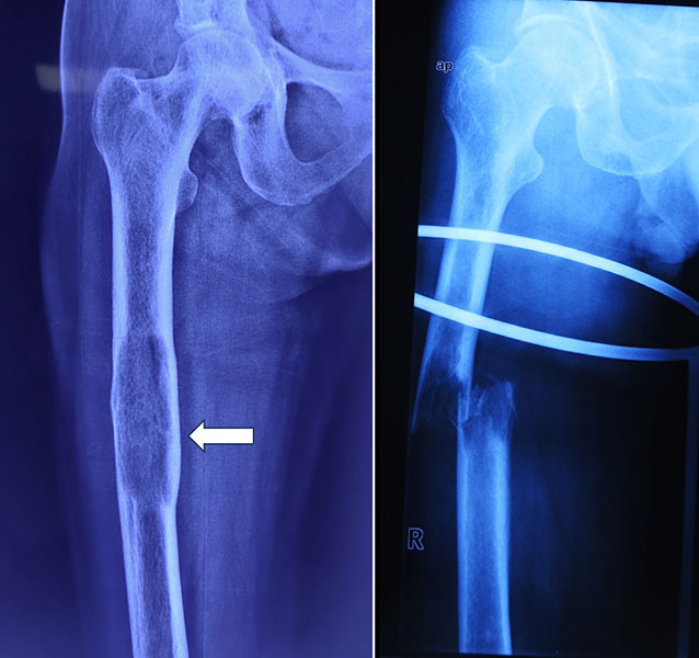
Figure 9. An impending fracture can become a complete pathological fracture if not treated in time.
Why Is Early Treatment Important?
Once a pathological fracture occurs, patients often experience sudden severe pain, loss of mobility, and the need for urgent medical care. Surgical treatment may also become more complex because the bone has already broken.
Treating the bone before it fractures can often:
- Prevent severe pain associated with a fracture
- Reduce the likelihood of emergency surgery
- Preserve mobility and independence
- Allow planned surgery under optimal conditions
- Simplify reconstruction in selected patients
- Shorten rehabilitation and hospital stay
- Improve overall quality of life
For many patients, the goal is not only to treat the disease but also to maintain function and prevent avoidable complications.
Clinical Pearl
Whenever feasible, preventing a pathological fracture generally provides better functional outcomes than treating the fracture after it occurs.
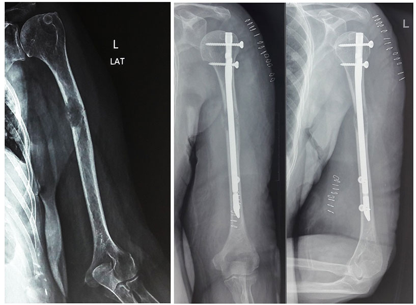
Figure 10. Preventive fixation of a weakened bone can stop an impending pathological fracture.
How Do Doctors Assess the Risk of Fracture?
Not every weakened bone will fracture, and not every patient requires preventive surgery.
Doctors assess fracture risk by combining information from your symptoms, physical examination, and imaging studies. Important factors include:
- Persistent pain, especially pain during weight-bearing
- The size of the bone lesion
- The amount of bone destruction
- The location of the lesion
- Whether the affected bone bears body weight
- The type of underlying disease
- Your overall health, mobility, and expected activity level
In some situations, doctors also use validated fracture risk scoring systems, such as the Mirel’s score, to help estimate the likelihood of fracture in long bones affected by metastatic disease. The Mirel’s score considers four factors: the location of the lesion, the severity of pain, the type of bone destruction (lytic, blastic, or mixed), and the size of the lesion relative to the bone diameter. While the score is a useful guide, it is not used in isolation. Treatment decisions also depend on imaging findings, the underlying disease, your overall health, and the opinion of the multidisciplinary team.
Did You Know?
Persistent pain during walking or standing may indicate that a weakened bone is at risk of fracturing—even before a break is visible on imaging.
When Is Preventive Surgery Recommended?
If a bone is considered to be at high risk of fracture, your doctor may recommend preventive (prophylactic) surgery before a break occurs.
The decision is individualized and depends on whether the expected benefits outweigh the risks of surgery.
Preventive surgery is more likely to be considered when:
- The bone has extensive structural damage.
- There is significant pain during normal activities.
- The affected bone is essential for walking or daily function.
- The underlying disease is unlikely to improve bone strength quickly with medical treatment alone.
- Stabilizing the bone is expected to improve mobility and quality of life.
Not every patient with a weakened bone requires surgery. Some patients may be managed with medications, radiotherapy, activity modification, and close follow-up, particularly when the fracture risk is low.
Table 6. Observation versus Preventive Surgery
Patient Tip
If you have been told that a bone is “weak” or “at risk of breaking,” ask your doctor whether preventive treatment is appropriate. In many cases, addressing the problem before a fracture occurs may reduce pain, preserve independence, and simplify recovery.
Reassurance Box
Being told that you have an impending pathological fracture does not mean that a fracture is inevitable. Many patients receive timely treatment that successfully stabilizes the bone and prevents it from breaking. Your healthcare team will recommend the approach that best balances safety, function, and treatment of the underlying disease.
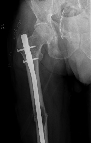
Figure 11. Early treatment of an impending fracture can prevent the pain and disability of a complete fracture.
Recovery and Rehabilitation
Recovery after a pathological fracture involves much more than allowing the bone to heal. Because the fracture occurs in a bone weakened by an underlying disease, rehabilitation focuses on restoring mobility, treating the underlying condition, reducing pain, and helping patients return to their daily lives as safely and independently as possible.
The recovery plan varies from person to person and depends on several factors, including:
- The underlying diagnosis
- Whether surgery was performed
- The bone involved
- Overall health
- Ongoing cancer treatment, if applicable
- Pre-fracture mobility and activity level
Your treatment team will develop an individualized rehabilitation program based on these factors.
What Can You Expect During Recovery?
Recovery usually occurs in stages.
Immediately After Treatment
The initial priorities are:
- Effective pain control
- Protecting the operated or fractured limb
- Preventing complications such as blood clots and pressure injuries
- Beginning gentle movement as soon as it is is safe
Many patients are encouraged to sit up, stand, or begin walking—with assistance—much earlier than they expect. Early mobilization helps reduce complications associated with prolonged bed rest and promotes faster functional recovery.
Physiotherapy
Physiotherapy is a structured rehabilitation program that uses exercises and movement techniques to improve strength, flexibility, balance, and function.
The goals of physiotherapy include:
- Restoring joint movement
- Rebuilding muscle strength
- Improving balance
- Training safe walking
- Reducing stiffness
- Increasing confidence during movement
Depending on the location of the fracture, therapy may involve gait training, stair climbing, upper limb exercises, or functional activities such as getting in and out of a chair.
Walking After Surgery
One of the most common questions patients ask is:
“When will I be able to walk again?”
The answer depends on several factors, including the type of surgery performed and the stability of the reconstruction.
Many modern surgical reconstructions are designed to allow early weight-bearing, meaning patients may begin standing or walking with support within a few days after surgery. Others may require a period of partial or protected weight-bearing while healing progresses.
Your surgeon and physiotherapist will explain exactly how much weight can safely be placed on the operated limb.
Patient Tip
Avoid comparing your recovery with that of other patients. Recovery timelines vary depending on the underlying disease, the complexity of surgery, and your overall health. Following your rehabilitation program consistently is more important than recovering quickly.
Returning to Daily Activities
As recovery progresses, most patients gradually resume everyday activities such as:
- Walking independently
- Personal care
- Household activities
- Driving (when medically appropriate)
- Returning to work
- Recreational activities suited to their condition
Your healthcare team will advise when it is safe to resume each activity.
Follow-Up Care
Regular follow-up appointments are an essential part of treatment.
These visits allow your healthcare team to:
- Monitor fracture healing or implant function
- Assess recovery of mobility
- Review imaging studies
- Detect recurrence or progression of disease when applicable
- Adjust medications
- Modify rehabilitation goals
The frequency of follow-up depends on your diagnosis and treatment plan.
Reassurance Box
Recovery from a pathological fracture is often gradual rather than immediate. Many patients worry if progress seems slow during the first few weeks. Improvement usually continues over several months, and rehabilitation remains an important part of achieving the best possible outcome.
Prognosis
Prognosis refers to the expected course and outcome of a medical condition.
The prognosis after a pathological fracture depends primarily on the disease that caused the fracture, rather than the fracture itself.
For example:
- A fracture caused by osteoporosis often heals well with appropriate treatment.
- Benign bone tumors usually have an excellent outlook after definitive management.
- Fractures related to metastatic cancer are managed with the goals of relieving pain, restoring function, and improving quality of life alongside treatment of the underlying cancer.
- Primary bone cancers require coordinated multidisciplinary treatment that may include chemotherapy, surgery, and occasionally radiotherapy.
Because every patient’s situation is unique, it is rarely helpful to compare outcomes with statistics found online. Your treating team is best placed to discuss your individual prognosis.
Doctor Insight
Modern orthopaedic oncology has transformed the management of pathological fractures. Advances in imaging, surgical reconstruction, implant design, systemic cancer therapies, and rehabilitation now allow many patients to maintain mobility and independence that would not have been possible a few decades ago.
Frequently Asked Questions
1. Is every pathological fracture caused by cancer?
No. Although cancer is an important cause, pathological fractures can also occur because of osteoporosis, benign bone tumors, bone cysts, infections, and several metabolic bone disorders.
2. Can a pathological fracture be the first sign of cancer?
Yes. Occasionally, a fracture is the first indication of an underlying cancer that has not yet been diagnosed. This is one reason why careful investigation is essential.
3. Will every patient need surgery?
No. Treatment depends on the underlying diagnosis, fracture stability, symptoms, and overall health. Some patients can be managed without surgery, while others benefit from surgical stabilization or reconstruction.
4. Can a pathological fracture heal?
Many pathological fractures can heal, particularly when the underlying disease is appropriately treated. Healing may be slower or less predictable in some cancers, making surgical stabilization an important part of treatment.
5. What is the purpose of a biopsy?
A biopsy identifies the exact disease responsible for weakening the bone. This information guides treatment and helps avoid inappropriate surgery.
6. Is a biopsy painful?
Most core needle biopsies are performed under local anaesthesia with imaging guidance. Patients usually experience only mild discomfort, and serious complications are uncommon.
7. Why can’t every pathological fracture simply be fixed with a metal rod or plate?
The treatment depends on why the bone fractured. In some conditions, simple fixation is appropriate. In others, the diseased bone must first be removed or treated to achieve the best long-term result.
8. Can I walk after treatment?
Many patients regain the ability to walk after treatment. The timeline depends on the location of the fracture, the type of surgery performed, and the underlying disease.
9. Will I need chemotherapy or radiotherapy?
Not everyone does. These treatments are recommended only when appropriate for the specific diagnosis.
10. What is a tumor prosthesis?
A tumor prosthesis (also called a megaprosthesis or endoprosthesis) is a specially designed artificial implant used to replace bone removed during surgery for certain tumors or extensive bone destruction.
11. Can pathological fractures be prevented?
In some situations, yes. Early diagnosis of bone disease, treatment of osteoporosis, medications that strengthen bone, and surgery for bones at high risk of breaking may reduce the likelihood of a pathological fracture.
12. Which specialist should manage a pathological fracture?
Whenever possible, pathological fractures should be evaluated by an orthopaedic oncologist working within a multidisciplinary team experienced in bone tumors and metastatic bone disease.
References
- Coleman RE, Croucher PI, Padhani AR, et al. Bone metastases. Nat Rev Dis Primers. 2020;6(1):83. doi:10.1038/s41572-020-00216-3.
- Damron TA, Morgan H, Prakash D, Grant W, Aronowitz J, Heiner J. Critical evaluation of Mirels’ rating system for impending pathological fractures. Clin Orthop Relat Res. 2003;(415 Suppl):S201-S207.
- Mirels H. Metastatic disease in long bones: A proposed scoring system for diagnosing impending pathologic fractures. Clin Orthop Relat Res. 1989;(249):256-264.
- National Comprehensive Cancer Network (NCCN). NCCN Clinical Practice Guidelines in Oncology: Bone Cancer. Version current at the time of access. Available at: https://www.nccn.org.
- National Comprehensive Cancer Network (NCCN). NCCN Clinical Practice Guidelines in Oncology: Multiple Myeloma. Version current at the time of access. Available at: https://www.nccn.org.
- Coleman R, Hadji P, Body JJ, et al. Bone health in cancer: ESMO Clinical Practice Guidelines. Ann Oncol. 2020;31(12):1650-1663.
- American Academy of Orthopaedic Surgeons (AAOS). OrthoInfo: Bone Tumors and Metastatic Bone Disease. Available at: https://orthoinfo.aaos.org.
Medical Disclaimer
This article is intended for educational purposes only and should not replace professional medical advice, diagnosis, or treatment. Every patient is unique, and treatment decisions should always be made in consultation with a qualified healthcare professional familiar with your individual medical condition.
 +91 93558 33005