Quick Summary
- A tumor prosthesis replaces bone removed during bone tumor surgery.
- It is commonly used during limb salvage surgery for primary bone cancers and selected metastatic bone tumors.
- Unlike a routine hip or knee replacement, a tumor prosthesis replaces both bone and the adjacent joint, when required.
- Most patients can walk, perform daily activities, and enjoy a good quality of life after recovery.
- Regular follow-up is important to monitor both the implant and cancer recovery.
Introduction
Advances in orthopaedic oncology have transformed the treatment of bone tumors over the past few decades. In the past, removing a malignant bone tumor often meant amputation because surgeons had limited options for reconstructing the large segment of bone removed during cancer surgery.
Today, that is no longer the case for most patients.
With improvements in imaging, chemotherapy for selected cancers, surgical techniques, and implant technology, limb salvage surgery has become the preferred treatment for many bone tumors affecting the arms and legs. Instead of removing the entire limb, surgeons can often remove the tumor with a margin of healthy tissue and reconstruct the resulting bone defect using a tumor prosthesis.
A tumor prosthesis is much more than a metal implant. It is a carefully engineered reconstruction designed to restore the structure of the limb, recreate joint function when necessary, and help patients regain independence after major surgery.
Although the terms tumor prosthesis, megaprosthesis, and endoprosthesis are often used interchangeably, they simply describe artificial implants used to replace large sections of bone following tumor removal. These implants are very different from the hip or knee replacements commonly performed for arthritis.
For many patients, hearing that they need a tumor prosthesis raises understandable concerns.
“Will I lose my leg?”
“Will I be able to walk again?”
“How long will the implant last?”
“Can I return to work or normal life?”
These are important questions, and understanding the role of a tumor prosthesis can make treatment decisions less overwhelming.
This guide explains:
- what a tumor prosthesis is,
- when it is recommended,
- how surgeons choose the appropriate implant,
- what recovery involves,
- the expected long-term outcomes, and
- the possible complications and future advances in this rapidly evolving field.
Rather than focusing on the technical details of surgery, this article aims to help patients and their families understand what to expect before, during, and after endoprosthetic reconstruction, so they can participate confidently in treatment decisions alongside their healthcare team.
Doctor Explains
A tumor prosthesis is not a treatment for cancer itself. The cancer is treated by completely removing the tumor. The prosthesis is then used to reconstruct the bone and joint that had to be removed, helping restore function and preserve the limb whenever possible.
Clinical Pearl
The success of a tumor prosthesis depends first on safe and complete tumor removal. The implant is the reconstruction that follows cancer surgery—not a substitute for it.
Reassurance Box
Being advised to undergo reconstruction with a tumor prosthesis does not necessarily mean your cancer is more advanced. It usually means that removing the tumor safely will leave a gap in the bone that needs to be reconstructed. In many cases, this approach allows patients to keep their limb while achieving the same cancer control as amputation.
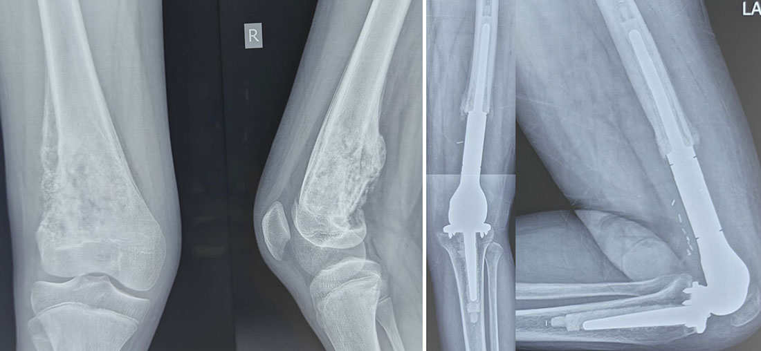
Figure 1. From Bone Tumor to Limb Salvage
Tumor prosthesis after removal of a bone tumor, restoring the affected bone and joint and allowing limb-salvage surgery.
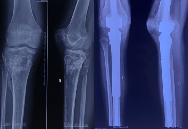
Figure 2. Proximal Tibia Tumor Reconstruction
Proximal tibia tumor removed and reconstructed with a tumor endoprosthesis to restore the knee and limb function.
What Is a Tumor Prosthesis?
A tumor prosthesis is a specially designed artificial implant used to reconstruct a limb after a bone tumor has been surgically removed. It replaces the section of bone that has been excised (removed), and when the tumor involves a nearby joint, it also replaces that joint to restore stability and movement.
Unlike routine hip or knee replacements, which replace only the worn-out joint surfaces affected by arthritis, a tumor prosthesis is designed to replace large segments of bone, adjacent joints, and sometimes the attachment sites of important muscles and ligaments. For this reason, these implants are often referred to as megaprostheses, reflecting the amount of bone they are designed to replace.
The primary goals of a tumor prosthesis are to:
- Restore the structural strength of the limb.
- Allow early mobilization and rehabilitation.
- Preserve limb function after tumor removal.
- Improve independence and quality of life.
For many patients, a tumor prosthesis makes it possible to undergo limb salvage surgery, avoiding amputation while achieving the same oncological principle of complete tumor removal.
Understanding the Terminology
Several terms are commonly used when discussing reconstruction after bone tumor surgery. Although they may sound different, they are closely related and are often used interchangeably.
Tumor Prosthesis
A tumor prosthesis is the broadest and most commonly used term. It refers to any artificial implant designed specifically to reconstruct a bone defect created after surgical removal of a bone tumor.
Megaprosthesis
A megaprosthesis is a type of tumor prosthesis used when a large segment of bone must be replaced. The prefix “mega” simply means large. It does not indicate that the cancer is larger, more aggressive, or more advanced.
Megaprostheses are commonly used after removal of tumors involving the:
- Distal femur (lower end of the thigh bone)
- Proximal tibia (upper end of the shin bone)
- Proximal femur (upper end of the thigh bone near the hip)
- Proximal humerus (upper arm bone near the shoulder)
- Pelvis
- Entire femur (in selected patients)
Endoprosthesis
The word endoprosthesis means an implant placed inside the body (endo means “within”). In orthopaedic oncology, it refers to an internal prosthetic reconstruction used after tumor resection.
While “endoprosthesis” is the technically correct medical term, patients are more likely to hear their surgeon use the terms tumor prosthesis or megaprosthesis.
Endoprosthetic Reconstruction
Endoprosthetic reconstruction describes the entire reconstructive procedure, not just the implant itself.
It includes:
- Removal of the tumor with adequate surgical margins.
- Reconstruction of the missing bone using a tumor prosthesis.
- Restoration of muscles, tendons, and soft tissues around the implant whenever possible.
- Rehabilitation to regain function.
Doctor Explains
Think of a tumor prosthesis as a custom-engineered replacement for a missing section of the skeleton. After removing the diseased bone, the surgeon reconstructs the limb with an implant that restores its length, alignment, and stability, allowing the patient to regain movement and function.
What Is a Tumor Prosthesis Made Of?
Modern tumor prostheses are manufactured using highly durable biocompatible materials. Biocompatible means the material is designed to function safely inside the human body without causing harmful reactions.
Common materials include:
- Titanium alloys – lightweight, strong, corrosion-resistant, and highly compatible with bone.
- Cobalt-chromium alloys – extremely durable metals used in areas subjected to high mechanical stress.
- Highly cross-linked polyethylene – a specialized medical-grade plastic used where moving surfaces articulate (move against each other), reducing wear.
- Porous metal coatings – textured surfaces that encourage bone to grow onto the implant, improving long-term fixation in selected designs.
These materials have been used safely in orthopaedic surgery for many years and are selected because they combine strength, durability, and excellent biological compatibility.
How Does a Tumor Prosthesis Work?
A tumor prosthesis acts as an internal structural framework that replaces the section of bone removed during surgery.
The implant typically has three main components:
- Stem – the portion inserted into the remaining healthy bone above and below the resection.
- Body – the central segment that replaces the missing bone.
- Joint Component – when the nearby joint has also been removed, this recreates the joint and allows movement.
Together, these components restore:
- Limb length.
- Alignment.
- Stability.
- Weight-bearing ability.
- Joint movement, where applicable.
Unlike biological reconstruction, which depends on transplanted bone healing over several months, a tumor prosthesis provides immediate mechanical stability, allowing rehabilitation to begin much earlier in many patients.
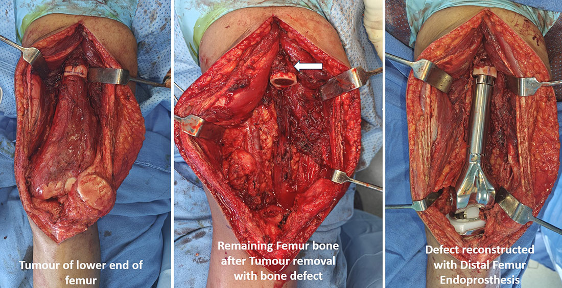
Figure 3. Distal Femur Tumor – Limb-Salvage Reconstruction
Removal of a distal femur tumor creates a bone defect that is reconstructed with a distal femur megaprosthesis, restoring limb length and alignment and allowing early weight bearing and mobilization.
Modular Versus Custom-Made Prostheses
Most modern tumor prostheses are modular, meaning they are assembled from standardized components available in different sizes. During surgery, the surgeon selects and combines these components to match the amount of bone removed and the patient’s anatomy.
The advantages of modular implants include:
- Greater flexibility during surgery.
- Reduced manufacturing time.
- Easier replacement of individual components during future revision surgery.
- Proven long-term clinical outcomes.
In selected complex cases—particularly tumors involving the pelvis or unusual anatomical locations—a custom-made prosthesis may be designed using advanced CT-based planning and, increasingly, 3D printing technology.
Custom implants are tailored to the individual patient’s anatomy and may provide better reconstruction when standard modular implants are not suitable.
Are Tumor Prostheses Permanent?
Tumor prostheses are designed to function for many years, but no artificial implant is permanent.
The lifespan of a prosthesis depends on several factors, including:
- The patient’s age.
- Activity level.
- Tumor location.
- Type of implant.
- Presence of complications such as infection or loosening.
Many modern implants function successfully for well over a decade, while some continue to perform well for much longer. However, because these implants are subjected to repeated mechanical stress over many years, some patients may eventually require revision surgery, which involves repairing or replacing part or all of the prosthesis.
The goal is not to create an implant that lasts forever, but one that provides durable function, allows independence, and can often be revised if necessary.
Did You Know?
Modern modular megaprostheses are available in a wide range of sizes and can replace almost any major long bone, including the entire femur in carefully selected patients.
Clinical Pearl
A tumor prosthesis is not simply a larger joint replacement. It is a specialized reconstruction designed to replace large sections of bone removed during cancer surgery while restoring limb function and maintaining oncological safety.
Reassurance Box
Many patients are concerned when they hear the word “metal implant.” Modern tumor prostheses are manufactured from materials that have been extensively tested and used in orthopaedic surgery worldwide. Although long-term follow-up is necessary, these implants have enabled thousands of patients to preserve their limbs and maintain an active, independent lifestyle after bone tumor surgery.
Why Is a Tumor Prosthesis Needed?
One of the first questions many patients ask after being told they need a tumor prosthesis is:
“Why can’t the surgeon simply remove the tumor and leave the rest of the bone?”
The answer lies in the principles of cancer surgery.
When treating a malignant (cancerous) bone tumor, the goal is not only to remove the visible tumor but also to remove a margin of healthy tissue around it. A surgical margin is a small cuff of normal tissue removed along with the tumor to reduce the risk of leaving behind microscopic cancer cells that cannot be seen with the naked eye.
Depending on the size and location of the tumor, this may require removal of a substantial segment of bone and, in some cases, the nearby joint. Once this section has been removed, the remaining bone alone is often too short or too weak to support the limb or allow normal movement.
When this defect is extensive, reconstruction becomes necessary to:
- Restore the length of the limb.
- Provide immediate structural support.
- Recreate the nearby joint, if it has been removed.
- Allow standing and walking.
- Protect the remaining healthy bone from excessive stress.
Without reconstruction, the limb would not function normally.
A tumor prosthesis reconstructs this defect, restoring the continuity of the skeleton and allowing the limb to function again.
Doctor Explains
Think of the skeleton as the framework of a building. If a damaged section of the framework must be removed to eliminate the disease, the missing segment has to be replaced before the structure can safely support weight again. A tumor prosthesis serves as that replacement framework.
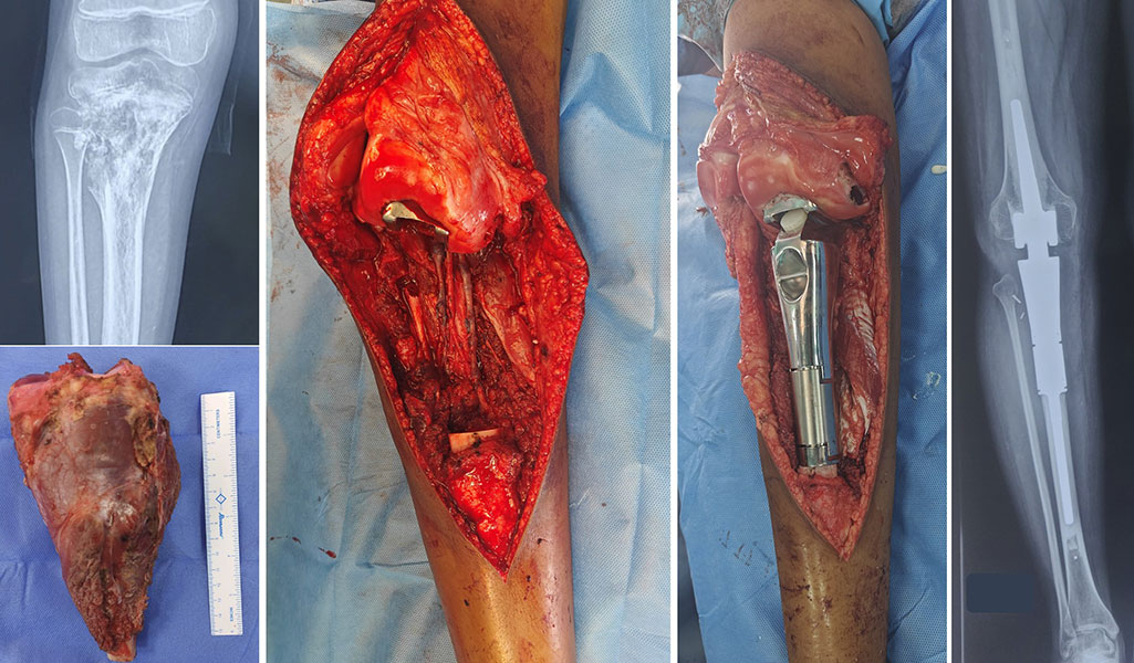
Figure 4. Proximal Tibia Tumor – Limb-Salvage Reconstruction
Which Patients May Need a Tumor Prosthesis?
A tumor prosthesis is not required for every bone tumor.
The decision depends on:
- The type of tumor.
- The amount of bone that must be removed.
- The location of the tumor.
- The patient’s age and expected function.
- Whether other reconstruction options are appropriate.
The procedure is most commonly performed after removal of tumors affecting the ends of long bones around major joints.
A tumor prosthesis is generally considered when removal of the tumor will leave a large bone defect that cannot heal on its own and is unlikely to be reconstructed effectively using biological techniques alone.
In general, patients are suitable candidates when:
- The tumor can be completely removed while preserving the major nerves and blood vessels supplying the limb.
- The expected functional outcome after reconstruction is good.
- The patient is medically fit to undergo major surgery.
- The remaining muscles and soft tissues are adequate to support the reconstruction.
- Limb preservation can be achieved without compromising cancer treatment.
The decision is therefore based on whether limb salvage is safe and functional, rather than simply whether it is technically possible.
Common Conditions That May Require a Tumor Prosthesis
Most of these conditions are discussed in greater detail on their respective pages.
Adults Are the Most Common Candidates
Most tumor prostheses are implanted in adults, particularly because many primary bone tumors requiring reconstruction occur after skeletal maturity.
In adults, modular prostheses provide several advantages:
- Immediate mechanical stability.
- Early rehabilitation.
- Reliable restoration of limb length.
- Excellent long-term functional outcomes in appropriately selected patients.
Age alone is not a contraindication to surgery. A healthy older adult may benefit significantly from reconstruction if it improves mobility and independence.
What About Children?
Children present unique challenges because their bones are still growing.
If a conventional prosthesis is implanted in a young child, the reconstructed limb will not grow at the same rate as the opposite limb. Over time, this may lead to a limb length discrepancy, meaning one leg or arm becomes shorter than the other.
To address this problem, surgeons may consider:
- Expandable prostheses, which can be lengthened as the child grows.
- Biological reconstruction, in selected situations.
- Other limb-preserving procedures, depending on the child’s age and the location of the tumor.
The choice depends on:
- Remaining growth potential.
- Tumor location.
- Expected survival.
- Functional goals.
- Family preferences after detailed counselling.
Because these decisions are complex, treatment should ideally be undertaken in specialist centres experienced in pediatric orthopaedic oncology.
Doctor Insight
Children are not simply “small adults.” Their ongoing growth means reconstruction must consider both today’s surgery and tomorrow’s development.
Why Is a Tumor Prosthesis Often Preferred?
Several methods are available to reconstruct bone after tumor removal, including biological reconstruction, where bone grafts from the patient or a donor are used to restore the defect.
A tumor prosthesis is often preferred because it offers several practical advantages.
These include:
- Immediate structural stability.
- Earlier rehabilitation.
- Earlier weight-bearing for many lower limb reconstructions.
- No need for transplanted bone to heal before the limb can be used.
- Predictable restoration of limb length and alignment.
- Proven long-term functional outcomes.
However, a tumor prosthesis is not always the best option. Younger patients, children, or individuals with certain tumor locations may benefit from alternative reconstructive techniques.
The choice is individualized and made after careful discussion within a multidisciplinary sarcoma team—a group of specialists that typically includes orthopaedic oncologists, medical oncologists, radiation oncologists, radiologists, pathologists, physiotherapists, and specialist nurses.
Doctor Insight
The best reconstruction is not necessarily the most complex one. It is the option that provides the safest cancer treatment while giving the patient the best long-term function based on their age, tumor type, anatomy, and lifestyle.
Patient Tip
During your consultation, ask your surgeon why a tumor prosthesis has been recommended instead of another reconstruction method. Understanding the reasoning behind the decision often makes the treatment plan much easier to understand and accept.
Tumor Prosthesis vs Knee Replacement: What Is the Difference?
A common misconception is that a tumor prosthesis is simply a larger version of a knee or hip replacement. Although both procedures use artificial implants made from medical-grade materials, they are performed for very different reasons.
A routine joint replacement (arthroplasty) is done to relieve pain caused by arthritis or joint damage. Only the worn joint surfaces are replaced, while most of the surrounding bone is preserved. Its primary goal is to reduce pain and improve mobility.
In contrast, a tumor prosthesis (megaprosthesis) is used after surgical removal of a bone tumor. To ensure complete cancer removal, surgeons often have to remove a substantial segment of bone, and sometimes the adjacent joint. The prosthesis replaces the missing bone and joint, restoring limb length, stability, and function.
The priorities of these two operations are also different. Joint replacement focuses on improving function in a diseased joint, whereas tumor prosthesis surgery prioritizes complete tumor removal with safe surgical margins. Once the cancer has been adequately treated, reconstruction is performed to preserve the limb and maximize function.
In simple terms, a joint replacement restores a damaged joint, while a tumor prosthesis reconstructs a missing part of the skeleton after bone tumor surgery. Tumor prosthesis surgery is therefore a far more complex procedure that requires meticulous planning, advanced imaging, multidisciplinary cancer care, and expertise in musculoskeletal oncology.
Does a Tumor Prosthesis Function Like a Normal Joint?
A tumor prosthesis restores movement, stability, and limb function, but it cannot completely replicate a natural joint. The final outcome depends on the amount of bone and muscle removed, preservation of tendons and ligaments, rehabilitation, and the patient’s overall health. Most patients can walk independently, climb stairs, drive, return to work, and perform everyday activities. However, high-impact activities such as running, jumping, or heavy manual labor may not be recommended. The goal is to achieve a stable, pain-free, and functional limb, rather than a perfectly normal joint.
Clinical Pearl
Success is measured not only by implant function but also by safe cancer control, preserved independence, and long-term quality of life.
Is Recovery Similar to a Knee Replacement?
Recovery after tumor prosthesis surgery is generally longer and more complex than after routine joint replacement because it involves extensive bone resection, soft tissue reconstruction, and recovery from cancer treatment. Rehabilitation is individualized and often takes several months.
Patient Tip
Avoid comparing your recovery with someone who has had a routine hip or knee replacement. Your rehabilitation is unique and should be judged against your own progress.
Did You Know?
A routine knee replacement replaces only a few millimetres of bone, whereas a tumor prosthesis may replace 10–30 cm or more of bone after tumor removal.
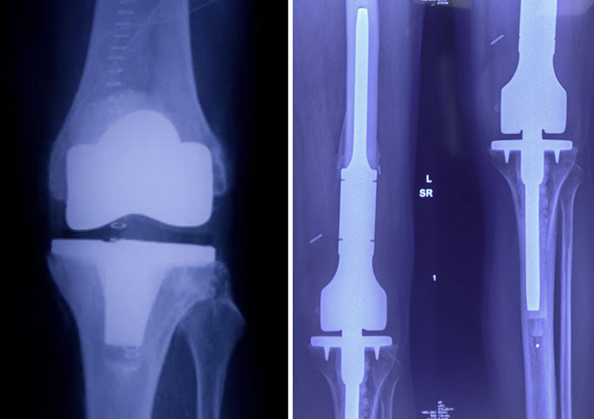
Figure 5. Standard Knee Replacement vs Tumor Prosthesis
A standard knee replacement replaces the damaged joint, while a tumor prosthesis replaces the bone and joint removed during bone tumor surgery
Types of Tumor Prostheses
Modern tumor prostheses are available for almost every major long bone, allowing reconstruction to be tailored to the exact location of the tumor while preserving limb function whenever possible.
Distal Femur Tumor Prosthesis
The distal femur (lower end of the thigh bone) is the most common site for tumor prosthetic reconstruction because bone tumors frequently occur around the knee. After tumor removal, the missing bone and knee joint are replaced using a distal femur megaprosthesis with a hinged or rotating knee mechanism.
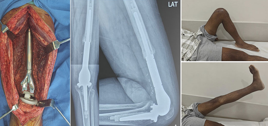
Figure 6. Distal Femur Tumor Prosthesis After Limb Salvage
Distal femur megaprosthesis used to reconstruct the knee after removal of a bone tumor, with good postoperative knee movement.
Proximal Tibia Tumor Prosthesis
The proximal tibia forms the lower half of the knee joint. Reconstruction is technically demanding because the patellar tendon, essential for straightening the knee, must also be reconstructed. Although recovery may be slower than distal femur reconstruction, dedicated physiotherapy usually results in good function.
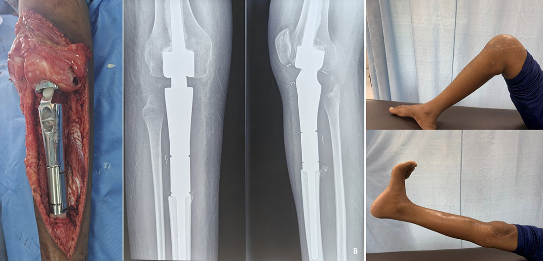
Figure 7. Proximal Tibia Megaprosthesis After Limb Salvage
Proximal tibia megaprosthesis reconstructing the knee after tumor removal, with good recovery of knee movement.
Clinical Pearl
Successful proximal tibia reconstruction depends as much on extensor mechanism reconstruction as on the prosthesis itself.
Proximal Femur Tumor Prosthesis
The proximal femur forms the hip joint. When tumors involve this region, the prosthesis replaces the upper femur while restoring hip stability, limb length, and walking ability. Preserving or reconstructing the hip abductor muscles is crucial to minimize limping.
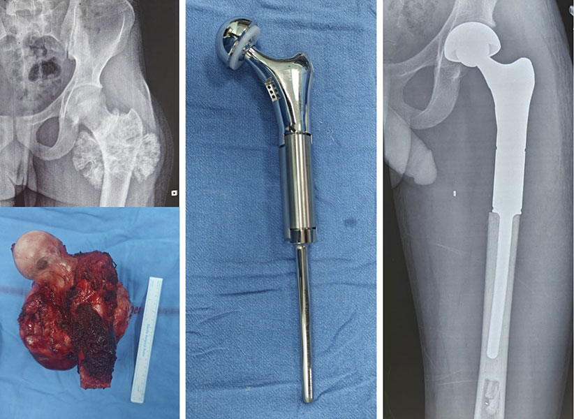
Figure 8. Proximal Femur Tumor Prosthesis After Limb Salvage
Proximal femur tumor removed and reconstructed with a tumor megaprosthesis to preserve the hip and limb.
Total Femur Prosthesis
When the entire femur is affected, surgeons may replace the whole thigh bone with a total femur prosthesis instead of performing an amputation. Although rehabilitation is demanding, many carefully selected patients regain independent mobility.
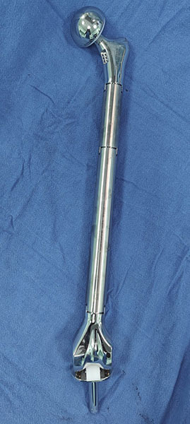
Figure 9. Total Femur Megaprosthesis
Total femur replacement with a megaprosthesis following extensive bone tumor removal, providing reconstruction of the entire thigh bone.
Did You Know?
A total femur prosthesis can replace the entire thigh bone, allowing limb preservation in situations that previously required amputation.
Proximal Humerus Tumor Prosthesis
The proximal humerus forms the shoulder joint. The aim of reconstruction is to provide a stable, painless upper limb that preserves useful hand and elbow function. Shoulder movement may be limited if important muscles are removed, but most patients remain independent in daily activities.
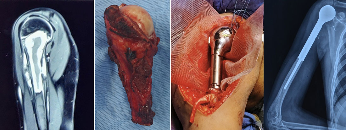
Figure 10. Proximal Humerus Megaprosthesis After Limb Salvage
Total Proximal humerus tumor removed and reconstructed with a megaprosthesis as part of limb-salvage surgery.
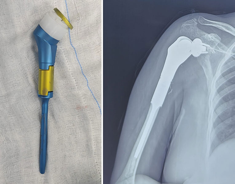
Figure 11. Reverse Shoulder Megaprosthesis After Limb Salvage
Reverse shoulder megaprosthesis used to reconstruct the shoulder after removal of a proximal humerus bone tumor.
Pelvic Tumor Prosthesis
Pelvic reconstruction is among the most complex procedures in orthopaedic oncology because tumors are often close to major nerves, blood vessels, and pelvic organs. Selected patients may benefit from custom-made or 3D-printed pelvic prostheses, with reconstruction planned individually.
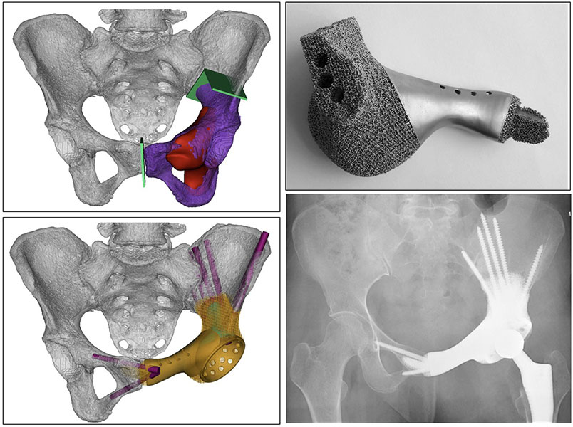
Figure 12. Patient-specific 3D-printed pelvic implant designed for reconstruction after pelvic bone tumor removal.
Doctor Insight
Advances in patient-specific implants and 3D printing have significantly improved reconstruction options for complex pelvic tumors.
Expandable Prosthesis for Children
Children with growing bones may require expandable prostheses, which can be lengthened as the child grows to reduce future limb-length differences. Modern designs can often be lengthened using minimally invasive or magnetic techniques.
Patient Tip
Ask your surgeon to show you your MRI or a diagram of the planned reconstruction. Understanding which part of the bone will be replaced often makes the surgery easier to visualize.
Reassurance Box
Modern tumor prostheses are available for most major bones of the body. With careful planning and specialist surgical expertise, they help restore movement, independence, and quality of life while maintaining the principles of safe cancer surgery.
Different Types of Tumor Prostheses Explained (3–4 minute animation with site-by-site overview)
How Is the Right Tumor Prosthesis Chosen?
Choosing the right tumor prosthesis is a highly individualized process. There is no single implant that suits every patient. The goal is to achieve complete tumor removal, durable reconstruction, and the best possible long-term function. Rather than selecting the newest or strongest implant, surgeons choose the reconstruction that offers the safest cancer treatment and the best functional outcome for each patient.
Doctor Explains
Selecting a tumor prosthesis is like designing a bridge after a damaged section has been removed. The replacement must fit precisely, restore stability, and withstand years of use while matching the patient’s anatomy and needs.
Tumor Factors
The tumor is the most important factor in planning reconstruction. MRI and CT scans determine the size and location of the tumor, involvement of the joint, muscles, nerves, and blood vessels, and the amount of bone that must be removed. The prosthesis is selected after defining the extent of safe tumor resection.
Patient Factors
Patient-related factors are equally important. Age influences whether a standard or expandable prosthesis is needed. Bone quality, activity level, overall health, and personal goals—such as returning to work, independent walking, or maintaining an active lifestyle—help determine the most suitable reconstruction.
Patient Tip
Tell your surgeon about your occupation, hobbies, and daily activities. These details help tailor the reconstruction to your lifestyle, not just your X-rays.
Implant Factors
Modern tumor prostheses come in different sizes and designs. Surgeons consider the length of bone to be replaced, fixation method, soft tissue reconstruction, and the possibility of future revision surgery. Long-term planning is especially important in younger patients.
Modular or Custom Prosthesis?
Most patients receive a modular prosthesis, which is assembled during surgery from standardized components. These implants are versatile, readily available, and easier to revise if needed. Custom-made prostheses are reserved for complex situations—such as pelvic tumors, unusual bone defects, or revision surgery—and are increasingly designed using 3D computer planning and printing to match the patient’s anatomy precisely.
Why Is Detailed Preoperative Planning So Important?
Successful tumor prosthetic reconstruction begins well before surgery. The multidisciplinary team reviews clinical findings, MRI, CT, biopsy, pathology, staging, and chemotherapy response to plan the extent of bone resection, implant size, soft tissue reconstruction, and potential challenges. Many centres also use digital templating and 3D surgical planning to improve accuracy.
Did You Know?
Modern surgeons often determine the size and position of the implant weeks before surgery using advanced imaging and computer planning.
Doctor Insight
Careful planning and appropriate implant selection are as important as the surgery itself.
There Is No “Best” Tumor Prosthesis
No single implant is best for every patient. Long-term success depends more on complete tumor removal, correct implant selection, surgical expertise, rehabilitation, and regular follow-up than on the implant brand.
Reassurance Box
Modern tumor prostheses provide excellent outcomes when used by experienced orthopaedic oncology teams following meticulous planning.
How Orthopaedic Oncologists Plan Tumor Prosthetic Reconstruction (3-minute educational animation)
What Happens During Tumor Prosthesis Surgery?
Tumor prosthesis surgery is performed as part of limb salvage surgery, aiming to completely remove the tumor while preserving the limb whenever it is oncologically safe.
Step 1: Tumor Removal
The operation begins with wide resection, where the tumor and a margin of healthy tissue are removed as one specimen to minimize the risk of recurrence. The amount of bone removed depends on the tumor’s size and location.
Clinical Pearl
Complete tumor removal with adequate surgical margins is the priority. Reconstruction is performed only after this objective is achieved.
Step 2: Reconstruction
The bone defect is reconstructed using a tumor prosthesis, which restores limb length, skeletal continuity, joint stability (when required), and mechanical strength. Muscles and tendons are reattached whenever possible to optimize function.
Step 3: Wound Closure
The wound is closed in layers, and temporary drains may be placed to reduce fluid collection during healing.
After the Operation
Recovery focuses on pain control, wound care, prevention of infection and blood clots, early physiotherapy, and gradual mobilization. Many patients begin assisted movement within a few days, depending on the reconstruction.
Reassurance Box
Tumor prosthesis surgery is a complex procedure performed by an experienced multidisciplinary team. Careful planning, precise surgery, and structured rehabilitation help maximize both cancer control and long-term limb function.
Learn More
If you would like to understand the operation in greater detail, including surgical approaches, reconstruction techniques, possible risks, and postoperative rehabilitation, read our comprehensive guide on Limb Salvage Surgery.
Recovery After Tumor Prosthesis Surgery
Recovery after tumor prosthesis surgery is a gradual process that continues well beyond the hospital stay. While the prosthesis restores the structural integrity of the limb, regaining strength, mobility, and confidence requires time, physiotherapy, and active participation. Recovery varies depending on the site of reconstruction, the amount of soft tissue removed, age, overall health, additional cancer treatments, and commitment to rehabilitation. In general, recovery is more extensive than after a routine hip or knee replacement.
Immediately After Surgery
During the first few days, the healthcare team focuses on pain control, wound care, prevention of infection and blood clots, monitoring nerve and blood vessel function, and starting gentle physiotherapy. Depending on the procedure, drains are removed within 1–2 days, and assisted sitting, standing, and walking begin as recovery progresses.
When Can I Start Walking?
One advantage of a tumor prosthesis is that it usually provides immediate structural stability, allowing rehabilitation to begin early. However, the timing of weight-bearing depends on the reconstruction site, soft tissue healing, and your surgeon’s advice. Some patients walk with a walker or crutches within a few days, while others require temporary protection of the limb.
Clinical Pearl
Early mobilization is encouraged, but rehabilitation should always follow your surgeon’s and physiotherapist’s guidance.
The Role of Physiotherapy
Physiotherapy is essential for achieving the best functional outcome. Rehabilitation focuses on restoring joint movement, strengthening muscles, improving balance, retraining walking, and gradually returning to daily activities. The program is individualized according to the type of reconstruction.
Returning to Everyday Life
Most patients progressively regain independence over several months. Many return to walking, climbing stairs, driving (after medical clearance), office-based work, and routine household activities. High-impact sports and heavy manual work may not be advisable for some patients.
How Long Does Recovery Take?
Recovery varies between individuals, but the following timeline provides a general guide.
Table. Typical Recovery Timeline After Tumor Prosthesis Surgery
Follow-Up After Surgery
Regular follow-up is essential to monitor both cancer recurrence and prosthesis function. Visits typically include clinical examination, X-rays, and additional imaging when required. Your follow-up schedule will be individualized according to your diagnosis.
Patient Tip
Recovery is a marathon, not a sprint. Consistent physiotherapy and celebrating small milestones often have a greater impact on long-term function than trying to progress too quickly.
Reassurance Box
Although recovery takes time, most patients improve steadily over several months. With dedicated rehabilitation, regular follow-up, and guidance from your healthcare team, excellent long-term function and independence are achievable.
What Are the Risks and Complications of a Tumor Prosthesis?
Like any major operation, tumor prosthesis surgery carries potential risks. However, most patients recover without serious complications, especially when surgery is performed at an experienced orthopaedic oncology centre. Your individual risk depends on factors such as the tumor location, overall health, previous chemotherapy or radiotherapy, and the complexity of the procedure. Early recognition and regular follow-up are key to successful management.
Doctor Explains
Every major surgery involves risks, but these must be balanced against the benefits of removing the tumor and preserving the limb. Your surgical team takes extensive measures to minimize complications before, during, and after surgery.
Infection
Infection is one of the most important complications because the prosthesis is a foreign implant. Symptoms include increasing pain, redness, swelling, wound discharge, or fever. Treatment may involve antibiotics, wound cleaning, or, in severe cases, revision surgery.
Implant Loosening
Over time, the bond between the implant and bone may weaken due to wear, mechanical stress, bone loss, or infection. Symptoms include increasing pain, instability, or difficulty bearing weight. Some patients may require revision surgery.
Implant Wear or Mechanical Failure
Although modern prostheses are designed for long-term use, mechanical wear, component failure, or, rarely, implant fracture can occur. Regular follow-up X-rays help detect problems before they become severe.
Dislocation or Instability
Hip and shoulder reconstructions have a small risk of instability or dislocation. Careful rehabilitation and adherence to postoperative precautions help minimize this risk.
Soft Tissue Problems
Delayed wound healing, muscle weakness, tendon problems, scar stiffness, or skin breakdown may occur and sometimes require additional wound care, physiotherapy, or further surgery.
Limb Length Difference and Functional Limitations
Some patients may experience a mild limb-length difference, reduced joint movement, muscle weakness, or a slight limp. These often improve with rehabilitation and, when necessary, shoe modifications or walking aids.
Warning Signs: When Should You Contact Your Doctor?
Seek medical attention if you develop fever, increasing redness or wound drainage, sudden pain, inability to bear weight, a new lump, chest pain, shortness of breath, or a feeling that the prosthesis is unstable.
Patient Tip
Do not ignore new symptoms, even years after surgery. Early medical evaluation often prevents more serious problems.
Clinical Pearl
Regular follow-up is as important as the surgery itself. Many complications can be detected on routine examination and X-rays before they significantly affect function.
Reassurance Box
Although complications are possible, they are not inevitable. With experienced surgical care, modern implants, structured rehabilitation, and regular follow-up, most patients enjoy many years of good function and an excellent quality of life.
How Long Does a Tumor Prosthesis Last?
One of the most common questions patients ask is, “Will this prosthesis last for the rest of my life?” The answer varies. Modern tumor prostheses are designed to be durable, and many function well for 10–20 years or longer. However, like any mechanical implant, they may eventually require revision because of wear, loosening, infection, or other complications. The aim is to provide a long-lasting reconstruction that supports an active and independent life.
Doctor Explains
A tumor prosthesis is like an artificial heart valve or a car—it is built to last for many years but experiences ongoing mechanical stress. Regular follow-up acts as routine maintenance, helping detect problems early.
What Determines How Long a Tumor Prosthesis Lasts?
Several factors influence implant longevity:
- Age: Younger patients often place more years of stress on the prosthesis and are more likely to require revision during their lifetime.
- Tumor Location: Implants around the knee, hip, and shoulder experience different mechanical demands.
- Activity Level: Everyday activities are encouraged, but repetitive high-impact activities such as running or heavy manual labour may shorten implant lifespan.
- Implant Design and Surgical Technique: Advances in implant materials, fixation methods, and accurate surgical placement have improved long-term durability.
- Complications: Infection, loosening, mechanical failure, fractures around the implant, or tumor recurrence may require additional surgery.
Will I Need Another Operation in the Future?
Possibly. Some patients never require further surgery, while others may need revision surgery to repair or replace part or all of the prosthesis because of wear, loosening, infection, fracture, or mechanical failure. Needing a revision does not necessarily mean the original surgery has failed; it often reflects the expected lifespan of a mechanical implant.
Clinical Pearl
Implant longevity depends not only on the prosthesis but also on patient age, activity level, tumor biology, and long-term survival.
Can Anything Be Done to Make the Prosthesis Last Longer?
Yes. You can help protect your reconstruction by attending follow-up appointments, participating in physiotherapy, maintaining a healthy weight, avoiding high-impact activities and tobacco use, and promptly reporting new pain, swelling, or signs of infection.
How Will My Surgeon Know If the Implant Is Wearing Out?
Regular follow-up is essential. Your surgeon will assess your symptoms, examine the limb, and obtain periodic X-rays or other imaging to detect early signs of wear or loosening before major problems develop.
Did You Know?
Many patients continue using the same tumor prosthesis for well over a decade, with regular surveillance helping maintain long-term function.
Patient Tip
Treat follow-up visits like routine maintenance for your prosthesis. Even if you feel well, regular check-ups help monitor both your implant and cancer recovery.
Reassurance Box
Modern tumor prostheses have transformed limb salvage surgery, allowing many patients to enjoy years of pain-free mobility and independence. If problems develop, revision surgery is often possible, and continuing advances in implant design are improving long-term outcomes.
X-ray series showing a well-functioning tumor prosthesis over several years of follow-up.
Life After a Tumor Prosthesis
Many patients worry more about life after surgery than the operation itself. Questions about walking, returning to work, travelling, and independence are common. The good news is that most patients regain a high level of independence after successful tumor prosthesis surgery. Although the reconstructed limb may not function exactly like a natural joint, modern prostheses allow many people to return to work, enjoy family life, travel, and perform everyday activities. Long-term outcomes depend on the site of reconstruction, muscle preservation, rehabilitation, and overall health.
Doctor Explains
The aim of tumor prosthetic reconstruction is not to create a perfect limb, but to provide a stable, pain-free, and functional limb that allows you to live as independently as possible.
Walking and Daily Activities
Walking is usually the first major milestone after surgery. Following rehabilitation, many patients can walk independently, climb stairs, perform household tasks, shop, and care for themselves. Some may require a walking stick for longer distances or have a mild limp, particularly after extensive hip or pelvic reconstruction. The focus is on safe and functional mobility rather than a completely normal gait.
Returning to Work
Most patients can return to work, although timing depends on the type of job and recovery. Desk-based and administrative work can often be resumed within a few months, whereas heavy manual labour may require permanent modifications. Your surgeon and physiotherapist will advise when it is safe to return.
Can I Exercise?
Yes. Regular low-impact exercise helps maintain muscle strength, flexibility, cardiovascular fitness, and a healthy weight. Walking, cycling, swimming, stretching, and physiotherapy exercises are recommended, while running, jumping, contact sports, and heavy weightlifting are generally discouraged.
Patient Tip
Choose exercises you can perform regularly and safely. Consistency is more important than intensity.
Can I Drive?
Most patients can drive again once they regain adequate strength and control of the operated limb, stop taking sedating medications, and receive clearance from their surgeon. Recovery time varies with the type of surgery and vehicle.
Can I Travel?
Yes. Travel is usually safe after recovery. During long journeys, stay hydrated, move regularly, and follow your doctor’s advice regarding blood clot prevention. Carrying a brief medical summary is helpful when travelling internationally.
Will Airport Security Detect My Implant?
Possibly. Because tumor prostheses contain metal, they may trigger airport scanners. Simply inform security personnel if this occurs.
Can I Sleep Normally?
Yes. Once healing is complete, most patients can sleep comfortably in any position unless advised otherwise during the early recovery period.
What About Pregnancy?
Pregnancy is usually possible after recovery, although women should discuss family planning with both their orthopaedic oncologist and obstetrician, especially after chemotherapy or radiotherapy.
Looking After Your Prosthesis
Protect your prosthesis by maintaining a healthy weight, exercising regularly, following physiotherapy, avoiding high-impact activities, treating infections promptly, and attending regular follow-up appointments.
Doctor Insight
Successful recovery depends on both physical rehabilitation and emotional resilience. Rebuilding confidence is as important as rebuilding strength.
Living Well with a Tumor Prosthesis
For most patients, the prosthesis gradually becomes part of everyday life. Many return to work, travel, hobbies, and family activities, enjoying a significant improvement in independence and quality of life.
Did You Know?
Many people live active lives with a tumor prosthesis for decades while maintaining regular follow-up with their orthopaedic oncology team.
Reassurance Box
Recovery is gradual, but with rehabilitation, patience, and ongoing medical care, most patients regain meaningful independence and an excellent quality of life after tumor prosthesis surgery.
Tumor Prosthesis vs Biological Reconstruction: Which Is Better?
Many patients ask, “Can my own bone be used instead of an artificial implant?” The answer is yes—in selected cases. While a tumor prosthesis is the most common method of reconstruction after bone tumor surgery, biological reconstruction is another important option. The best choice depends on factors such as the patient’s age, tumor location, expected survival, activity level, and the amount of bone removed. Reconstruction is always individualized and planned by a multidisciplinary sarcoma team.
What Is Biological Reconstruction?
Biological reconstruction restores the bone defect using living or donor bone. Options include autografts (the patient’s own bone), allografts (donor bone), vascularized fibular grafts, and, in selected centres, recycled bone techniques where the patient’s treated bone is reimplanted. Unlike a prosthesis, these methods rely on the body’s ability to heal and incorporate the graft.
Advantages of a Tumor Prosthesis
Tumor prostheses provide immediate stability, allow earlier rehabilitation and weight-bearing, do not depend on bone healing, and offer predictable restoration of limb length and function. They are particularly suitable for adults and for reconstructing large bone defects.
Advantages of Biological Reconstruction
Biological reconstruction preserves living bone, has no artificial joint components to wear out, and may provide excellent long-term durability if healing is successful. It is often considered for selected children and young adults but requires several months for bone healing.
Limitations of Each Approach
Tumor prostheses may be associated with infection, implant wear, loosening, and possible revision surgery. Biological reconstruction has a longer recovery and carries risks such as delayed union, non-union, graft fracture, and additional procedures. Neither technique is perfect, making careful patient selection essential.
Which Patients Benefit More?
Tumor prostheses are generally preferred when large bone segments or major joints require reconstruction and early rehabilitation is important. Biological reconstruction may be suitable for younger patients with reconstructable defects who can tolerate a longer healing period.
Clinical Pearl
There is no universally superior reconstruction. The best option is the one that provides safe cancer treatment with the best long-term functional outcome for the individual patient.
Can Both Techniques Be Combined?
Yes. In selected cases, surgeons combine prosthetic and biological techniques, such as using tendon or muscle grafts around a prosthesis or combining donor bone with a prosthetic joint. These complex procedures are usually performed in specialist bone tumor centres.
Which Option Is Better?
Rather than asking, “Which operation is better?”, the more appropriate question is, “Which reconstruction is best for my tumor, anatomy, age, and lifestyle?” The decision is based on tumor characteristics, bone quality, soft tissue involvement, functional goals, rehabilitation potential, and long-term durability.
Patient Tip
If more than one reconstruction option is suitable, ask your surgeon about the expected recovery, long-term function, potential complications, and the likelihood of future surgery before making a decision.
Reassurance Box
Modern orthopaedic oncology offers multiple effective reconstruction options. Whether your surgeon recommends a tumor prosthesis or biological reconstruction, the choice is based on achieving the safest cancer treatment and the best possible long-term outcome for your individual situation.
Frequently Asked Questions (FAQs)
1. What is a tumor prosthesis?
A tumor prosthesis (megaprosthesis or endoprosthesis) is a specialized implant used to replace bone—and sometimes a nearby joint—removed during bone tumor surgery. It helps preserve the limb, restore function, and improve mobility.
2. Is a tumor prosthesis the same as a knee or hip replacement?
No. Joint replacements replace only damaged joint surfaces due to arthritis, whereas tumor prostheses replace large segments of bone, often with the adjacent joint, after tumor removal.
3. Why do I need a tumor prosthesis?
It reconstructs the bone defect left after tumor removal, restoring limb stability, length, and function.
4. Will I be able to keep my limb?
In many cases, yes. Limb salvage is possible when the tumor can be safely removed without sacrificing critical nerves or blood vessels.
5. How long does tumor prosthesis surgery take?
Most operations take 3–6 hours, although complex reconstructions may take longer.
6. How long will I stay in the hospital?
Hospital stay is usually 5–10 days, depending on recovery and rehabilitation.
7. When can I start walking?
Many patients begin standing or walking with assistance within a few days, depending on the reconstruction.
8. Will I need physiotherapy?
Yes. Physiotherapy is essential to regain strength, mobility, and function.
9. Will I walk normally again?
Many patients walk independently, although some may have a mild limp depending on the surgery and rehabilitation.
10. How long does a tumor prosthesis last?
Modern implants often last 10–20 years or longer, depending on age, activity level, and complications.
11. Will I need another operation in the future?
Possibly. Revision surgery may be required for wear, loosening, infection, or mechanical failure.
12. Can I play sports after surgery?
Low-impact activities are encouraged. High-impact sports and heavy lifting are generally discouraged.
13. Can I drive after tumor prosthesis surgery?
Yes, once you have regained adequate strength and your surgeon considers it safe.
14. Can I travel with a tumor prosthesis?
Yes. Stay hydrated, move regularly during long journeys, and follow advice on blood clot prevention.
15. Will airport security scanners detect my implant?
They may. Inform security personnel if your implant triggers the scanner.
16. Is a tumor prosthesis safe during MRI scans?
Yes. Modern implants are MRI-compatible but may cause image distortion near the prosthesis.
17. Can the tumor come back after surgery?
Yes. Local recurrence depends on the tumor type and treatment, not the prosthesis itself. Regular follow-up is essential.
18. What are the most common complications?
Potential complications include infection, implant loosening or wear, instability, wound problems, blood clots, and local recurrence. Early diagnosis often allows successful treatment.
19. How often will I need follow-up?
Follow-up is frequent initially and then less often. Clinical examination and imaging monitor both the prosthesis and cancer recurrence.
20. Will I be able to return to work?
Most patients return to work after rehabilitation. Timing depends on recovery and job demands.
21. Is a tumor prosthesis better than biological reconstruction?
Neither is universally superior. Tumor prostheses offer earlier rehabilitation, while biological reconstruction may be preferable in selected younger patients. The choice is individualized.
22. Can children receive tumor prostheses?
Yes. Expandable prostheses can accommodate growth, although biological reconstruction may be preferred in some children.
23. Will I be able to live a normal life?
Most patients return to independent, active lives and can work, travel, drive, and enjoy everyday activities after recovery.
24. How do I know if something is wrong with my prosthesis?
Seek medical attention if you develop increasing pain, fever, wound drainage, swelling, difficulty walking, instability, or sudden inability to bear weight.
25. Where should tumor prosthesis surgery be performed?
Ideally, surgery should be performed at a specialist musculoskeletal oncology centre by an experienced multidisciplinary team with expertise in bone tumor surgery and rehabilitation.
References
- Schwartz HS, Holt GE. Musculoskeletal Cancer Surgery: Treatment of Sarcomas and Allied Diseases.
- Malawer MM, Sugarbaker PH. Musculoskeletal Cancer Surgery: Treatment of Sarcomas and Allied Diseases.
- Simon MA, Springfield DS. Surgery for Bone and Soft-Tissue Tumors.
- Henderson ER, Groundland JS, Pala E, et al. Failure mode classification for tumor endoprostheses: retrospective review of five institutions and a literature review. J Bone Joint Surg Am. 2011.
- Jeys LM, Kulkarni A, Grimer RJ, et al. Endoprosthetic reconstruction for the treatment of musculoskeletal tumors. J Bone Joint Surg Br.
- Grimer RJ, Carter SR, Tillman RM. Endoprosthetic replacement of the proximal tibia. J Bone Joint Surg Br.
- Mankin HJ, Hornicek FJ, et al. Long-term results of limb salvage surgery. Clinical Orthopaedics and Related Research.
- Healey JH, Morris CD, Athanasian EA, Boland PJ. Operative management of primary bone tumors.
- Aponte-Tinao LA, Farfalli GL, et al. Current concepts in endoprosthetic reconstruction after bone tumor resection.
 +91 93558 33005