13 October, 2026
If you have persistent pain or swelling around a joint such as the knee, wrist, hip, or shoulder, your doctor may recommend an X-ray or other imaging tests. Sometimes these investigations reveal a Giant Cell Tumor of Bone (GCTB).
Being told that you may have a bone tumor can naturally be worrying. The good news is that GCTB has characteristic features on imaging, and a systematic approach using X-rays, MRI, CT when required, and biopsy can help establish the diagnosis and plan appropriate treatment.
What Is the First Step in Diagnosing Giant Cell Tumor of Bone?
The evaluation usually begins with your medical history and clinical examination, followed by plain X-rays of the affected bone.
Your doctor will want to know:
- How long you have had pain or swelling
- Whether the pain is getting worse
- Whether you have difficulty moving the nearby joint
- Whether there has been a sudden increase in pain, which could indicate a pathological fracture
- Whether there has been any previous treatment or surgery for a bone lesion
A careful examination is then combined with the imaging findings to determine whether the lesion appears benign, locally aggressive, or potentially malignant.
Importantly, no single test should be interpreted in isolation. The clinical findings, imaging and pathology need to be considered together when diagnosing a bone tumor.
X-ray: Usually the First and Most Important Investigation
A plain X-ray is usually the first imaging investigation for a suspected bone tumor. It provides important information about the location, size, pattern of bone destruction and aggressiveness of the lesion.
A typical Giant Cell Tumor of Bone appears as a well-defined, purely lytic lesion near the end of a long bone. It commonly extends from the metaphysis into the epiphysis and approaches the joint surface. A sclerotic rim or mineralized tumor matrix is generally absent.
GCTB commonly occurs around the knee, particularly in the lower end of the femur and upper end of the tibia. It can also occur in areas such as the distal radius, proximal humerus, pelvis and other bones.
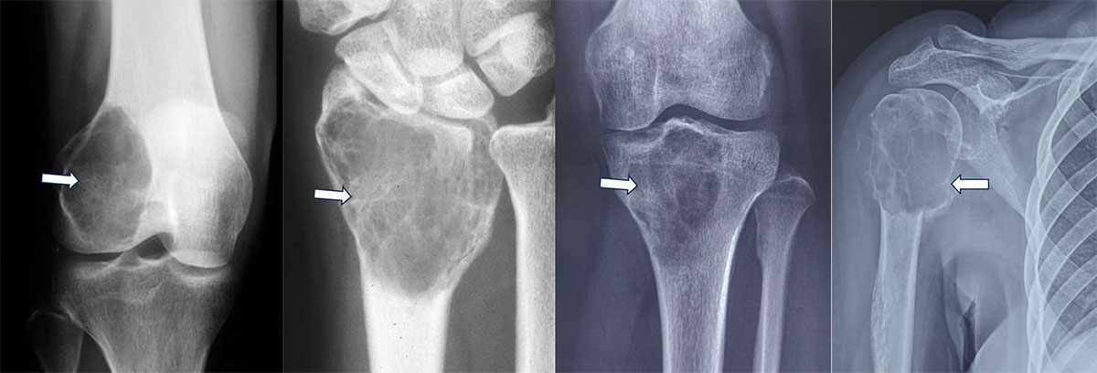
Figure 1. X-rays showing giant cell tumors of bone at different sites, with the characteristic expansile bone destruction highlighted.
The X-ray can also provide clues about how aggressively the tumor is behaving. Expansion and thinning of the cortex, cortical destruction, or a soft-tissue component may indicate more extensive disease.
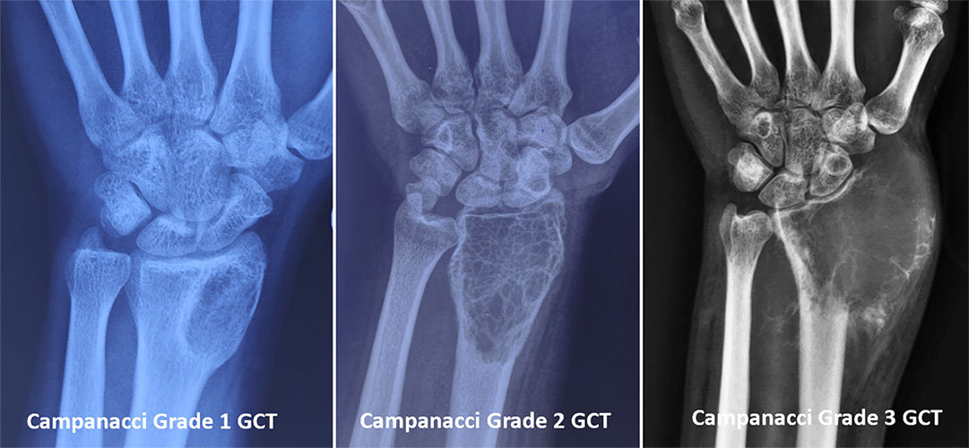
Figure 2. Campanacci grading of giant cell tumor of bone, ranging from Grade 1 with limited bone involvement to Grade 3 with cortical destruction and soft-tissue extension.
However, an X-ray does not always tell us the complete extent of the tumor. This is where MRI becomes particularly important.
MRI: Understanding the Full Extent of the Tumor
An MRI is an important part of evaluating GCTB, particularly when the tumor is close to a joint.
While an X-ray shows us the overall pattern of bone destruction, MRI provides much greater detail about:
- How far the tumor extends within the bone
- Whether the tumor has broken through the cortex
- Extension into surrounding soft tissues
- Relationship to muscles, tendons and neurovascular structures
- Involvement of the joint or subchondral bone
- The presence of additional areas of tumor within the same bone
MRI is particularly valuable for local staging and surgical planning because it provides detailed images in multiple planes and is better suited to assessing soft-tissue and intramedullary tumor extension.
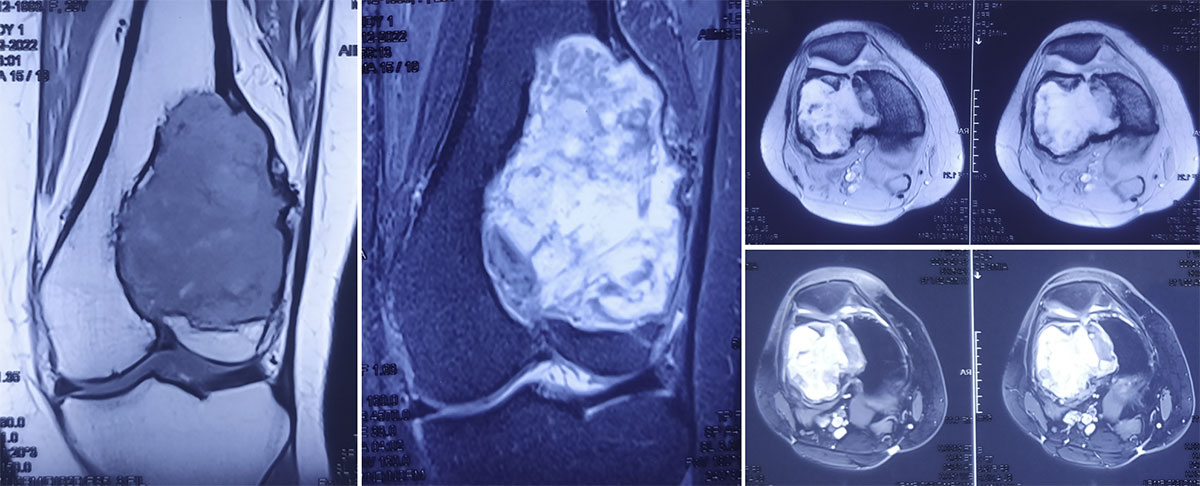
Figure 3. MRI provides a detailed view of the giant cell tumor, showing its extent within the bone and surrounding soft tissues.
For a patient, this information can be very important. Before deciding between a joint-preserving procedure and a more extensive resection, the surgeon needs to understand exactly where the tumor is and what structures are involved.
What Is the Role of CT Scan in GCTB?
CT is not always necessary, but it can provide useful additional information in selected cases.
CT is particularly helpful for assessing:
- The condition of the cortical bone
- The degree of cortical thinning or destruction
- The presence of a pathological fracture
- Complex bony anatomy
- The relationship of the tumor to important structures in certain locations
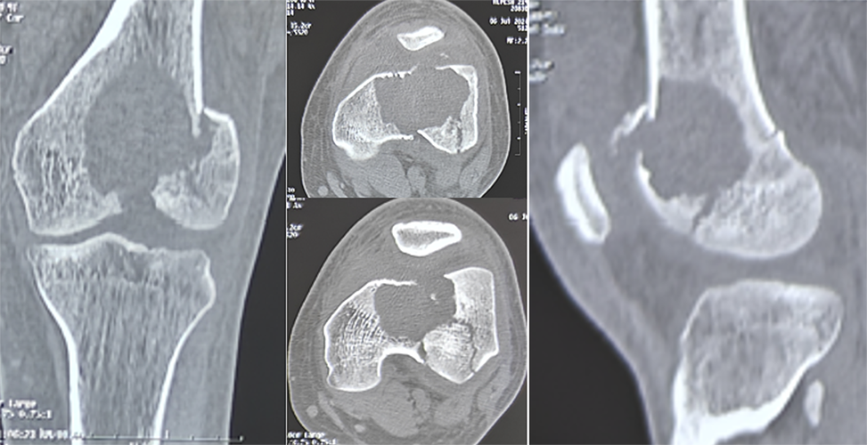
CT can sometimes demonstrate cortical destruction more clearly than conventional X-rays.
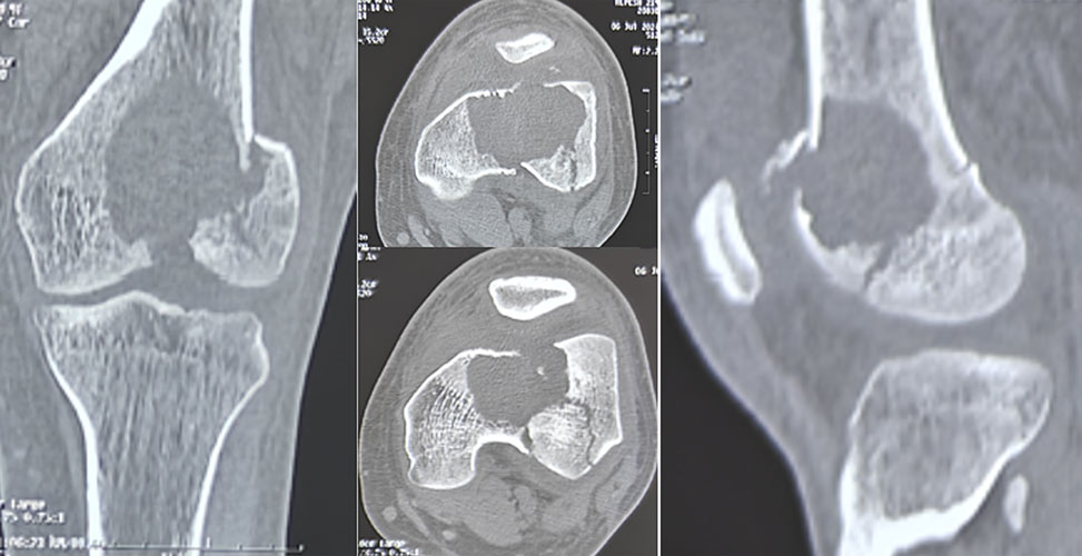
Figure 4. CT scan showing the extent of bone destruction caused by a giant cell tumor of bone, helping plan the appropriate surgical treatment.
Therefore, X-ray, MRI and CT are complementary investigations rather than competing tests. The combination required depends on the location and characteristics of the tumor.
Is a Biopsy Necessary?
Imaging can strongly suggest a Giant Cell Tumor of Bone, but a biopsy is generally required to confirm the diagnosis.
This is important because giant cells can be seen in several different bone lesions. Some of these can look similar on imaging or under the microscope. Conditions such as aneurysmal bone cyst, chondroblastoma, brown tumor related to hyperparathyroidism, and certain malignant bone tumors may need to be considered depending on the clinical and radiological findings.
A biopsy allows a pathologist to examine the tumor tissue and establish the diagnosis in conjunction with the clinical and imaging findings.
What Happens After the Diagnosis?
Once the diagnosis of GCTB has been established, the next step is to understand how extensive the tumor is and what treatment would be appropriate.
Your orthopaedic oncologist will consider factors such as:
- Location of the tumor
- Extent of bone destruction
- Involvement of the joint surface
- Soft-tissue extension
- Presence of a pathological fracture
- Whether this is a newly diagnosed or recurrent tumor
These factors help determine whether a joint-preserving procedure such as curettage and reconstruction may be appropriate or whether a more extensive resection and reconstruction may be necessary.
The objective is not simply to remove the tumor. The treatment needs to provide appropriate tumor control while preserving as much useful bone, joint function and limb function as safely possible.
Why Consult Dr. Love Kapoor for Giant Cell Tumor?
When you are told that you may have a Giant Cell Tumor of Bone, it is natural to have questions: Is this definitely a tumor? Do I really need a biopsy? How extensive is it? Can my joint be preserved? What will the surgery involve?
These questions are best answered by looking at your clinical examination, X-rays, MRI and biopsy together, rather than interpreting any one report in isolation.
As an AIIMS-trained Orthopaedic Oncologist specializing in bone tumors and soft tissue sarcomas, Dr. Love Kapoor focuses on the diagnosis and surgical management of musculoskeletal tumors.
If you have been diagnosed with or are suspected to have GCTB, the aim of consultation is to understand your individual tumor, confirm that the diagnostic work-up is appropriate, and discuss the treatment options based on the extent of disease.
For many patients, one of the most important questions is whether the natural joint can be preserved. When it is oncologically safe and technically feasible, treatment can be planned with preservation of the joint and limb function in mind.
If you have an abnormal X-ray or MRI, a biopsy report suggesting GCTB, or have been advised surgery for a suspected Giant Cell Tumor, a specialist evaluation can help you understand exactly what the diagnosis means and what options are available for you.
This article is intended for general educational purposes and does not replace an individual medical consultation, examination, imaging review or pathology assessment.
Category:
Giant Cell Tumor of Bone
Tags:
Bone Biopsy
Bone Tumor Imaging
GCTB Diagnosis
MRI
X-ray
 +91 93558 33005